. 2013 Dec 19;8(12):e80336.
doi: 10.1371/journal.pone.0080336. eCollection 2013.

EBV infection is common in gingival epithelial cells of the periodontium and worsens during chronic periodontitis

Affiliations

EBV infection is common in gingival epithelial cells of the periodontium and worsens during chronic periodontitis

Séverine Vincent-Bugnas et al. PLoS One. .

Abstract

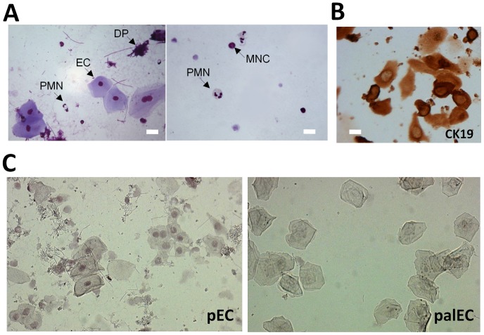
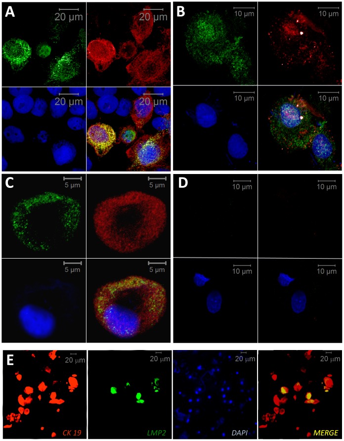
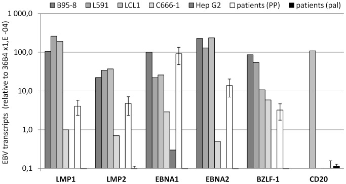
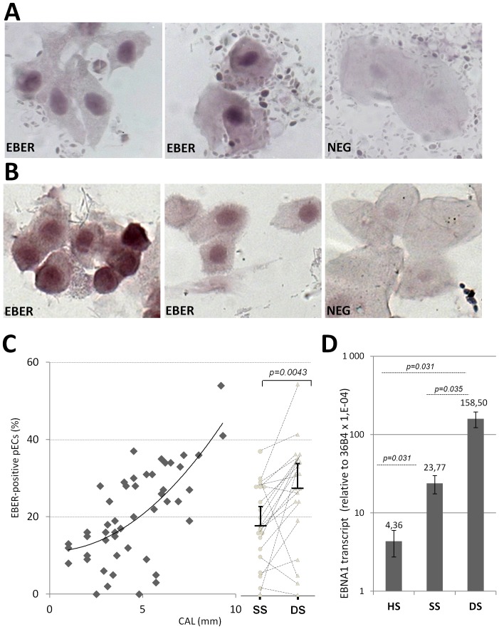
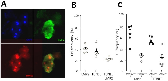
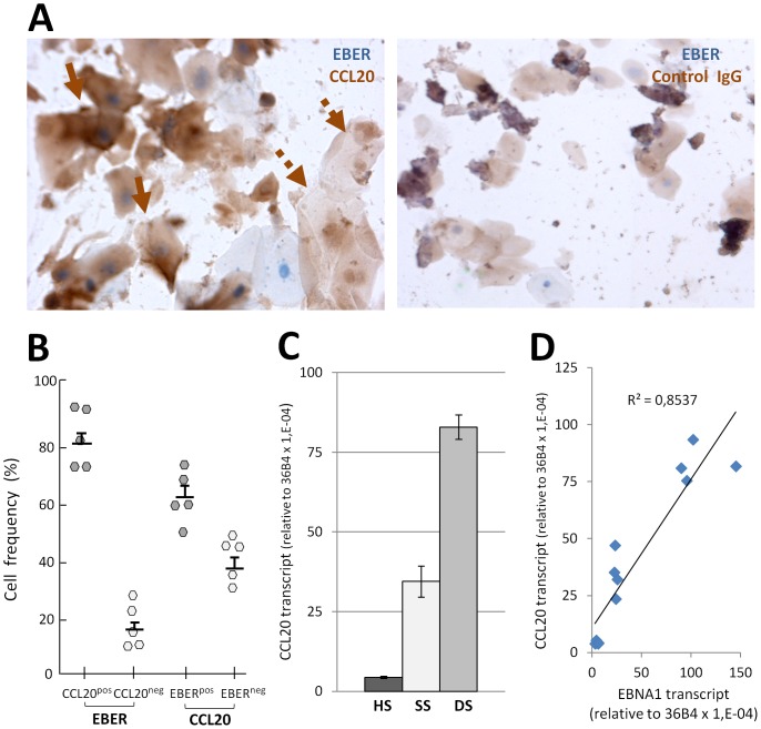
An amplifying role for oral epithelial cells (ECs) in Epstein-Barr Virus (EBV) infection has been postulated to explain oral viral shedding. However, while lytic or latent EBV infections of oro/nasopharyngeal ECs are commonly detected under pathological conditions, detection of EBV-infected ECs in healthy conditions is very rare. In this study, a simple non-surgical tissue sampling procedure was used to investigate EBV infection in the periodontal epithelium that surrounds and attaches teeth to the gingiva. Surprisingly, we observed that the gingival ECs of the periodontium (pECs) are commonly infected with EBV and may serve as an important oral reservoir of latently EBV-infected cells. We also found that the basal level of epithelial EBV-infection is significantly increased in chronic periodontitis, a common inflammatory disease that undermines the integrity of tooth-supporting tissues. Moreover, the level of EBV infection was found to correlate with disease severity. In inflamed tissues, EBV-infected pECs appear to be prone to apoptosis and to produce larger amounts of CCL20, a pivotal inflammatory chemokine that controls tissue infiltration by immune cells. Our discovery that the periodontal epithelium is a major site of latent EBV infection sheds a new light on EBV persistence in healthy carriers and on the role of this ubiquitous virus in periodontitis. Moreover, the identification of this easily accessible site of latent infection may encourage new approaches to investigate and monitor other EBV-associated disorders.


Conflict of interest statement

Competing Interests: The authors have declared that no competing interests exist.

Figures

Figure 1
Figure 1. EBER-ISH staining of liquid-based periodontal samples reveals EBV-Infected epithelial cells.
(A) Representative MGG staining of cytospin cells collected from PP samples (n = 10). Large spread epithelial-like cells (EC), polymorphonuclear leucocytes (PMN), mononuclear cells (MNC) as well as traces of dental plaque (DP) are indicated with arrows. Size bar represents 15 µM. (B) Representative CK19 staining of pECs from 5 CP patients. Coverslips were processed for IHC with DAB chromogen staining, CK19 specific staining was assessed by comparing with background staining observed using non specific mouse IgGs (not shown). Size bar represents 15 µM. (C) Nuclear EBER-ISH staining in pECs and palECs. EBER-ISH was used to detect EBER in periodontal and palatal cells sampled from 3 patients with chronic periodontitis. Two representative fields (x20) of EBER staining (EBER) are shown for the same selected CP patient with pECs (left panel) and palECs (right panel).
Figure 2
Figure 2. LMP1 and LMP2 IF co-staining in oral epithelial cells.
(A–D) Detection of LMP2 and LMP1 by IF double staining in oral epithelial cells, LMP2 (green, upper left), LMP1 (red, upper right), DAPI (blue, below left), merge (below right). (A) Oral TR146 ECs infected with Ad5F35 recombinant adenoviruses expressing LMP2 and/or ΔLMP1 were used to assess IF-based co-staining of LMP1 and LMP2 in oral epithelial cells. Results show representative specific co-detection of LMP1 and LPM2 in TR146. (B–D) pECs and palECs were sampled from 3 patients with CP and results are shown for the same selected patient. (B, C) Representative LMP1 and LMP2 co-staining in pECs. (D) Lack of LMP1 and LMP2 specific staining in palECs. TR146 were handled and fixed in a manner similar to pECs and palECs. Processing of all cell preparation with two different rat LMP2-specific antibodies (clone 14B7 clone 15F9) showed very similar results, only results obtained with clone 15F9 are presented. Specific staining was assessed by comparing with LMP1 and LMP2 negative ECs, i.e., TR146 cells infected with adenoviral vectors expressing either inactive-LMP1 (Ad5F35-ΔLMP1) or LMP2 (Ad5F35-LMP2) (not presented), and EBV-negative palECs. Background staining was also assessed in presence of unspecific rat primary antibodies (not shown). Size-bar is indicated for each panel.
Figure 3
Figure 3. Specific real-time RT-PCR quantification of EBV gene expression in periodontal material.
Whole RNA was isolated from 12 periodontal paired-samples (PP) from 6 CP patients, 3 palatal sites from 3 additional CP patients (pal), 4 EBV-infected cell lines (B95-8, L591, LCL1, C666-1) and an EBV-negative cell line (HepG2). The average levels of EBNA1, EBNA2, LMP1, LMP2, and BZLF1 EBV transcripts in clinical samples (shown as mean and SD) were compared with the expression of these EBV-genes in the different cell lines. Values obtained with an EBV-negative cell line were used to determine background level (HDLM2). The average levels of the B-cell marker CD20 transcript were also compared between LCL1, pal, and PP samples. The 36B4 housekeeping gene was used for normalization, with results presented as relative to 36B4 using a LOG10 scale. The results shown are from one representative experiment of two RT-PCR experiments performed, each in triplicate.
Figure 4
Figure 4. EBV infection increases with disease severity.
(A–B) EBER-ISH staining was performed on paired periodontal pocket DS and SS samples (n = 40) from CP patients (n = 20), and from HS samples from healthy gingival sulcus from HDs (n = 10). Two representative fields (x40) of EBER staining (EBER) is shown for (A) one selected CP patient and (B) for one selected HD. Negative controls (NEG) were processed using a random PNA probe, and for each individual, the same cell sampling was used for positive and negative staining. (C) EBER-ISH-based determination of the frequency of EBV-infected pECs (EBERpos) in periodontal samples from healthy sites (HS), swallow sites (SS), and deep sites (DS). The graph (left part) shows the tendency curve of EBERpos pECs and the clinical attachment level (CAL) in 40 paired-samples from 20 CP patients and 10 samples from healthy donors (same as in A and B). The dot-plot analysis (right part) shows the comparative analysis of the frequency of EBERpos pECs in paired-samples (SS circles, DS triangles) collected from 20 CP patients (same samples as on graft). (D) The average levels of EBNA1 transcript were also compared between whole RNA from HS (n = 10) and 12 paired-samples (SS and DS, n = 6 for each) from 6 CP patients (same samples as in C). The 36B4 housekeeping gene was used for normalization, with results presented as relative to 36B4 using a LOG10 scale. The p values were calculated using Wilcoxon signed-rank test.
Figure 5
Figure 5. Infection of periodontal epithelial cells by EBV is associated with apoptotic cell death.
TUNEL assay associated with IF LMP2 detection was used to identify apoptotic cells and EBV-infected cells in DS samples from 5 CP patients. (A) Representative double fluorescent staining with antibodies specific for LMP2 (upper right panel), TUNEL staining (below left panel), with the latter two merged (below right panel). The upper-left panel shows DAPI staining of nuclei in the same field (x20). (B) Quantitative evaluations of A. Cell counting of LMP2pos (LMP2), TUNELpos (TUNEL) and double positive (LMP2 & TUNEL) pECs (n = 5). Data represent the frequency of positive pECs identified in each category. (C) Calculations from B showing the frequency of apoptotic pECs (TUNELpos) among EBV-infected pECs (LMP2) (left part) and the frequency of EBV-infected pECs (LMP2pos) among apoptotic pECs (TUNEL) (right part). Mean and standard deviation is shown for each group.
Figure 6
Figure 6. Production of the inflammatory chemokine CCL20 by EBV-infected periodontal epithelial cells.
(A) Left panel shows representative CCL20 staining of pECs from 5 CP patients (DS samples) (HIS, x40) of EBV-infected cells (EBER-ISH) (solid arrows) and of EBV-negative pECs (dotted arrows). Right panel shows background staining observed using nonspecific goat IgGs. (B) Quantitation of (A) (n = 5). The frequency (%) of CCL20pos pECs among EBV-infected pECs (EBER, left part) and of EBERpos pECs among CCL20-producing pECs (CCL20, right part) is shown. Mean and standard deviations are shown for each group. (C) Real-time RT-PCR quantification of CCL20 transcripts in whole RNA isolated from 12-paired RNA samples from 6 CP patients (SS n = 6, DS n = 6) and 2 RNA samples from healthy donors. (D) Real-time RT-PCR quantification of EBNA1 and CCL20 transcripts in whole RNA isolated from CP patients (n = 8) and from healthy donors (n = 2). Graph shows the linear tendency curve of CCL20 related to EBNA1. Simple linear regression analysis showed a positive correlation. Data are representative of 2 independent experiments, each performed in triplicate. The 36B4 housekeeping gene was used for normalization. The results are presented as relative to 36B4 (×10−6).

Similar articles

Cited by

References

    1. Young LS, Rickinson AB (2004) Epstein-Barr virus: 40 years on. Nat Rev Cancer 4: 757–768. - PubMed
    1. Chen MR (2011) Epstein-barr virus, the immune system, and associated diseases. Front Microbiol 2: 5. - PMC - PubMed
    1. Raab-Traub N (2012) Novel mechanisms of EBV-induced oncogenesis. Curr Opin Virol 2: 453–458. - PMC - PubMed
    1. Hadinoto V, Shapiro M, Sun CC, Thorley-Lawson DA (2009) The dynamics of EBV shedding implicate a central role for epithelial cells in amplifying viral output. PLoS Pathog 5: e1000496. - PMC - PubMed
    1. Sixbey JW, Nedrud JG, Raab-Traub N, Hanes RA, Pagano JS (1984) Epstein-Barr virus replication in oropharyngeal epithelial cells. N Engl J Med 310: 1225–1230. - PubMed

Publication types

LinkOut - more resources