

Characterization of neuronal populations in the human trigeminal ganglion and their association with latent herpes simplex virus-1 infection
- PMID: 24367603
- PMCID: PMC3868591
- DOI: 10.1371/journal.pone.0083603
Characterization of neuronal populations in the human trigeminal ganglion and their association with latent herpes simplex virus-1 infection
Abstract
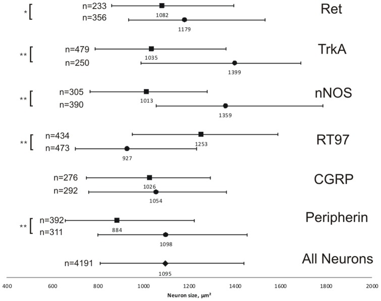
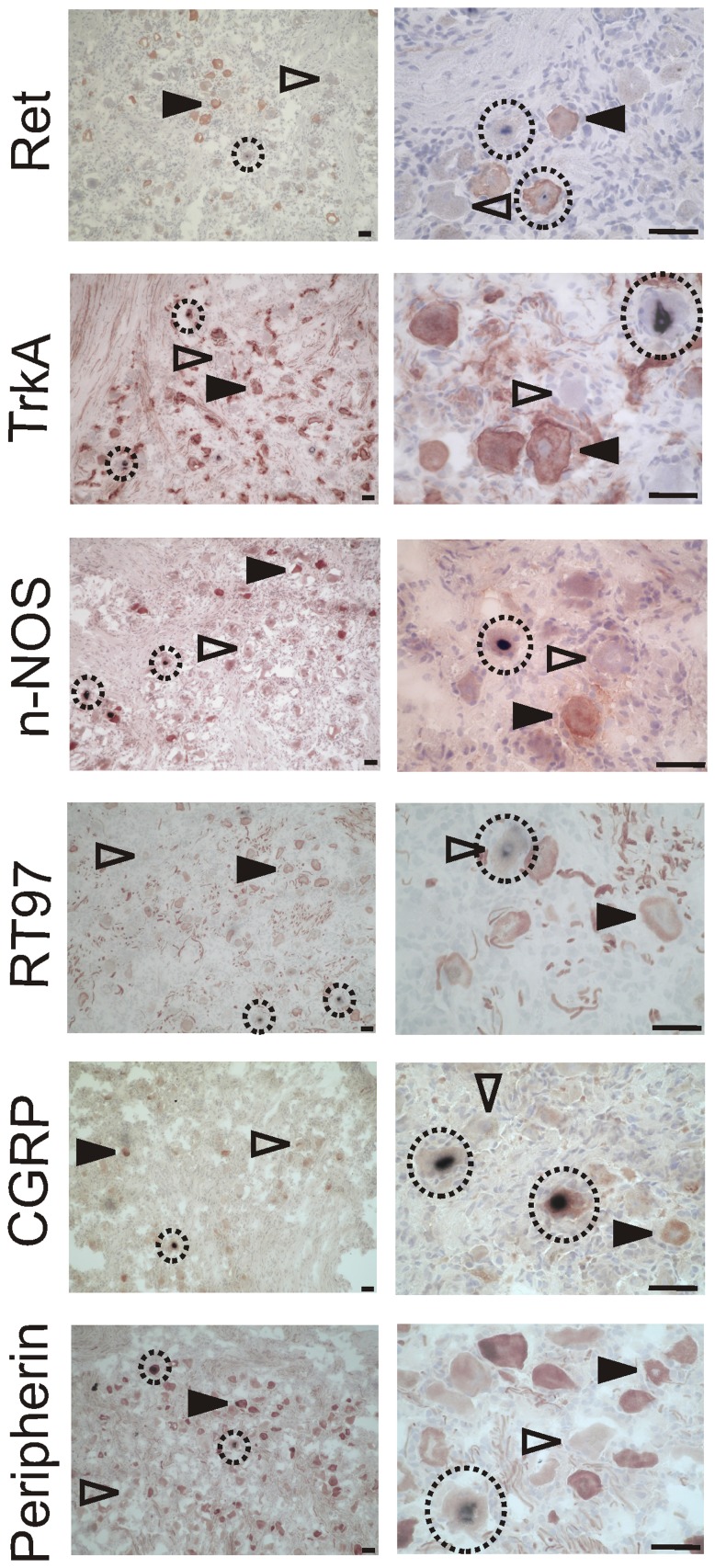
Following primary infection Herpes simplex virus-1 (HSV-1) establishes lifelong latency in the neurons of human sensory ganglia. Upon reactivation HSV-1 can cause neurological diseases such as facial palsy, vestibular neuritis or encephalitis. Certain populations of sensory neurons have been shown to be more susceptible to latent infection in the animal model, but this has not been addressed in human tissue. In the present study, trigeminal ganglion (TG) neurons expressing six neuronal marker proteins were characterized, based on staining with antibodies against the GDNF family ligand receptor Ret, the high-affinity nerve growth factor receptor TrkA, neuronal nitric oxide synthase (nNOS), the antibody RT97 against 200 kDa neurofilament, calcitonin gene-related peptide and peripherin. The frequencies of marker-positive neurons and their average neuronal sizes were assessed, with TrkA-positive (61.82%) neurons being the most abundant, and Ret-positive (26.93%) the least prevalent. Neurons positive with the antibody RT97 (1253 µm(2)) were the largest, and those stained against peripherin (884 µm(2)) were the smallest. Dual immunofluorescence revealed at least a 4.5% overlap for every tested marker combination, with overlap for the combinations TrkA/Ret, TrkA/RT97 and Ret/nNOS lower, and the overlap between Ret/CGRP being higher than would be expected by chance. With respect to latent HSV-1 infection, latency associated transcripts (LAT) were detected using in situ hybridization (ISH) in neurons expressing each of the marker proteins. In contrast to the mouse model, co-localization with neuronal markers Ret or CGRP mirrored the magnitude of these neuron populations, whereas for the other four neuronal markers fewer marker-positive cells were also LAT-ISH+. Ret and CGRP are both known to label neurons related to pain signaling.
Conflict of interest statement
Figures

Similar articles
-
Herpes Simplex Virus 1 Infection of Tree Shrews Differs from That of Mice in the Severity of Acute Infection and Viral Transcription in the Peripheral Nervous System.J Virol. 2015 Oct 28;90(2):790-804. doi: 10.1128/JVI.02258-15. Print 2016 Jan 15. J Virol. 2015. PMID: 26512084 Free PMC article.
-
Two alphaherpesvirus latency-associated gene products influence calcitonin gene-related peptide levels in rat trigeminal neurons.Neurobiol Dis. 2007 Mar;25(3):553-60. doi: 10.1016/j.nbd.2006.10.016. Epub 2006 Dec 20. Neurobiol Dis. 2007. PMID: 17184994 Free PMC article.
-
A5-positive primary sensory neurons are nonpermissive for productive infection with herpes simplex virus 1 in vitro.J Virol. 2011 Jul;85(13):6669-77. doi: 10.1128/JVI.00204-11. Epub 2011 Apr 20. J Virol. 2011. PMID: 21507969 Free PMC article.
-
A comparison of herpes simplex virus type 1 and varicella-zoster virus latency and reactivation.J Gen Virol. 2015 Jul;96(Pt 7):1581-602. doi: 10.1099/vir.0.000128. Epub 2015 Mar 20. J Gen Virol. 2015. PMID: 25794504 Free PMC article. Review.
-
Control of HSV-1 latency in human trigeminal ganglia--current overview.J Neurovirol. 2011 Dec;17(6):518-27. doi: 10.1007/s13365-011-0063-0. Epub 2011 Dec 3. J Neurovirol. 2011. PMID: 22139603 Review.
Cited by
-
An Immortalized Human Dorsal Root Ganglion Cell Line Provides a Novel Context To Study Herpes Simplex Virus 1 Latency and Reactivation.J Virol. 2017 May 26;91(12):e00080-17. doi: 10.1128/JVI.00080-17. Print 2017 Jun 15. J Virol. 2017. PMID: 28404842 Free PMC article.
-
Chromatin-mediated epigenetic regulation of HSV-1 transcription as a potential target in antiviral therapy.Antiviral Res. 2021 Aug;192:105103. doi: 10.1016/j.antiviral.2021.105103. Epub 2021 Jun 1. Antiviral Res. 2021. PMID: 34082058 Free PMC article. Review.
-
Novel mutations in UL24 and gH rescue efficient infection of an HSV vector retargeted to TrkA.Mol Ther Methods Clin Dev. 2023 Jul 3;30:208-220. doi: 10.1016/j.omtm.2023.06.012. eCollection 2023 Sep 14. Mol Ther Methods Clin Dev. 2023. PMID: 37519407 Free PMC article.
-
Herpes Simplex Virus Establishment, Maintenance, and Reactivation: In Vitro Modeling of Latency.Pathogens. 2017 Jun 23;6(3):28. doi: 10.3390/pathogens6030028. Pathogens. 2017. PMID: 28644417 Free PMC article. Review.
-
Latent herpes simplex virus 1 infection does not induce apoptosis in human trigeminal Ganglia.J Virol. 2015 May;89(10):5747-50. doi: 10.1128/JVI.03481-14. Epub 2015 Mar 11. J Virol. 2015. PMID: 25762734 Free PMC article.
References
-
- Griffin BD, Verweij MC, Wiertz EJ (2010) Herpesviruses and immunity: the art of evasion. VetMicrobiol 143: 89–100. - PubMed
-
- Held K, Derfuss T (2011) Control of HSV-1 latency in human trigeminal ganglia-current overview. JNeurovirol 17: 518–527. - PubMed
-
- Stevens JG, Wagner EK, vi-Rao GB, Cook ML, Feldman LT (1987) RNA complementary to a herpesvirus alpha gene mRNA is prominent in latently infected neurons. Science 235: 1056–1059. - PubMed
Publication types
MeSH terms
Substances
LinkOut - more resources
Full Text Sources
Other Literature Sources
Research Materials
Miscellaneous
