. 2013 Dec 19;8(12):e84147.
doi: 10.1371/journal.pone.0084147. eCollection 2013.

Structural context of disease-associated mutations and putative mechanism of autoinhibition revealed by X-ray crystallographic analysis of the EZH2-SET domain

Affiliations

Structural context of disease-associated mutations and putative mechanism of autoinhibition revealed by X-ray crystallographic analysis of the EZH2-SET domain

Stephen Antonysamy et al. PLoS One. .

Abstract

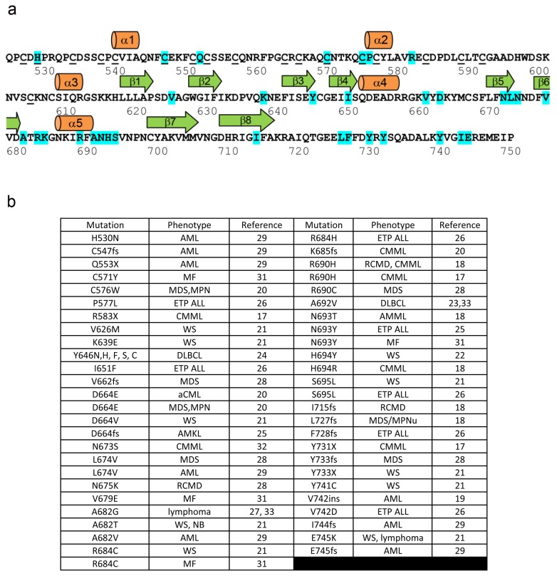
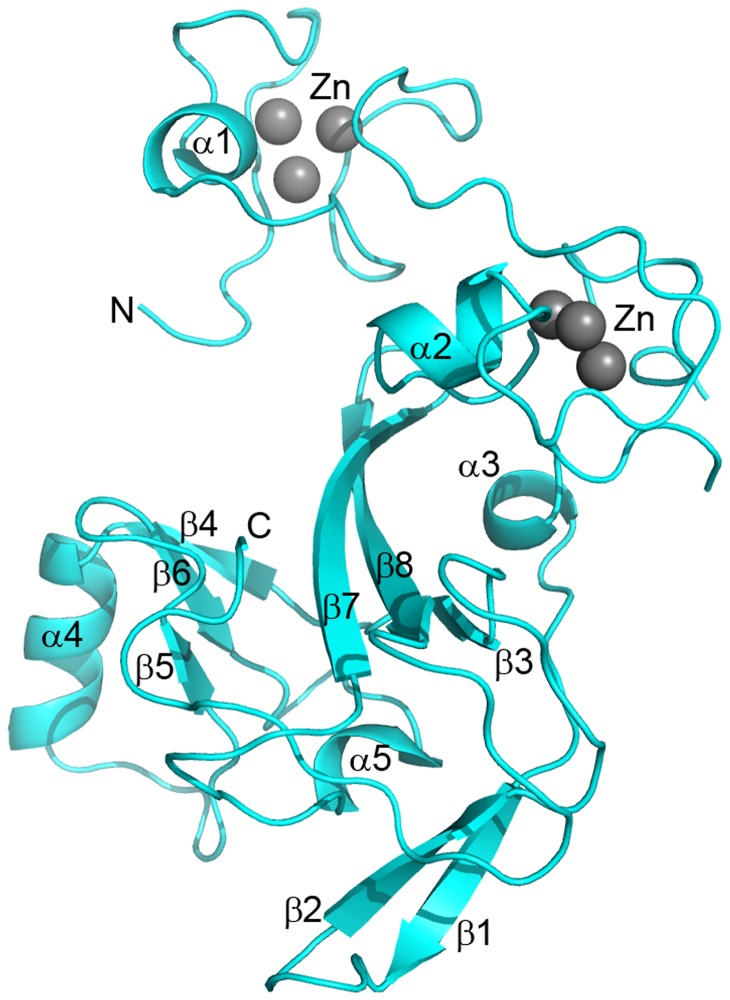
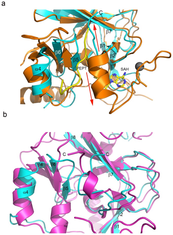
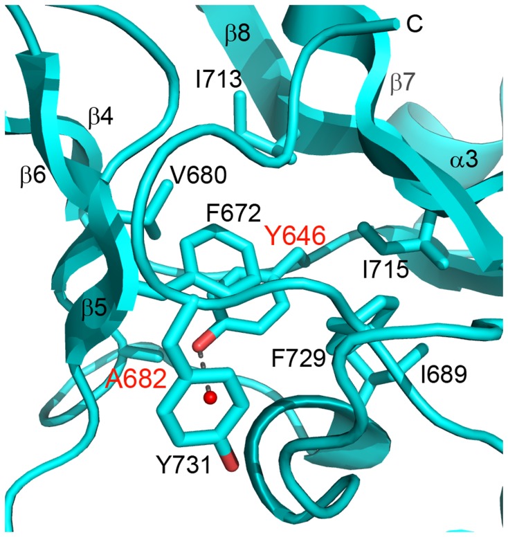
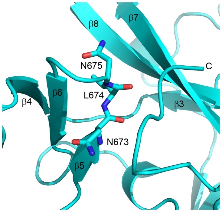
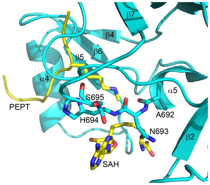
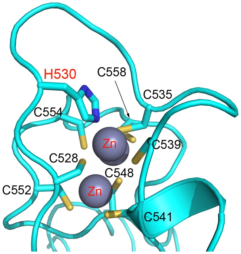
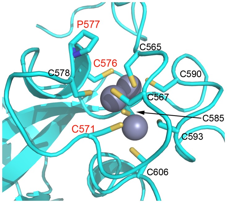
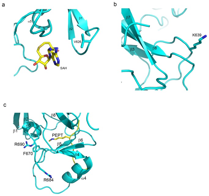
The enhancer-of-zeste homolog 2 (EZH2) gene product is an 87 kDa polycomb group (PcG) protein containing a C-terminal methyltransferase SET domain. EZH2, along with binding partners, i.e., EED and SUZ12, upon which it is dependent for activity forms the core of the polycomb repressive complex 2 (PRC2). PRC2 regulates gene silencing by catalyzing the methylation of histone H3 at lysine 27. Both overexpression and mutation of EZH2 are associated with the incidence and aggressiveness of various cancers. The novel crystal structure of the SET domain was determined in order to understand disease-associated EZH2 mutations and derive an explanation for its inactivity independent of complex formation. The 2.00 Å crystal structure reveals that, in its uncomplexed form, the EZH2 C-terminus folds back into the active site blocking engagement with substrate. Furthermore, the S-adenosyl-L-methionine (SAM) binding pocket observed in the crystal structure of homologous SET domains is notably absent. This suggests that a conformational change in the EZH2 SET domain, dependent upon complex formation, must take place for cofactor and substrate binding activities to be recapitulated. In addition, the data provide a structural context for clinically significant mutations found in the EZH2 SET domain.


Conflict of interest statement

Competing Interests: SA, BC, ZD, TG, FZ, AZ, SA, LR, MR and JGL are employed by Lilly Biotechnology Center. There are no patents, products in development or marketed products to declare. This does not alter the authors' adherence to all the PLOS ONE policies on sharing data and materials, as detailed online in the guide for authors.

Figures

Figure 1
Figure 1. Mutations of the EZH2-SET domain.
(a) The amino acid sequence of the EZH2-SET domain is shown with the secondary structure assignments depicted above. Residues which coordinate zinc are underlined. Mutated amino acids identified in association with disease are highlighted cyan. The specific mutations are annotated below with the disease-associated with each mutation, the nature of the mutation, and the reference in which the mutation is described. The sequence is numbered in accordance with EZH2 isoform A and the numbering for some mutations has been transposed from the original references so that all mutations can be referred to relative to the same sequence. (b) Details of mutations delineated in Figure 1a. (Abbreviations: AMKL, acute megakaryoblastic leukemia; AML, acute myeloid leukemia; AMML, acute myelomonocytic leukemia; CMML, chronic myelomonocytic leukemia; DLBCL, diffuse large B-cell lymphoma; ETP ALL, early T-cell precursor acute lymphoblastic leukemia; MDS, myelodysplastic syndrome; MPN, myeloproliferative neoplasms; MPNu, myeloproliferative neoplasms unclassifiable; NB, neuroblastoma; MF, myelofibrosis; RCMD, refractory cytopenia with multilineage dysplasia; WS, Weaver Syndrome; fs, frameshift; X, nonsense).
Figure 2
Figure 2. The crystal structure of the EZH2-SET domain.
The crystal structure of the EZH2-SET domain is represented as a ribbon model colored cyan. Bound zinc atoms are represented by spheres and colored gray. Secondary structure elements are labeled. The crystal structure contains two N-terminal zinc binding domains each of which binds three zinc molecules. The core of the domain is formed by β-strands 3, 7, and 8. This core is flanked on one side by a three stranded antiparallel β-sheet (β-4, β-6, β-5) and an accessory α-helix (α-4) and bounded below by α-5, β-1, and β-2. The C-terminus turns upward insinuating through the substrate binding cleft between the β-5/β-6 loop and β-7.
Figure 3
Figure 3. The EZH2-SET domain C-terminus partially occupies the substrate binding groove.
(a) The EZH2-SET (cyan) and hEHMT1-SET (orange) (PDB ID:3HNA) domains are superimposed and represented by ribbons. Zinc bound by hEHMT-SET is represented as a gray sphere. The substrate peptide bound by hEHMT1 is a yellow ribbon with the lysine side chain represented as sticks. The SAH bound by hEHMT1-SET is represented by sticks and colored by atom (carbon, yellow; oxygen, red; nitrogen, blue; sulfur, sienna). The C-terminal tail of EZH2-SET turns upwards and occupies the upper region of the substrate binding groove (red arrow pointing up). The C-terminus of hEHMT1-SET turns downward (red arrow pointing downward) forming the lower lobe of the cofactor binding pocket and coordinating one zinc atom. (b) The EZH2-SET (cyan) and SUV39H2 SET domain (magenta) (PDB ID:2R3A) crystal structures are superimposed and represented by ribbons. The C-termini in both structures occupy the collapsed substrate binding groove.
Figure 4
Figure 4. Structural context of Y646 and A682 mutations.
The crystal structure of the EZH2-SET domain is represented as a ribbon model (cyan). Side chains are represented as sticks colored by atom (carbon, cyan; oxygen, red). Secondary structure elements are labeled. Y646 is completely buried in a hydrophobic cluster except for the solvent exposed tip of the phenyl ring where the phenyl oxygen forms a hydrogen bond with a water molecule. A682 is packed against the Y646 side chain distal to the catalytic site. Mutation of A682 likely indirectly affects substrate specificity by influencing the conformation of Y646 in the active state. Y646 and A682 mutations have been found in lymphoma [24,27,33], WS [21], and AML [29].
Figure 5
Figure 5. Mutations in the β-5/β-6 loop of EZH2-SET are contiguous with the putative substrate binding cleft.
The crystal structure of the EZH2-SET domain is represented as a ribbon model (cyan). Side chains are represented as sticks colored by atom (carbon, cyan; oxygen, red; nitrogen, blue). Secondary structure elements are labeled. N673, L674, and N675 all interact directly with the C-terminal tail which occupies the substrate binding groove. Mutation of these residues could potentially affect substrate binding in the active state as well as the transition from the inactive to active state. An N673S mutation has been identified in CMML [32]. L674V mutations have been found in both MDS [28] and AML [29]. An N675K mutation was discovered in RCMD [28].
Figure 6
Figure 6. EZH2-SET mutations that may affect cofactor binding.
The crystal structure of the EZH2-SET domain is represented as a ribbon model (cyan) with the hypothetical positions of cofactor and substrate (sticks colored by atom: carbon, yellow; oxygen, red; nitrogen, blue) extracted from the superimposed structure of EHMT1/PEPTIDE/SAH (PDB ID: 3HNA). EZH2-SET amino acid side chains are represented as sticks colored by atom (C, cyan; O, red; N, nitrogen). Secondary structure elements are labeled. Mutations at positions A692 {(DLBCL) (A>V) [23,33]}, N693 {(AMML) (N>T) [18]; (ETP ALL) (N>Y) [26]; (MF) (N>Y) [31]}, and H694 {(WS) (H>Y) [22]; (CMML) (H>R) [18]} have been found in association with numerous diseases. All three mutations likely affect cofactor binding. An S695L mutation was identified in both WS [21] and ETP ALL [26]. This mutation may affect cofactor and substrate binding indirectly by influencing the conformation of residues in direct contact with these ligands.
Figure 7
Figure 7. Location of mutation in the first zinc binding domain of EZH2-SET.
EZH2-SET (cyan) is represented as a ribbon diagram with zinc atoms shown as gray spheres and side chain represented as sticks (carbon, cyan; nitrogen, blue; sulfur, sienna) A H530N mutation was identified in AML [29]. This mutation disrupts coordination of zinc in the first zinc binding domain likely having a strong destabilizing effect on the protein.
Figure 8
Figure 8. Location of mutation in the second zinc binding domain of EZH2-SET.
EZH2-SET (cyan) is represented as a ribbon diagram with zinc atoms shown as gray spheres and side chain represented as sticks (carbon, cyan; nitrogen, blue; sulfur, sienna) A C571Y mutation was identified in MF [31] and a C576W mutation was identified in MDS [20]. These mutations disrupt coordination of zinc in the second zinc binding domain likely destabilizing the protein. Additionally, a P577L mutation was observed in ETP ALL [26].
Figure 9
Figure 9. Additional disease-associated mutations outside this active site.
The crystal structure of the EZH2-SET domain is represented as a ribbon model (cyan) with the hypothetical positions of cofactor and substrate (sticks colored by atom: carbon, yellow; oxygen, red; nitrogen, blue) extracted from the superimposed structure of EHMT1/PEPTIDE/SAH (PDB ID: 3HNA). EZH2-SET amino acid side chains are represented as sticks colored by atom (carbon, cyan; oxygen, red; nitrogen, blue). Secondary structure elements are labeled. (a) A V626M mutation was identified in WS [21]. This residue is located in the loop connecting β-1 and β-2 and may indirectly affect cofactor binding. (b) A K639E mutation was identified in WS [21]. This residue is located in the loop connecting β-2 and β-3. (c) R684 mutations were identified in WS (R>C) [21], ETP ALL (R>H) [26], and MF (R>C) [31]. This residue does not participate in cofactor or substrate binding; however, its side chain does pack against α-4 which does participate in substrate binding in homologous SET domains. R690 mutations were identified in CMML (R>H) [17,18] and MDS (R>C) [28]. This residue packs against F670 which in homologous SET domains contributes to the substrate lysine binding channel.

References

    1. Diffley JF, Stillman B (1989) Similarity between the transcriptional silencer binding proteins ABF1 and RAP1. Science 246: 1034-1038. doi:10.1126/science.2511628. PubMed: 2511628. - DOI - PubMed
    1. Wedeen C, Harding K, Levine M (1986) Spatial regulation of Antennapedia and bithorax gene expression by the Polycomb locus in Drosophila. Cell 44: 739-748. doi:10.1016/0092-8674(86)90840-8. PubMed: 3081265. - DOI - PubMed
    1. Simon J, Chiang A, Bender W, Shimell MJ, O'Connor M (1993) Elements of the Drosophila bithorax complex that mediate repression by Polycomb group products. Dev Biol 158(1): 131-144. doi:10.1006/dbio.1993.1174. PubMed: 8101171. - DOI - PubMed
    1. Umlauf D, Goto Y, Cao R, Cerqueira F, Wagschal A et al. (2004) Imprinting along the Kcnq1 domain on mouse chromosome 7 involves repressive histone methylation and recruitment of Polycomb group complexes. Nat Genet 36: 1296-3100. doi:10.1038/ng1467. PubMed: 15516932. - DOI - PubMed
    1. Plath K, Fang J, Mlynarczyk-Evans SK, Cao R, Worringer KA et al. (2003) Role of histone H3 lysine 27 methylation in X inactivation. Science 300: 131-135. doi:10.1126/science.1084274. PubMed: 12649488. - DOI - PubMed

Publication types

Substances