. 2013 Dec 19;8(12):e84269.
doi: 10.1371/journal.pone.0084269. eCollection 2013.

Exposure to inflammatory cytokines IL-1β and TNFα induces compromise and death of astrocytes; implications for chronic neuroinflammation

Affiliations

Exposure to inflammatory cytokines IL-1β and TNFα induces compromise and death of astrocytes; implications for chronic neuroinflammation

Christa van Kralingen et al. PLoS One. .

Abstract

Background: Astrocytes have critical roles in the human CNS in health and disease. They provide trophic support to neurons and are innate-immune cells with keys roles during states-of-inflammation. In addition, they have integral functions associated with maintaining the integrity of the blood-brain barrier.

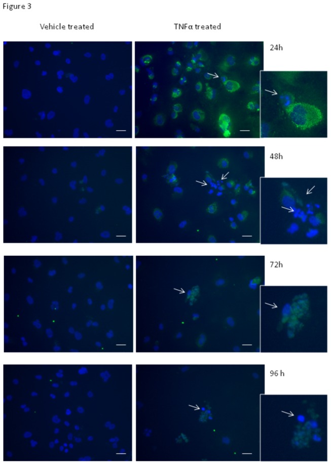
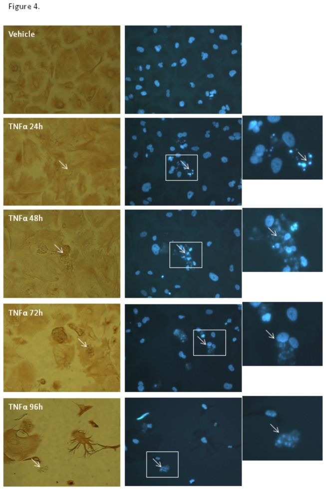
Methods: We have used cytometric bead arrays and xCELLigence technology to monitor the to monitor the inflammatory response profiles and astrocyte compromise in real-time under various inflammatory conditions. Responses were compared to a variety of inflammatory cytokines known to be released in the CNS during neuroinflammation. Astrocyte compromise measured by xCELLigence was confirmed using ATP measurements, cleaved caspase 3 expression, assessment of nuclear morphology and cell death.

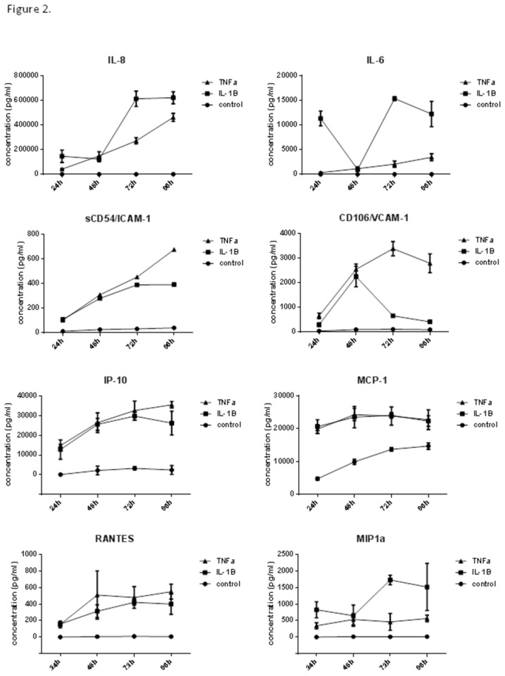
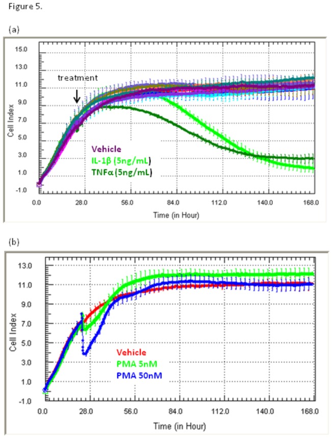
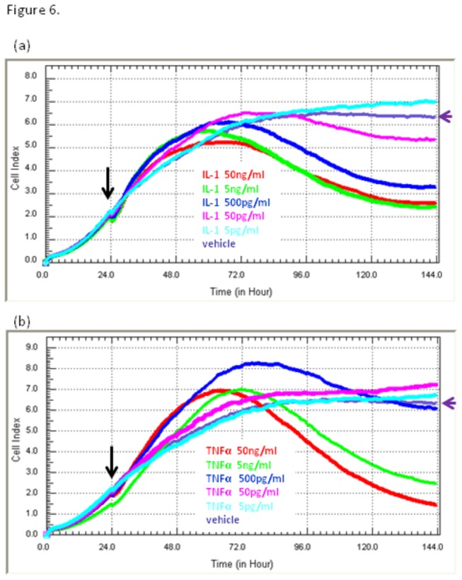
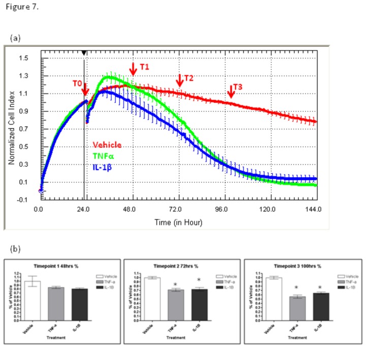
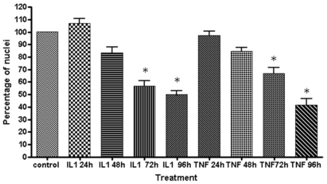
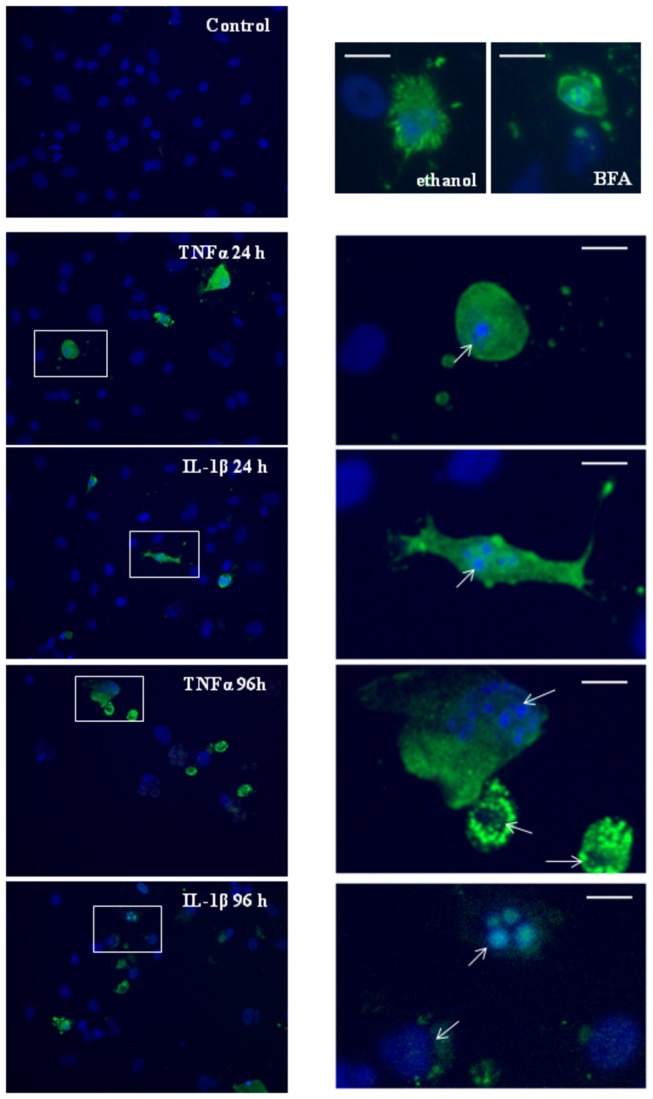
Results: Inflammatory activation (IL-1β or TNFα) of astrocytes results in the transient production of key inflammatory mediators including IL-6, cell surface adhesion molecules, and various leukocyte chemoattractants. Following this phase, the NT2-astrocytes progressively become compromised, which is indicated by a loss of adhesion, appearance of apoptotic nuclei and reduction in ATP levels, followed by DEATH. The earliest signs of astrocyte compromise were observed between 24-48 h post cytokine treatment. However, significant cell loss was not observed until at least 72 h, where there was also an increase in the expression of cleaved-caspase 3. By 96 hours approximately 50% of the astrocytes were dead, with many of the remaining showing signs of compromise too. Numerous other inflammatory factors were tested, however these effects were only observed with IL-1β or TNFα treatment.

Conclusions: Here we reveal direct sensitivity to mediators of the inflammatory milieu. We highlight the power of xCELLigence technology for revealing the early progressive compromise of the astrocytes, which occurs 24-48 hours prior to substantive cell loss. Death induced by IL-1β or TNFα is relevant clinically as these two cytokines are produced by various peripheral tissues and by resident brain cells.


Conflict of interest statement

Competing Interests: The authors have declared that no competing interests exist.

Figures

Figure 1
Figure 1. Expression of GFAP by NT2-astrocyte cultures.
GFAP and vimentin expression in NT2 astrocytes. Images show the varied morphology of the (a) GFAP and (b) vimentin stained NT2 astrocytes. Most are strongly positive for GFAP staining, with some having highly pronounced intermediate-filament structures radiating from around centre of the cell body. Note that GFAP filaments are not present throughout the entirety of the astrocyte cytoplasm. The vimentin filaments are typically much finer are present throughout the astrocytic volume. Scale bars represents 20 µm.
Figure 2
Figure 2. Time-course of astrocyte inflammatory response to IL-1β and TNFα (cytokine secretion).
The time-course of cytokine secretion following IL-1β and TNFα treatment shows pronounced inflammatory activation within the initial 48 hour period. In addition, the time-course reveals differential secretion of IL-6, MIP1α, IL-8 and VCAM liberation. The concentration of cytokine was measured using multiplex cytometric bead array. The temporal nature of the data is very powerful especially for IL-6 and VCAM-1, which both show pronounced time-dependent dynamic reductions in concentration, which would not have been identified with a single time-point analysis. All concentrations are represented as pg/mL. Data show the mean +- SE (N=4).
Figure 3
Figure 3. Analysis of intracellular expression of IP-10 following astrocyte activation.
Induction of IP-10 and intracellular localisation following treatment with IL-1β or TNFα reveals a subpopulation of NT2-astrocyte which fail to produce IP-10 and have severely compromised nuclei. Cells were stimulated with either IL-1β (data not shown) or TNFα (5 ng/mL), or control media (vehicle) for 24 hours to 96 hours. IP-10 was absent in vehicle treated cells. Intracellular staining for IP-10 (green) shows elevated IP-10 levels 24 hours after cytokine addition in the majority of NT2-astrocytes (>95%). 48 hours after cytokine addition, there is a marked increase in the number of astrocytes with compromised nuclei (severely disintegrated or multi-lobed; abnormal). Detailed inspection reveals that these astrocytes have little or no IP-10 staining. In contrast, astrocytes with healthy looking nuclei typically have an abundance of chemokine present (especially at 24-48hrs). Note the smaller condensed nuclei and multi-lobed nuclei (highlighted by white arrows) present in the cytokine treated astrocytes. The response to IL-1β was very similar (data not shown). Scale bars are 40 µm.
Figure 4
Figure 4. TNFα and IL-1β activation of astrocytes induces nuclear disintegration and cell compromise.
Analysis of nuclear and cellular morphology further corroborates the earlier observation that activation with TNFα or IL-1β induces astrocyte compromise. Cells were stained with Hoechst and vimentin to reveal integrity of the nucleus and cytoplasm, respectively. The white arrow points to the same compromised cells for comparison between the vimentin and nuclear staining. The enlarged region of interest within the white box shows an example of a highly compromised astrocyte with a severely disintegrated nucleus. Data are shown for TNFα treatment. Note also the loss of following cytokine treatment of the astrocytes with time (72 and 96 hours).
Figure 5
Figure 5. Astrocyte adhesion is compromised by treatment with TNFα and IL-1β, but not by other pro-inflammatory mediators.
NT2-astrocytes were treated with a range of pro-inflammatory mediators including IL-1β and TNFα (5 ng/mL) 24 hours after seeding into E96 xCELLigence biosensor plates (indicated by black arrow). The xCELLigence biosensor measures net adhesion (Cell Index) of living cells. (a) The individual profiles for IL-1β (green) and TNFα (dark green) reveal that total adhesion levels decline progressively with time. On set of loss usually occurs within the first 24-48 hours after stimulation with IL-1β or TNFα. This does not occur in cells treated with the following chemokines (all used at 50ng mL); SDF-1 (red), IL-8 (aqua), IP-10 (brown), MCP-1 (tan), MIP1α (blue), MIP1β (pink), RANTES (orange) or control media (purple). Furthermore, the cytokines IFNγ, IL-2 and IL-4 had no effect on astrocyte survival (curves not shown). The lower panel (b) shows the response to PMA (5nM and 50nM), where there is an immediate transient reduction in adhesion (Cell Index). This transient reduction rebounds back to the vehicle Cell Index values, indicating that the response is acute, but is not cytotoxic. The plotted data shows the mean +- SD (n=6 wells from a representative experiment). The progressive loss of adhesion following IL-1β and TNFα has been observed in more than 20 independent xCELLigence experiments.
Figure 6
Figure 6. Compromise of astrocyte adhesion induced by TNFα and IL-1β occurs in a concentration-dependent manner.
NT2-astrocytes were stimulated with a range of concentrations of IL-1β and TNFα (50 ng/mL to 5pg/mL) 24 hours after seeding into E96 plates (indicated by black arrow). The individual pharmacological profiles for (a) IL-1β and (b) TNFα reveal that the on-set of compromise (decline in adhesion levels, Cell Index) is concentration dependent. The cytokine concentrations are colour-coded to highlight the subtle difference in the responsiveness to IL-1β or TNFα. xCELLigence revealed that the responses were similar but not identical. The black arrow signifies addition of cytokines and the purple arrow labels the control treated cells for reference (purple curve). Each curve is the mean of 6 treatments. The SD bars have been omitted for simplification of the figure.
Figure 7
Figure 7. Progressive loss of adhesion correlates with astrocyte compromise.
NT2 astrocytes were prepared in parallel for quantification of ATP levels (mitochondrial function) and measurement of loss-of-adhesion with xCELLigence. The response to IL-1β and TNFα (5 ng/mL) was monitored in real-time to identify the time-points for conducting the ATP assays, which were conducted using parallel plates. Cytokines were added at T0. The time point 1 (T1) represents the early loss of adhesion, T2 represents significant reduction in Cell Index (i.e. reduction in astrocyte adhesion), T3 represents major sustained reduction in Cell Index. (b) quantification of astrocytes ATP levels using parallel plates terminated at the indicated time-points (T1 to T3). Significance level is P < 0.05. Data are from a single experiment, which is representative of 4 independent experiments.
Figure 8
Figure 8. IL-1β and TNFα induced astrocyte death.
Astrocytes were stimulated across a 96-hour time course to assess the extent of cell loss following IL-1β and TNFα treatment. Cell numbers were quantified by counting Hoechst stained nuclei. However, healthy and compromised nuclei were not discriminated by the automated software used. Data are from a single experiment quantified from 6 fields of view. Data are representative of three independent experiments. Significance where P < 0.05* .
Figure 9
Figure 9. Induction of cleaved caspase-3 and nuclear disintegration following IL-1β and TNFα treatment.
Expression of cleaved caspase 3 was investigated to assess whether it was present during the early stages of cytokine-induced compromise. Panels show cleaved caspase 3 expression in green and nuclei in blue. The white boxes highlight cleaved caspase 3 positive astrocytes, with the enlarged panel showing the severe disintegration of the nucleus. In many cases, the nucleus was visible only as a small (<5 µm) intense vesicle or series of vesicle like structures, consistent with apoptotic nuclei. Cleaved caspase 3 was not detected in vehicle treated cells. Ethanol (1%) and Brefeldin A (BFA 10 µM) were used as positive controls [46,47]. The scale bar represents 20 µm for reference. Data representative of 4 independent experiments.

Similar articles

Cited by

References

    1. Oberheim NA, Goldman SA, Nedergaard M (2012) Heterogeneity of astrocytic form and function. Methods Mol Biol 814: 23-45. doi:10.1007/978-1-61779-452-0_3. PubMed: 22144298. - DOI - PMC - PubMed
    1. Ransohoff RM, Engelhardt B (2012) The anatomical and cellular basis of immune surveillance in the central nervous system. Nat Rev Immunol 12: 623-635. doi:10.1038/nri3265. PubMed: 22903150. - DOI - PubMed
    1. Ransohoff RM, Brown MA (2012) Innate immunity in the central nervous system. J Clin Invest 122: 1164-1171. doi:10.1172/JCI58644. PubMed: 22466658. - DOI - PMC - PubMed
    1. Cartier L, Hartley O, Dubois-Dauphin M, Krause KH (2005) Chemokine receptors in the central nervous system: role in brain inflammation and neurodegenerative diseases. Brain research Brain research reviews 48: 16-42. - PubMed
    1. Krasowska-Zoladek A, Banaszewska M, Kraszpulski M, Konat GW (2007) Kinetics of inflammatory response of astrocytes induced by TLR 3 and TLR4 ligation. J Neurosci Res 85: 205-212. doi:10.1002/jnr.21088. PubMed: 17061254. - DOI - PubMed

Publication types

MeSH terms