. 2013 Dec 19;8(12):e84980.
doi: 10.1371/journal.pone.0084980. eCollection 2013.

Borrelia burgdorferi elicited-IL-10 suppresses the production of inflammatory mediators, phagocytosis, and expression of co-stimulatory receptors by murine macrophages and/or dendritic cells

Affiliations

Borrelia burgdorferi elicited-IL-10 suppresses the production of inflammatory mediators, phagocytosis, and expression of co-stimulatory receptors by murine macrophages and/or dendritic cells

Yutein Chung et al. PLoS One. .

Erratum in

  • PLoS One. 2014;9(1). doi:10.1371/annotation/2ce59bc4-fcf0-498f-86f0-376432428bf4
  • PLoS One. 2014;9(1). doi:10.1371/annotation/680090aa-3e1b-4135-94d6-8082c09180d4

Abstract

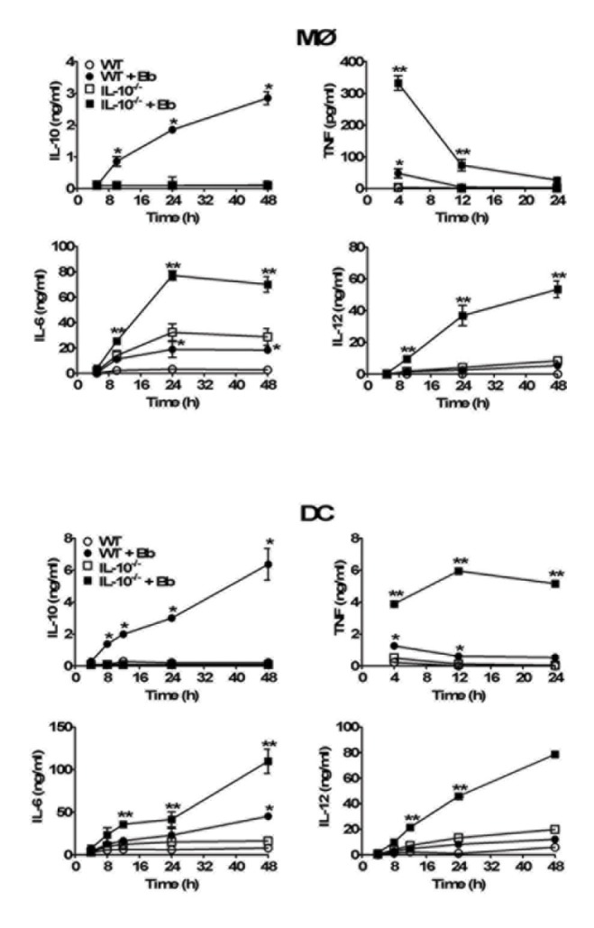
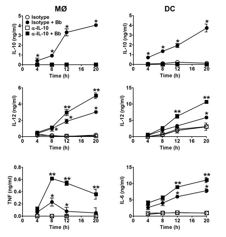
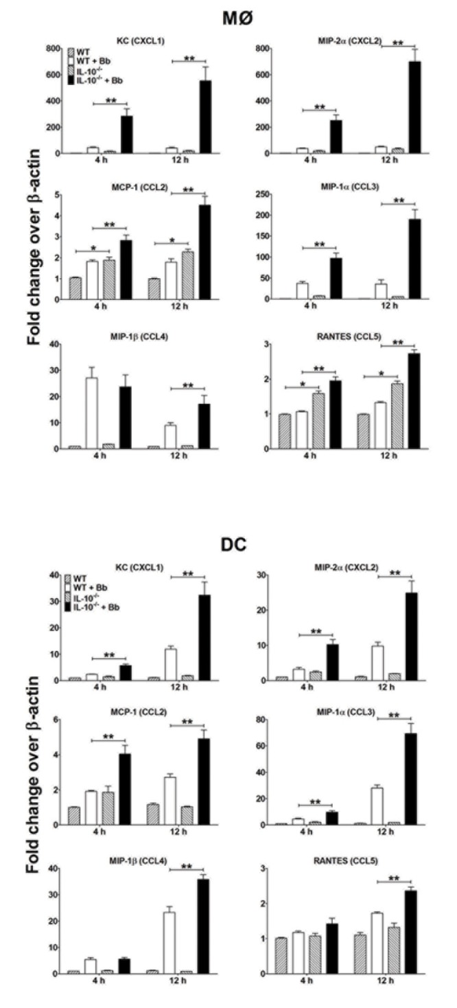
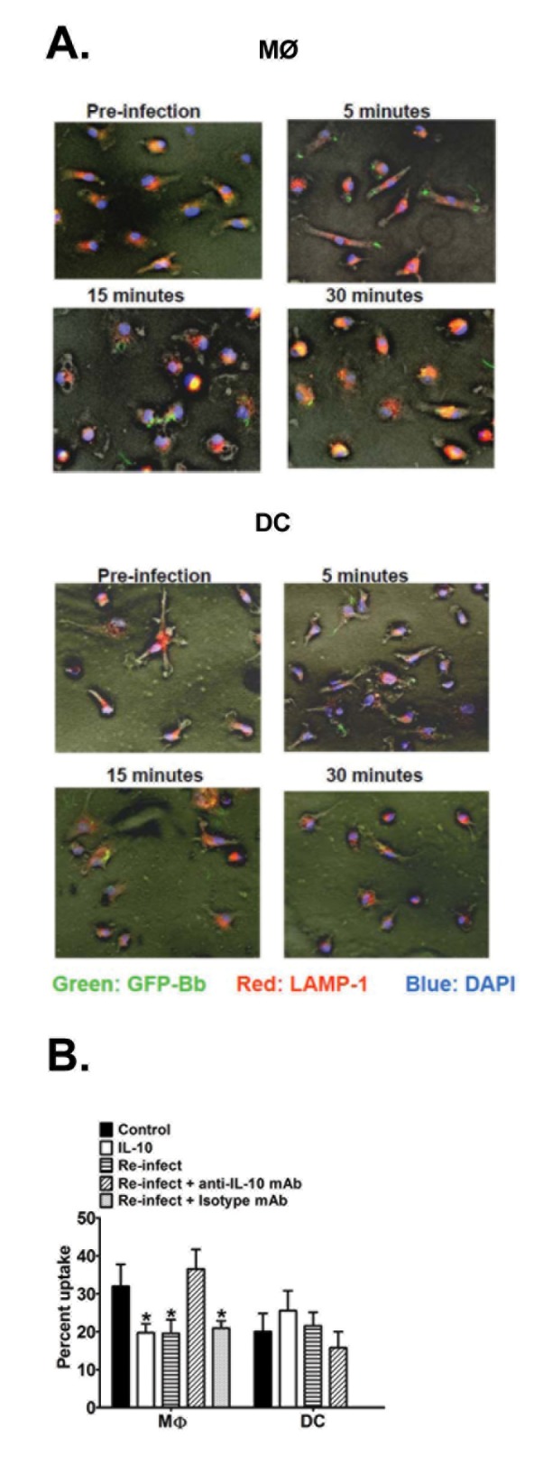
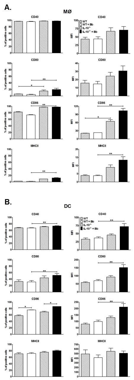
Borrelia burgdorferi (Bb) is a tick-borne spirochete that is the causative agent for Lyme disease. Our previous studies indicate that virulent Bb can potently enhance IL-10 production by macrophages (MØs) and that blocking IL-10 production significantly enhances bacterial clearance. We hypothesize that skin-associated APC types, such as MØs and dendritic cells (DCs) are potent producers of IL-10 in response to Bb, which may act in autocrine fashion to suppress APC responses critical for efficient Bb clearance. Our goal is to delineate which APC immune functions are dysregulated by Bb-elicited IL-10 using a murine model of Lyme disease. Our in vitro studies indicated that both APCs rapidly produce IL-10 upon exposure to Bb, that these levels inversely correlate with the production of many Lyme-relevant proinflammatory cytokines and chemokines, and that APCs derived from IL-10(-/-) mice produced greater amounts of these proinflammatory mediators than wild-type APCs. Phagocytosis assays determined that Bb-elicited IL-10 levels can diminish Bb uptake and trafficking by MØs, suppresses ROS production, but does not affect NO production; Bb-elicited IL-10 had little effect on phagocytosis, ROS, and NO production by DCs. In general, Bb exposure caused little-to-no upregulation of several critical surface co-stimulatory markers by MØs and DCs, however eliminating Bb-elicited IL-10 allowed a significant upregulation in many of these co-stimulatory receptors. These data indicate that IL-10 elicited from Bb-stimulated MØs and DCs results in decreased production of proinflammatory mediators and co-stimulatory molecules, and suppress phagocytosis-associated events that are important for mediating both innate and adaptive immune responses by APCs.


Conflict of interest statement
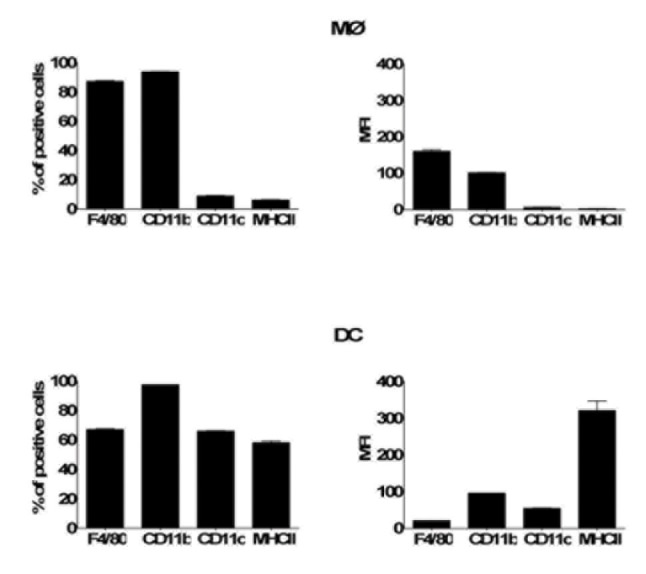
Competing Interests: The authors have declared that no competing interests exist.

Figures

Figure 1
Figure 1. Surface markers on naïve MØs and DCs.
Bone marrow-derived naïve MØs and DCs were collected prior to addition of Bb, stained with antibodies specific for the indicated surface markers, and analyzed by flow cytometry.
Figure 2
Figure 2. Effects of Bb-elicited IL-10 on the production of proinflammatory cytokines by MØs and DCs in vitro.
MØs (upper panel) or DCs (lower panel) derived from either wild type (wt) C57BL/6 or IL-10-/- mice were co-cultured with Bb (MOI=10) at 37Co. Culture supernatants were collected at the indicated times post-stimulation and cytokine content assessed by ELISA. Each symbol represents the average of triplicate samples from at least three separate experiments. Statistically significant (P<0.05) values are indicated between Bb-stimulated versus unstimulated APCs (*), or stimulated B6 versus IL-10-/- APCs (**).
Figure 3
Figure 3. Use of IL-10-blocking antibodies to assess IL-10 effects on MØs and DC cytokines.
Experiments were performed as in Figure 2, except MØs and DC from B6 mice were preincubated with 3μg/ml of either an IL-10-blocking (αIL-10) or isotype control antibody for 30 min before adding Bb (MOI=10) at 37°C. Each symbol represents the average of triplicate samples from at least three separate experiments. Statistically significant (P<0.05) values are indicated between Bb-stimulated versus unstimulated APCs (*), or stimulated isotype Ab-treated B6 versus αIL-10-treated APCs (**).
Figure 4
Figure 4. Effects of Bb-elicited IL-10 on chemokine expression by APCs: MØs (upper panel) and DCs (lower panel) from B6 and IL-10-/- mice co-cultured with Bb at MOI=10 at 37°C.
APCs were collected at the indicated times post-stimulation and total RNA was purified and reverse transcribed into cDNA for Q-PCR analyses. The transcript levels of each chemokine are normalized to levels of β-actin for each sample and reported as fold-induced over the baseline values for unstimulated MØs (upper panel) and DCs (lower panel). Each bar represents the average of triplicate samples from at least three separate experiments. Statistically significant values are indicated between unstimulated and Bb-stimulated APCs (*), or stimulated B6 versus IL-10-/- APCs (**).
Figure 5
Figure 5. Effect of IL-10 on Bb uptake and trafficking by APCs.
A. MØs (upper panel) and DCs (lower panel) from B6 mice were cultured on glass coverslips and infected with GFP-expressing (green) Bb (MOI=10) at 37°C. At 5, 15, and 30 min post-infection, APCs were fixed with 4% paraformaldehyde and permeablized for staining of LAMP-1 using TRITC-labeled (red) antibodies; nuclei are stained with DAPI (blue). Immunofluorescent images (200x) are representative of triplicate samples from three separate experiments, each with at least 3 different fields of views. (B) Quantitative analysis of Bb phagocytosis by APCs. MØs and DCs from B6 mice were infected with GFP-expressing Bb as in A (above) under five different conditions: GFP-Bb only (control), recombinant IL-10 (2ng/ml) administered overnight prior to GFP-Bb infection (rIL-10), infected with non-fluorescent Bb overnight prior to GFP-Bb infection (Re-infect), same as Re-infect, but initial infection was performed in the presence of anti-IL-10 antibody (Re-infect + anti-IL-10 mAb) or an isotype control (Re-infect + isotype mAb). Percent internalization was calculated as percentage of APCs per field of view containing internalized Bb at 30 min post-Bb infection. Data represents the average of ten separate fields of views, each containing 75-150 APCs, comprising at least three separate experiments. * Indicates statistically significant values (P≤0.05) compared to cells cultured with GFP-Bb only.
Figure 6
Figure 6. Effects of IL-10 on Bb-induced ROS and RNI production by APCs.
A. MØs (left panels) and DCs (right panels) were cultured in 96-well plates for 15 h before loading with DCF for 15 min. Cells were then pre-treated with or without rIL-10 (2ng/105 cells) or the NADPH oxidase inhibitor DPI (10μM) for an additional 30 min prior to Bb-stimulation; 0.01% H2O2 provided a positive control. ROS production was assessed by detecting the relative fluorescence intensity (RFU) values for up to 40 min post-stimulation. Top: Data are expressed as raw RFU values per min over a 40 min interval (Left: MØs, Right: DCs). Middle: Slope of RFU for each condition is calculated as changes in RFU over time for the first 30 min. Data are representative of three separate experiments. Statistically significant (P<0.05) values are indicated compared to unstimulated control (*) or Bb stimulation (**). B. MØs (left panels) and DCs (right panels) were stimulated with Bb for 24h. Supernatant nitric oxide (nitrite) levels were assessed using the Griess assay. Statistically significant (P<0.05) values are indicated compared to unstimulated controls (*) or Bb stimulation (**).
Figure 7
Figure 7. Effect of IL-10 on Bb-elicited upregulation of surface co-stimulation molecule expression on APCs.
MØs (A) and DCs (B) expanded from B6 and IL-10-/- mice were cultured with Bb for 24h before staining with fluorescence antibodies specific for the indicated surface molecules and analyzed by flow cytometry. Data are reported both as percentage of positive cells (left panels) and mean fluorescent intensity (MFI). Each bar represents triplicate samples from at least three separate experiments. Statistically significant (P<0.05) values are indicated compared to unstimulated (*) or Bb-stimulated APCs (**).

Similar articles

Cited by

References

    1. Wooten RM, Weis JJ (2001) Host-pathogen interactions promoting inflammatory Lyme arthritis: use of mouse models for dissection of disease processes. Curr Opin Microbiol 4: 274-279. doi:10.1016/S1369-5274(00)00202-2. PubMed: 11378478. - DOI - PubMed
    1. Radolf JD, Caimano MJ, Stevenson B, Hu LT (2012) Of ticks, mice and men: understanding the dual-host lifestyle of Lyme disease spirochaetes. Nat Rev Microbiol 10: 87-99. PubMed: 22230951. - PMC - PubMed
    1. Benach JL, Habicht GS, Gocinski BL, Coleman JL (1984) Phagocytic cell responses to in vivo and in vitro exposure to the Lyme disease spirochete. Yale J Biol Med 57: 599-605. PubMed: 6393611. - PMC - PubMed
    1. Montgomery RR, Nathanson MH, Malawista SE (1994) Fc- and non-Fc-mediated phagocytosis of Borrelia burgdorferi by macrophages. J Infect Dis 170: 890-893. doi:10.1093/infdis/170.4.890. PubMed: 7930732. - DOI - PubMed
    1. Montgomery RR, Lusitani D, Chevance Ad Ade B, Malawista SE (2002) Human phagocytic cells in the early innate immune response to Borrelia burgdorferi . J Infect Dis 185: 1773-1779. doi:10.1086/340826. PubMed: 12085324. - DOI - PubMed

Publication types

MeSH terms