. 2014 Feb 7;6(2):e00137.
doi: 10.1042/AN20120041.

PDGF-responsive progenitors persist in the subventricular zone across the lifespan

Affiliations

PDGF-responsive progenitors persist in the subventricular zone across the lifespan

Lisamarie Moore et al. ASN Neuro. .

Abstract

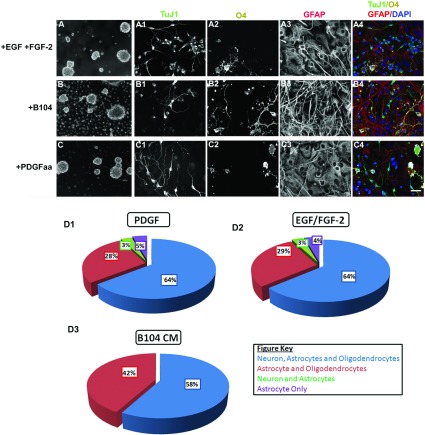
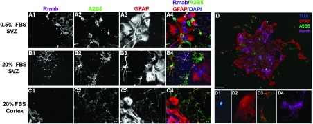
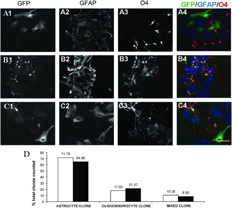
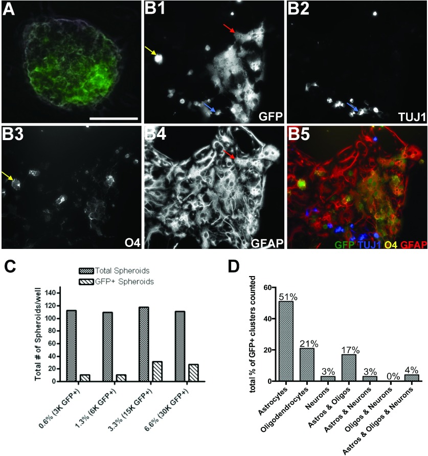
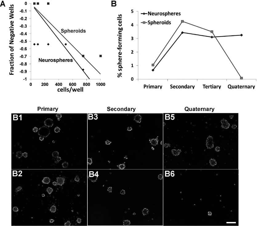
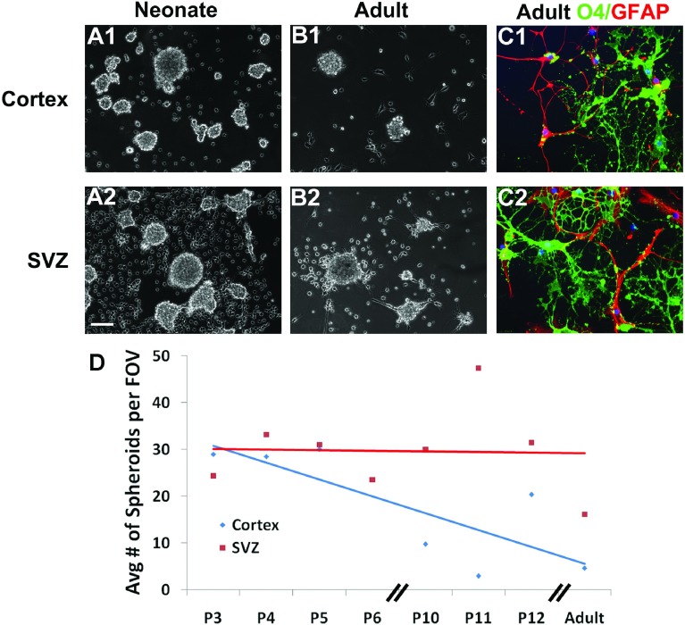
The SVZ (subventricular zone) contains neural stem cells and progenitors of various potentialities. Although initially parsed into A, B, and C cells, this germinal zone is comprised of a significantly more diverse population of cells. Here, we characterized a subset of postnatal PRPs (PDGF-AA-responsive precursors) that express functional PDGFα and β receptors from birth to adulthood. When grown in PDGF-AA, dissociated neonatal rat SVZ cells divided to produce non-adherent clusters of progeny. Unlike the self-renewing EGF/FGF-2-responsive precursors that produce neurospheres, these PRPs failed to self-renew after three passages; therefore, we refer to the colonies they produce as spheroids. Upon differentiation these spheroids could produce neurons, type 1 astrocytes and oligodendrocytes. When maintained in medium supplemented with BMP-4 they also produced type 2 astrocytes. Using lineage tracing methods, it became evident that there were multiple types of PRPs, including a subset that could produce neurons, oligodendrocytes, and type 1 and type 2 astrocytes; thus some of these PRPs represent a unique population of precursors that are quatropotential. Spheroids also could be generated from the newborn neocortex and they had the same potentiality as those from the SVZ. By contrast, the adult neocortex produced less than 20% of the numbers of spheroids than the adult SVZ and spheroids from the adult neocortex only differentiated into glial cells. Interestingly, SVZ spheroid producing capacity diminished only slightly from birth to adulthood. Altogether these data demonstrate that there are PRPs that persist in the SVZ that includes a unique population of quatropotential PRPs.


Figures

Figure 1
Figure 1. Spheroids are responsive to PDGF-AA and express PDGFRα and PDGFRβ
Spheroids and neurospheres were generated from neonatal SVZs and cultured for 7 DIV in 2% O2, 5% CO2, 93% N2. OPCs were generated from mixed glial cell cultures from neonatal rat brains. (A) Expression levels of pERK1/2 (44,42 kDa) and total ERK1/2 (44,42 kDa) in 6 h growth factor-starved spheroids untreated (T0) or treated with 10 ng/ml PDGF-AA stimulation for 15 min (T15) and 30 min (T30). (B) Expression levels of PDGFRα (195 kDa) and PDGFRβ (185 kDa) in OPCs, spheroids, neurospheres and meninges. Data are representative of two independent experiments.
Figure 2
Figure 2. Expression of PSA-NCAM and GFAP in spheroids and differentiation into immature oligodendrocytes and astrocytes under different growth conditions
(A) Spheroids were gently dissociated and plated on to PDL-coated chamber slides for PSA-NCAM and GFAP immunostaining. (B) Spheroids were dissociated into single cells, plated for 6 h with 2% serum and grown in an atmospheric incubator. Media was changed to N2B2 with 0.5% serum for 96 h. Cells were then stained for O4, followed by fixation and then for GFAP and DAPI. Data represent averages±S.E.M. from three independent experiments. * denote significant differences in (A) versus Pro-N/B104. n.s. denotes no statistical significance. For comparisons in both (A) and (B), P<0.05 using ANOVA followed by Fisher's PLSD post-hoc test.
Figure 3
Figure 3. Cell density affects spheroid generation while O2 tension affects oligodendrocyte differentiation, however neither O2 nor T3 affect glial specification
(A) Cultures were seeded at initial densities of 1.5×10⁵, 0.75×10⁵ and 0.375×10⁵ cells/ml and grown for 8–10 days in 2% O2 or 20% O2 in B104 CM. (B) Quantification of the average number of spheroids per field of view (FOV). (C) Cells were grown in 2% O2 and differentiated in either 2% O2 or 20% O2, then analyzed for positive staining of oligodendrocytes (O4) and astrocytes (GFAP+). (D) Fluorescence images of the cells differentiated in 2% O2 (D’) or 20% O2 (D”). (E) Cells were grown in 2% O2 and differentiated in the absence of T3 (N2B2) or presence of T3 (N2B3) at 20% O2. Scale bar represents 100 μm (A) and 10 μm (D). Error bars represent±S.E.M. from at least three independent experiments. * denote significant differences and n.s. denotes no statistical significance, where P<0.05 using Student's t test.
Figure 4
Figure 4. Greater than half of the spheroid-forming precursors are tripotential
Neurospheres (A) and spheroids (B and C) were grown for 8–10 days in 2% O2 ProN medium supplemented with 10 ng/ml EGF and 5 ng/ml FGF-2, 30% B104 CM, or 10 ng/ml PDGF-AA. The spheres were plated on to slides and differentiated for 5 days then stained for neuronal (TUJ1) (A1, B1, C1), oligodendrocyte (O4) (A2, B2, C2), and astrocyte (GFAP) (A3, B3, C3) markers. The pseudocolor images show an overlay of TUJI (green), O4 (white), GFAP (red), and DAPI (blue) (A4, B4, C4). At least 60 individual neurospheres (D1) and spheroids (D2, D3) were analyzed based on colony composition. Pie charts represent three independent experiments. Scale bar represents 100 μm (A, B, C) and 10 μm (A1A4, B1B4, C1C4).
Figure 5
Figure 5. Spheroids from the neonatal SVZ and neocortex generate colonies that contain neurons, type 1 astrocytes, type 2 astrocytes and oligodendrocytes
Spheroids were cultured with 10 ng/ml PDGF-AA, differentiated for 3 days in either 0.5% FBS or 20% FBS then stained for oligodendrocytes (Rmab) (A1, B1, C1), O-2A lineage (A2B5) (A2, B2, C2), and astrocytes (GFAP) (A3, B3, C3). The pseudocolor images show an overlay of Rmab (magenta), A2B5 (green), GFAP (red), and DAPI (blue) (A4, B4, C4). When differentiated in 0.5% FBS and 10 ng/ml BMP-4, a single spheroid produced neurons (TUJ1), type 1 astrocytes (A2B5−/GFAP+), type 2 astrocytes (A2B5+/GFAP+) and oligodendrocytes (Rmab) (D). Examples of the four cell types from a single spheroid neuron (D1), type 1 astrocyte (D2), type 2 astrocyte (D3) and oligodendrocyte (D4) within the spheroid. Scale bar represents 20 μm.
Figure 6
Figure 6. Clonal analyses of spheroids reveal three subclasses of progenitors
Spheroids were grown for 7–10 days in 2% O2, 5% CO2, 93% N2 and then gently dissociated into single cells and plated on to PDL-coated chamber slides. Cells were infected with 50 CFU pNIT retrovirus for 1 day followed by growth in N2B2+0.5% FBS for 7 days after which cells were fixed in 4% PFA and triple-stained for GFP-green (A1, B1, C1), GFAP-blue (A2, B2, C2) and O4-red (A3, B3, C3). Clones were classified as astrocyte - only clones (A4); oligodendrocyte - only clones (B4); or mixed clones (C4). Scale bar represents 40 μm (A and B) and 20 μm (C). (D) Quantification of two independent experiments in which clonal analyses were performed after infection with 1×10⁵ CFU of the pNIT reporter replication-deficient retrovirus in N2B2 differentiation media (white) or in ProN/B104 CM (black).
Figure 7
Figure 7. In vitro co-cultures reveal probability and potentially of single PDGF-responsive SVZ cells
SVZs from P3, GFP+/− and GFP−/−, littermates were grown at a low density of 5×10⁴ cells/ml in co-culture at a ratio of 1:150 for 8 days in serum-free preconditioned media and atmospheric gases of 2% O2, 5% CO2, 93% N2. Most spheroids were comprised of both GFP+ and GFP− cells (A). Spheroids were then plated on to PDL and laminin-coated chamber slides in the absence of growth factors and differentiated for 5 days. Clusters of GFP+ cells (B1) were analyzed for antigens TUJ1 (B2), O4 (B3) and GFAP (B4). Spheroids that contained ≥5 GFP+ cells, by fluorescence imaging followed by ImageJ analysis, were counted as a GFP+ spheroids (C). Colonies of GFP+ cells were classified as astrocyte-only, oligodendrocyte-only, neuron-only, astrocyte-oligodendrocyte, astrocyte-neuron, oligodendrocyte-neuron, astrocyte-oligodendrocyte-neuron (D). Scale bar represents 40 μm (A).
Figure 8
Figure 8. Neurospheres contain a greater number of self-renewing cells than spheroids, whose precursors lose their self-renewal capacity over serial passages
(A) Spheroids and neurospheres were generated from neonatal SVZs and cultured for 7 DIV in 2% O2, 5% CO2, 93% N2, passaged and plated in a 96-well plate. The limiting dilution assay was performed by plating cells at decreasing densities and then scoring the fraction of negative wells. The reciprocal of the natural logarithm (e) was used to measure the frequency of sphere-forming cells in neurospheres (black) and spheroids (gray). (B) Spheroids and neurospheres were dissociated into single cells and re-plated every 7 days to generate subsequent spheres. The percentage of sphere-forming cells to the total cells plated was determined. Representative images of neurospheres and spheroids from primary (B1, B2), secondary (B3, B4) and quaternary (B5, B6) spheres. Data represent the averaged from two independent experiments.
Figure 9
Figure 9. Neocortical spheroid formation decreases with age
Spheroids were generated from P5 neocortex (A1), P5 SVZ (A2), P70 neocortex (B1) and P70 SVZ (B2). Neonatal cells were grown for 7 days while the adult cells where grown for 16 days in 2% O2 in B104 CM. Spheroids were plated on to slides and differentiated for 5 days then stained for oligodendrocyte (O4-green) and astrocyte (GFAP-red) markers (C1, C2). The average number of spheroids from P3, P4, P5, P6, P10, P11, P12 and adult (P64–P70) rodents is plotted (D). Scale bar represents 100 μm (A1A2, B1B2) and 10 μm (C1C2).

Similar articles

Cited by

References

    1. Aguirre AA, Chittajallu R, Belachew S, Gallo V. NG2-expressing cells in the subventricular zone are type C-like cells and contribute to interneuron generation in the postnatal hippocampus. J Cell Biol. 2004;165:575–589. - PMC - PubMed
    1. Alagappan D, Balan M, Jiang Y, Cohen RB, Kotenko SV, Levison SW. Egr-1 is a critical regulator of EGF-receptor-mediated expansion of subventricular zone neural stem cells and progenitors during recovery from hypoxia-hypoglycemia. ASN NEURO. 2013;5 art:e00116, doi:10.1042/AN20120032. - PMC - PubMed
    1. Alvarez-Buylla A, Garcia-Verdugo JM. Neurogenesis in adult subventricular zone. J Neurosci. 2002;22:629–634. - PMC - PubMed
    1. Andrae J, Hansson I, Afink GB, Nister M. Platelet-derived growth factor receptor-alpha in ventricular zone cells and in developing neurons. Mol Cell Neurosci. 2001;17:1001–1013. - PubMed
    1. Assanah MC, Bruce JN, Suzuki SO, Chen A, Goldman JE, Canoll P. PDGF stimulates the massive expansion of glial progenitors in the neonatal forebrain. Glia. 2009;57:1835–1847. - PubMed

Publication types

MeSH terms

Substances

LinkOut - more resources