Prevention of allergic airway hyperresponsiveness and remodeling in mice by Astragaliradix antiasthmatic decoction
- PMID: 24367979
- PMCID: PMC3922862
- DOI: 10.1186/1472-6882-13-369
Prevention of allergic airway hyperresponsiveness and remodeling in mice by Astragaliradix antiasthmatic decoction
Abstract
Background: Astragali radix Antiasthmatic Decoction (AAD), a traditional Chinese medication, is found effective in treating allergic diseases and chronic cough. The purpose of this study is to determine whether this medication could suppress allergen-induced airway hyperresponsiveness (AHR) and remodeling in mice, and its possible mechanisms.
Methods: A mouse model of chronic asthma was used to investigate the effects of AAD on the airway lesions. Mice were sensitized and challenged with ovalbumin (OVA), and the extent of AHR and airway remodeling were characterized. Cells and cytokines in the bronchoalveolar lavage fluid (BALF) were examined.
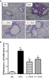
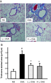
Results: AAD treatment effectively decreased OVA-induced AHR, eosinophilic airway inflammation, and collagen deposition around the airway. It significantly reduced the levels of IL-13 and TGF-β1, but exerted inconsiderable effect on INF-γ and IL-10.
Conclusions: AAD greatly improves the symptoms of allergic airway remodeling probably through inhibition of Th2 cytokines and TGF-β1.
Figures


Similar articles
-
Suhuang antitussive capsule at lower doses attenuates airway hyperresponsiveness, inflammation, and remodeling in a murine model of chronic asthma.Sci Rep. 2016 Feb 10;6:21515. doi: 10.1038/srep21515. Sci Rep. 2016. PMID: 26861679 Free PMC article.
-
Inhibition airway remodeling and transforming growth factor-β1/Smad signaling pathway by astragalus extract in asthmatic mice.Int J Mol Med. 2012 Apr;29(4):564-8. doi: 10.3892/ijmm.2011.868. Epub 2011 Dec 23. Int J Mol Med. 2012. Retraction in: Int J Mol Med. 2023 Oct;52(4):87. doi: 10.3892/ijmm.2023.5290. PMID: 22200784 Free PMC article. Retracted.
-
Huangqi-Fangfeng protects against allergic airway remodeling through inhibiting epithelial-mesenchymal transition process in mice via regulating epithelial derived TGF-β1.Phytomedicine. 2019 Nov;64:153076. doi: 10.1016/j.phymed.2019.153076. Epub 2019 Aug 23. Phytomedicine. 2019. PMID: 31473579
-
Descurainia sophia Ameliorates Eosinophil Infiltration and Airway Hyperresponsiveness by Reducing Th2 Cytokine Production in Asthmatic Mice.Am J Chin Med. 2019;47(7):1507-1522. doi: 10.1142/S0192415X19500770. Am J Chin Med. 2019. PMID: 31752525
-
Yan-Hou-Qing formula attenuates allergic airway inflammation via up-regulation of Treg and suppressing Th2 responses in Ovalbumin-induced asthmatic mice.J Ethnopharmacol. 2019 Mar 1;231:275-282. doi: 10.1016/j.jep.2018.11.038. Epub 2018 Nov 26. J Ethnopharmacol. 2019. PMID: 30496840
Cited by
-
Traditional Chinese medicine for treatment of liver diseases: progress, challenges and opportunities.J Integr Med. 2014 Sep;12(5):401-8. doi: 10.1016/S2095-4964(14)60039-X. J Integr Med. 2014. PMID: 25292339 Free PMC article. Review.
-
Beneficial Effects of Astragalus membranaceus (Fisch.) Bunge Extract in Controlling Inflammatory Response and Preventing Asthma Features.Int J Mol Sci. 2023 Jun 30;24(13):10954. doi: 10.3390/ijms241310954. Int J Mol Sci. 2023. PMID: 37446131 Free PMC article.
-
The Anti-Inflammatory Effects of Fermented Herbal Roots of Asparagus cochinchinensis in an Ovalbumin-Induced Asthma Model.J Clin Med. 2018 Oct 22;7(10):377. doi: 10.3390/jcm7100377. J Clin Med. 2018. PMID: 30360392 Free PMC article.
-
Suhuang antitussive capsule at lower doses attenuates airway hyperresponsiveness, inflammation, and remodeling in a murine model of chronic asthma.Sci Rep. 2016 Feb 10;6:21515. doi: 10.1038/srep21515. Sci Rep. 2016. PMID: 26861679 Free PMC article.
-
Herbal Medicines for Asthmatic Inflammation: From Basic Researches to Clinical Applications.Mediators Inflamm. 2016;2016:6943135. doi: 10.1155/2016/6943135. Epub 2016 Jul 20. Mediators Inflamm. 2016. PMID: 27478309 Free PMC article. Review.
References
Publication types
MeSH terms
Substances
LinkOut - more resources
Full Text Sources
Other Literature Sources
Medical

