

The cytotoxic effect of 2-acylated-1,4-naphthohydroquinones on leukemia/lymphoma cells
- PMID: 24368029
- PMCID: PMC4215946
- DOI: 10.1016/j.bmc.2013.12.007
The cytotoxic effect of 2-acylated-1,4-naphthohydroquinones on leukemia/lymphoma cells
Abstract
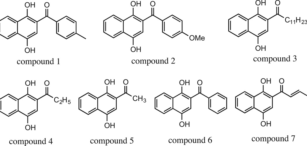
Here, we tested seven 2-acylated-1,4-hydronaphthoquinones for their cytotoxic effects on a panel of cancer lymphoma/leukemia cells and compared to a non-cancer origin cell line. Several naphthohydroquinones exhibited selective cytotoxic effects on lymphoma/leukemia cells with lowest activity on non-cancer cells. The mode of cell death induced by an acylated naphthohydroquinone, which has a long alkyl chain, was found to be via apoptosis. Furthermore, the naphthohydroquinone provoked mitochondria depolarization and activation of its downstream effector, caspase-3, thus implicating the intrinsic apoptotic pathway as its mechanism to exert cell death.
Keywords: Acylated hydroquinones; Anti-lymphoma; Apoptosis; Cytotoxicity; Photochemistry.
Copyright © 2013 Elsevier Ltd. All rights reserved.
Figures


References
-
- Maruyama K, Osuka A. In: The Chemistry of the Quinonoid Compounds. Patai S, Rappoport Z, editors. Vol. 2. New York: John Wiley & Sons; 1988. p. 759.
-
- Thomson RH. Naturally Occurring Quinones IV. Springer; 1997.
-
- Iyanagi T, Yamazaki I. Biochim. Biophy. Acta (BBA)—Bioenerg. 1970;216:282. - PubMed
-
- Kitamura S, Tatsumi K. Drug Metab. Dispos. 1999;27:98. - PubMed
-
- Lind C, Hochstein P, Ernster L. Arch. Biochem. Biophys. 1982;216:178. - PubMed
Publication types
MeSH terms
Substances
Grants and funding
LinkOut - more resources
Full Text Sources
Other Literature Sources
Medical
Research Materials
