. 2013 Dec 24:2:e01503.
doi: 10.7554/eLife.01503.

H3K4 mono- and di-methyltransferase MLL4 is required for enhancer activation during cell differentiation

Affiliations

H3K4 mono- and di-methyltransferase MLL4 is required for enhancer activation during cell differentiation

Ji-Eun Lee et al. Elife. .

Abstract

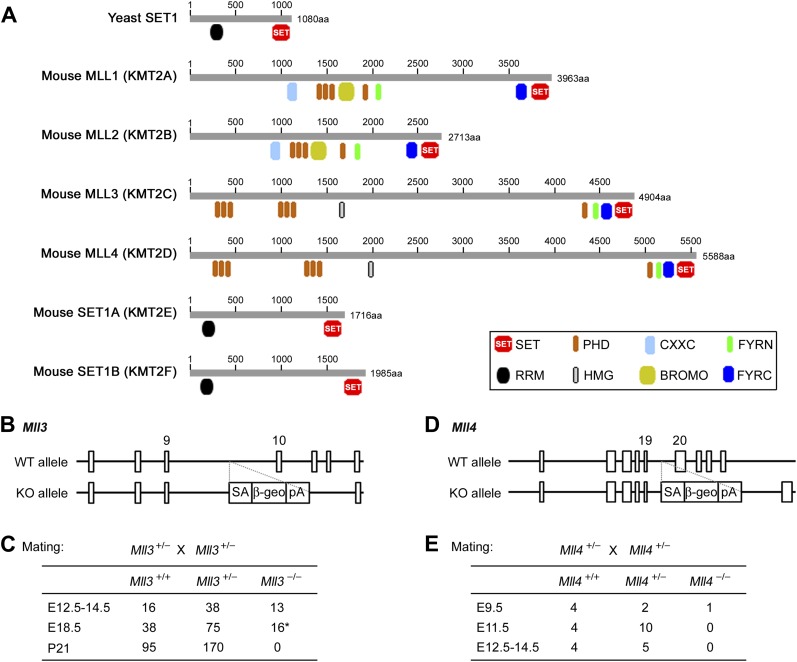
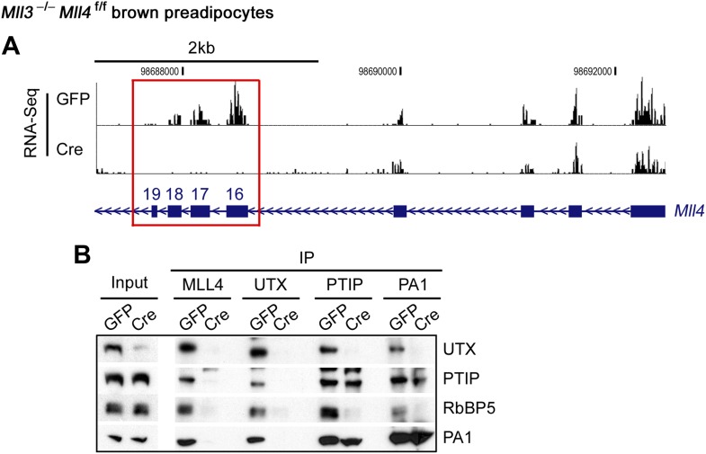
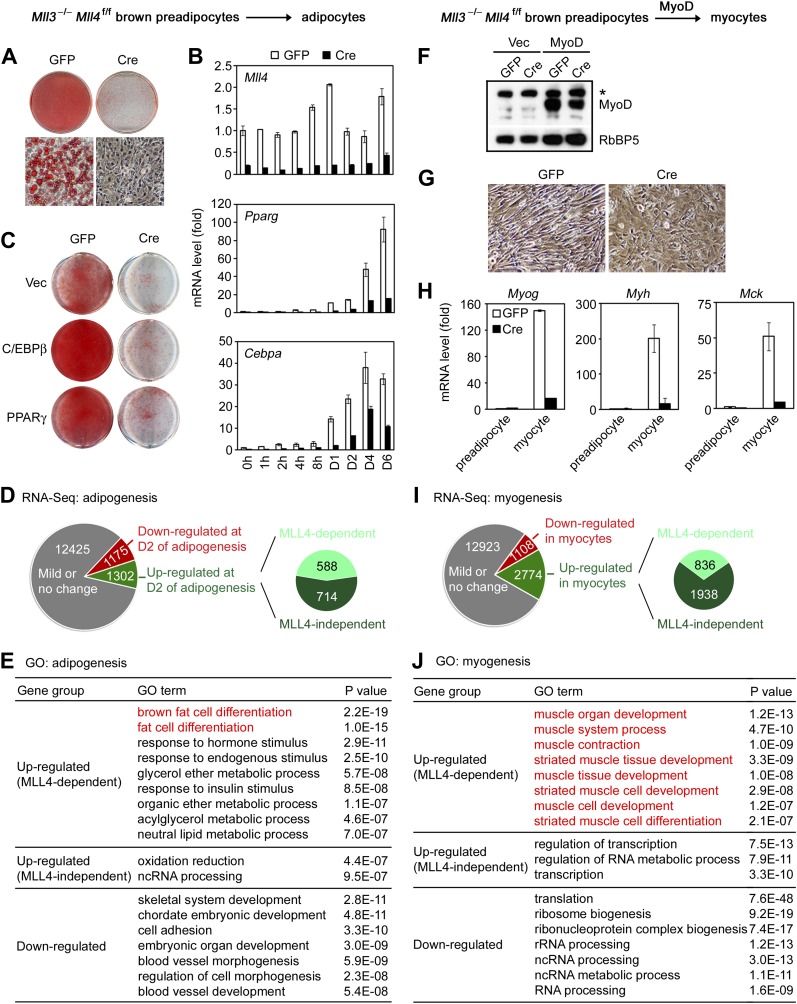
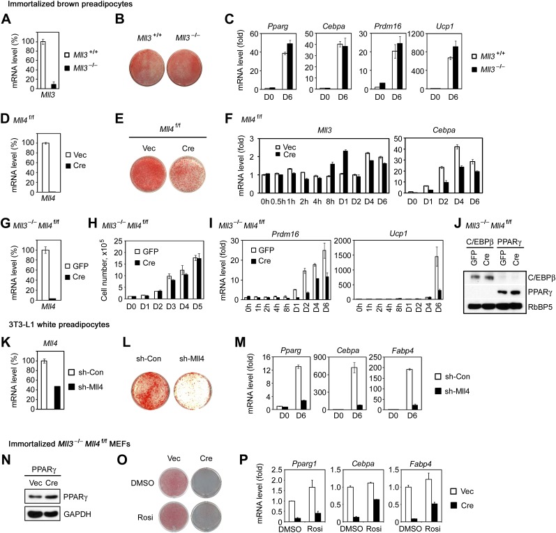
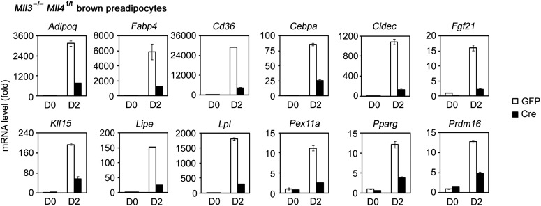
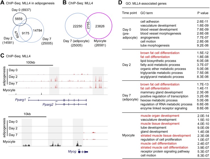
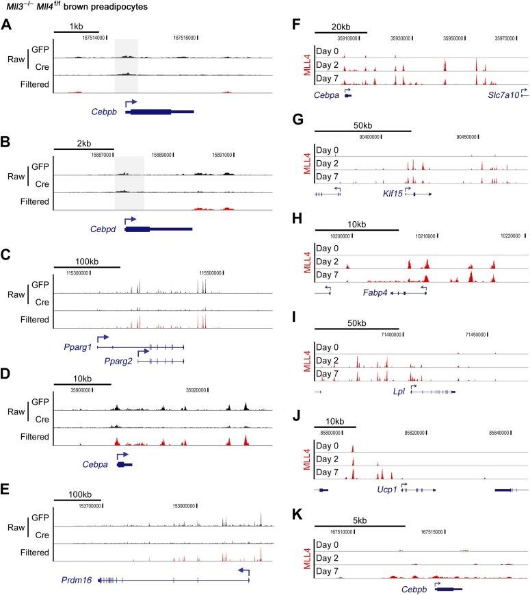
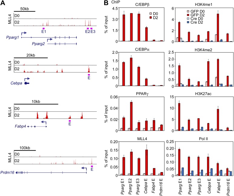
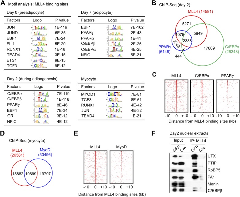
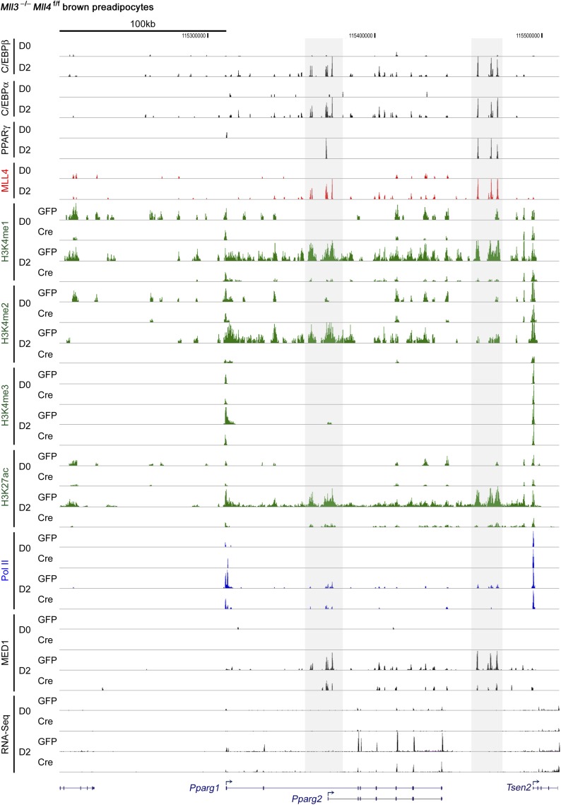
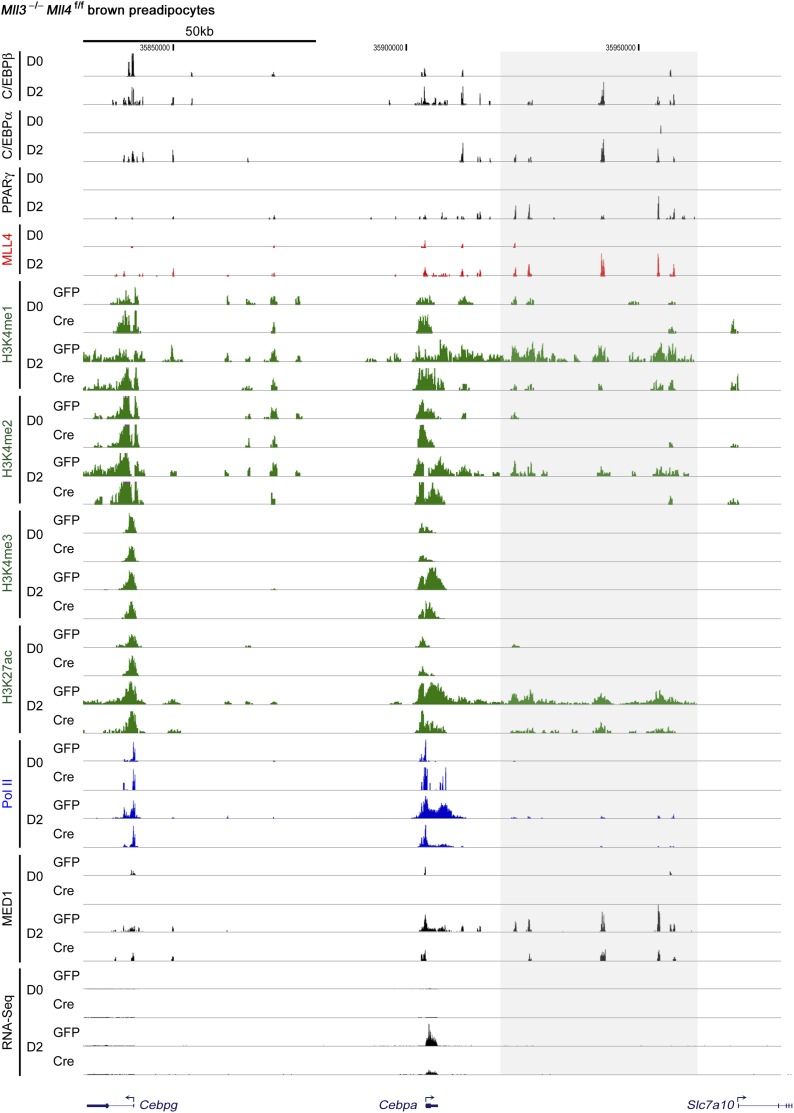
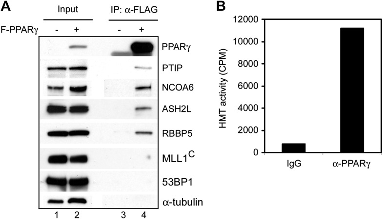
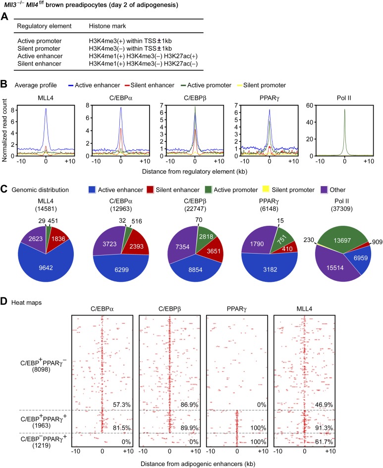
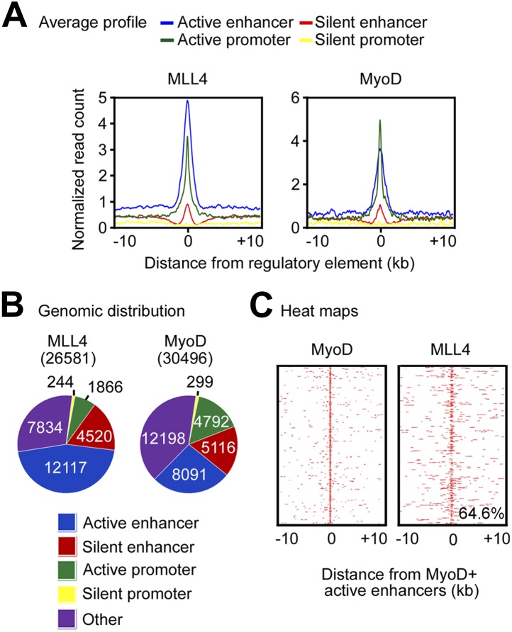
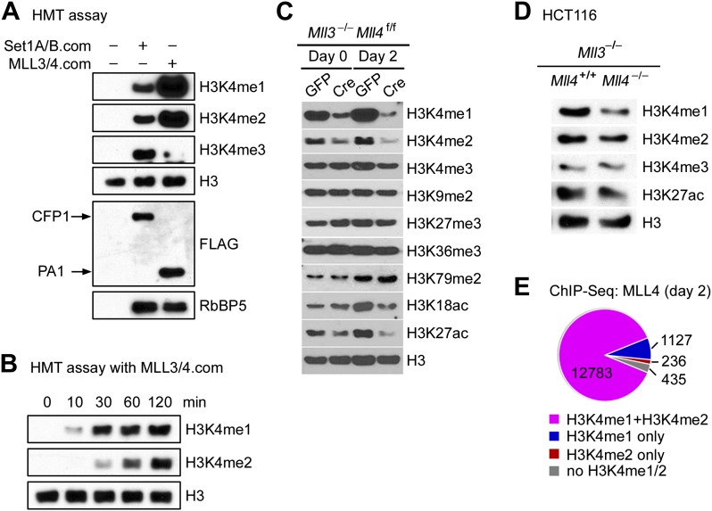
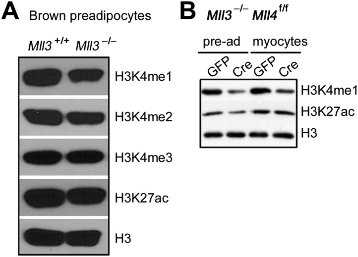
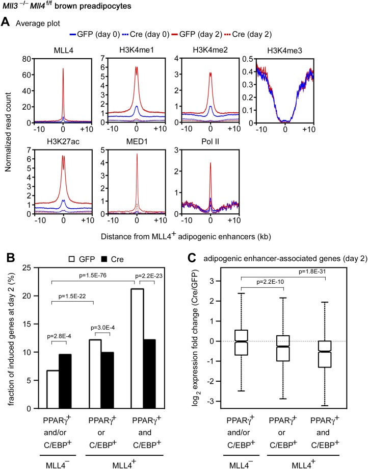
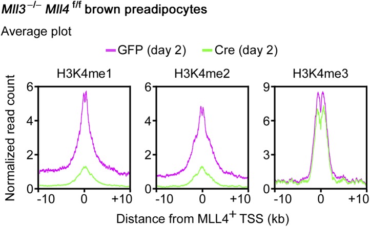
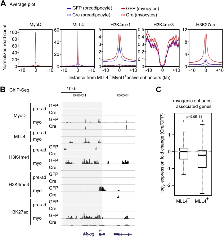
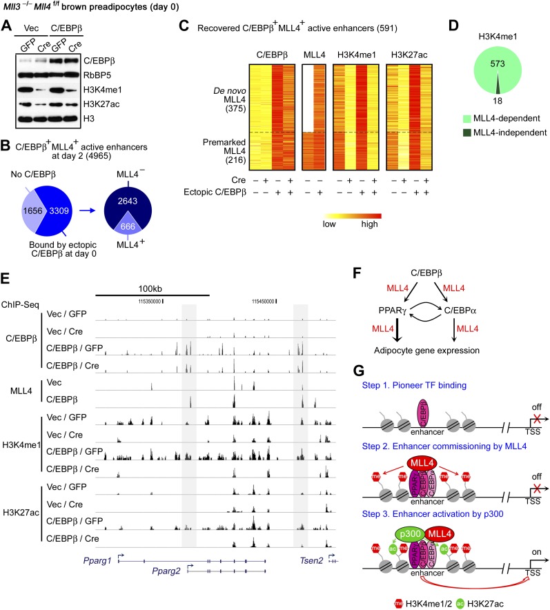
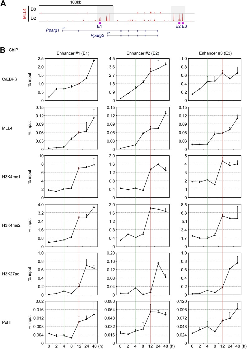
Enhancers play a central role in cell-type-specific gene expression and are marked by H3K4me1/2. Active enhancers are further marked by H3K27ac. However, the methyltransferases responsible for H3K4me1/2 on enhancers remain elusive. Furthermore, how these enzymes function on enhancers to regulate cell-type-specific gene expression is unclear. In this study, we identify MLL4 (KMT2D) as a major mammalian H3K4 mono- and di-methyltransferase with partial functional redundancy with MLL3 (KMT2C). Using adipogenesis and myogenesis as model systems, we show that MLL4 exhibits cell-type- and differentiation-stage-specific genomic binding and is predominantly localized on enhancers. MLL4 co-localizes with lineage-determining transcription factors (TFs) on active enhancers during differentiation. Deletion of Mll4 markedly decreases H3K4me1/2, H3K27ac, Mediator and Polymerase II levels on enhancers and leads to severe defects in cell-type-specific gene expression and cell differentiation. Together, these findings identify MLL4 as a major mammalian H3K4 mono- and di-methyltransferase essential for enhancer activation during cell differentiation. DOI: http://dx.doi.org/10.7554/eLife.01503.001.

Keywords: H3K27ac; H3K4me1; KMT2D; MLL4; adipogenesis; cell differentiation; enhancer activation; enhancer chromatin modification; myogenesis.


Conflict of interest statement

The authors declare that no competing interests exist.

Figures

Figure 1.
Figure 1.. MLL4 is required for brown adipose tissue and muscle development.
(A and B) Generation of Mll4 conditional KO mice (Mll4f/f). (A) Schematic representation of mouse Mll4 wild-type (WT) allele, targeted allele, conditional KO (flox) allele and KO allele. In the targeted allele, a single loxP site was inserted in the intron before exon 16. A neomycin (neo) selection cassette flanked by FRT sites and the second loxP site was inserted in the intron after exon 19. The locations of PCR genotyping primers P1, P2, and P3 are indicated by arrows. (B) PCR genotyping of cell lines using mixtures of P2 + P3 or P1 + P3 primers. The genotypes are indicated at the top. (C) Genotype of E18.5 embryos isolated from crossing Mll4f/+;Myf5-Cre with Mll4f/f mice. The expected ratios of the four genotypes are 1:1:1:1. Mll4f/f;Myf5-Cre mice died immediately after cesarean section because of breathing malfunction due to defects in muscles of the rib cage. (D) Representative pictures of E18.5 embryos of the indicated genotypes. (E) E18.5 embryos were sagittally sectioned along the midline. The sections of the cervical/thoracic area indicated in the schematic were stained with H&E (upper panels) or with antibodies recognizing BAT marker UCP1 (green) and skeletal muscle marker Myosin (red) (lower panels). Scale bar = 800 μm. DOI: http://dx.doi.org/10.7554/eLife.01503.003
Figure 1—figure supplement 1.
Figure 1—figure supplement 1.. Generation of Mll3 and Mll4 whole body KO mice.
(A) Protein domains in yeast SET1 and the homologous mouse SET1-like H3K4 methyltransferases. (BE) Generation of Mll3 and Mll4 whole body KO mice. Wild-type (WT) and KO alleles of Mll3 and Mll4 genomic loci are shown in (B) and (D), respectively. The exons flanking the gene trap vectors are numbered. Mll3 KO mice are perinatal lethal (C), while Mll4 KO mice are early embryonic lethal (E). DOI: http://dx.doi.org/10.7554/eLife.01503.004
Figure 1—figure supplement 2.
Figure 1—figure supplement 2.. Confirmation of Mll4 deletion.
Immortalized Mll3−/−Mll4f/f brown preadipocytes were infected with adenoviral GFP or Cre. (A) Genome browser view of RNA-Seq analysis on Mll4 gene locus. The targeted exons 16–19 are highlighted in red box. (B) Deletion of Mll4 disrupts MLL4 complex in cells. Nuclear extracts were incubated with MLL4, UTX, PTIP or PA1 antibody. The immunoprecipitates were analyzed by Western blotting using antibodies indicated on the right. DOI: http://dx.doi.org/10.7554/eLife.01503.005
Figure 2.
Figure 2.. MLL4 controls induction of cell-type-specific genes during differentiation.
(AE) Adipogenesis of Mll3−/−Mll4f/f brown preadipocytes. (A and B) MLL4 is required for adipogenesis. Immortalized Mll3−/−Mll4f/f brown preadipocytes were infected with adenoviral GFP or Cre, followed by adipogenesis assay. (A) 6 days after induction of differentiation, cells were stained with Oil Red O. Upper panels, stained dishes; lower panels, representative fields under microscope. (B) qRT-PCR of Mll4, Pparg and Cebpa expression at indicated time points of adipogenesis. Quantitative PCR data in all figures are presented as means ± SD. D1, day 1. (C) MLL4 is required for C/EBPβ- and PPARγ-stimulated adipogenesis. Mll3−/−Mll4f/f brown preadipocytes were infected with retroviruses expressing vector (vec), C/EBPβ or PPARγ. After hygromycin selection, cells were infected with adenoviral GFP or Cre, followed by adipogenesis assay. (DE) MLL4 is required for induction of cell-type-specific genes during adipogenesis. Adipogenesis was done as in (A). Cells were collected before (day 0) and during (day 2) adipogenesis for RNA-Seq. (D) Schematic of identification of MLL4-dependent and -independent up-regulated genes during adipogenesis. The threshold for up- or down-regulation is 2.5-fold. (E) Gene ontology (GO) analysis of gene groups defined in (D). (FJ) MLL4 is required for MyoD-stimulated myogenesis. Immortalized Mll3−/−Mll4f/f brown preadipocytes were infected with retroviruses expressing Vec or MyoD. After hygromycin selection, cells were infected with adenoviral GFP or Cre, followed by myogenesis assay. (F) Western blot analysis of MyoD expression before differentiation. RbBP5 was used as a loading control. The asterisk indicates a non-specific band. (G) 5 days after induction of differentiation, cell morphologies were observed under microscope. (H) qRT-PCR analysis of myogenic gene expression after differentiation. (I and J) MLL4 is required for induction of cell-type-specific genes during myogenesis. Brown preadipocytes and myocytes were collected for RNA-Seq. (I) Schematic of identification of MLL4-dependent and -independent up-regulated genes during myogenesis. The threshold for up- or down-regulation is 2.5-fold. (J) GO analysis of gene groups defined in (I). DOI: http://dx.doi.org/10.7554/eLife.01503.006
Figure 2—figure supplement 1.
Figure 2—figure supplement 1.. MLL4 is required for adipogenesis.
(AC) MLL3 is dispensable for adipogenesis of brown preadipocytes. Brown preadipocytes isolated from Mll3+/+ and Mll3−/− E18.5 embryos were immortalized by retroviruses expressing SV40T. Cells were induced for adipogenesis for 6–7 days. (A) qRT-PCR analysis of Mll3 expression before adipogenesis. (B) Oil Red O staining after adipogenesis. (C) qRT-PCR analysis of gene expression before (D0) and after (D6) adipogenesis. (DF) Single KO of Mll4 leads to reduced adipogenesis. (D) qRT-PCR analysis of Cre-mediated Mll4 deletion before adipogenesis. (E) Oil Red O staining after adipogenesis. (F) qRT-PCR analysis of gene expression at indicated time points during adipogenesis. (G and H) Deletion of Mll4 does not affect growth of immortalized preadipocytes. (G) qRT-PCR analysis of Mll4 deletion in Cre-infected Mll3−/−Mll4f/f preadipocytes. (H) Cell growth curves. (I) qRT-PCR of BAT-specific Prdm16 and Ucp1 expression during adipogenesis. (J) Western blot analyses of retroviral C/EBPβ and PPARγ expression in adenoviral GFP- or Cre-infected Mll3−/−Mll4f/f brown preadipocytes before adipogenesis. RbBP5 was used as a loading control. (KM) Knockdown of Mll4 inhibits adipogenesis of 3T3-L1 white preadipocytes. 3T3-L1 cells were infected with lentivirus shRNA targeting Mll4 or control (Con) virus, followed by adipogenesis assay. (K) qRT-PCR confirmation of Mll4 knockdown before adipogenesis. (L) Oil red O staining after adipogenesis. (M) qRT-PCR analysis of gene expression before (D0) and after (D6) adipogenesis. (NP) MLL4 is required for PPARγ-stimulated adipogenesis of MEFs. 3T3-immortalized Mll3−/−Mll4f/f MEFs were infected with retroviruses MSCVhyg-PPARγ, followed by infection of MSCVpuro-Cre. Adipogenesis was induced with PPARγ ligand Rosiglitasone (Rosi) or vehicle DMSO alone. (N) Western blot analysis of retroviral PPARγ expression before adipogenesis. GAPDH was used as a loading control. (O) Oil red O staining and (P) qRT-PCR analysis of gene expression after adipogenesis. DOI: http://dx.doi.org/10.7554/eLife.01503.007
Figure 2—figure supplement 2.
Figure 2—figure supplement 2.. Confirmation of RNA-Seq data by qRT-PCR.
DOI: http://dx.doi.org/10.7554/eLife.01503.008
Figure 3.
Figure 3.. Cell-type- and differentiation-stage-specific genomic binding of MLL4.
Adipogenesis and myogenesis were done as in Figure 2A,F,G, respectively. Cells were collected for ChIP-Seq analysis of MLL4. (A) Venn diagram of MLL4 binding regions at day 0 (preadipocytes), day 2 (during adipogenesis) and day 7 (adipocytes) of adipogenesis. (B) Venn diagram of MLL4 binding regions in adipocytes and myocytes. (C) ChIP-Seq profiles of MLL4 binding on gene loci encoding PPARγ and myogenin (Myog) at indicated time points and cell types. (D) GO analysis of genes associated with emergent MLL4 binding regions at indicated time points and cell types. DOI: http://dx.doi.org/10.7554/eLife.01503.009
Figure 3—figure supplement 1.
Figure 3—figure supplement 1.. MLL4 binds to adipogenesis genes.
ChIP-Seq of MLL4 was performed during adipogenesis as described in Figure 2. (AE) MLL4 binding on adipogenic genes at day 2 of adipogenesis. In each panel, the top 2 tracks show the raw data while the bottom track shows the filtered data. (FJ) MLL4 binding on Cebpa (F), Klf15 (G), Fabp4 (H), Lpl (I) and Ucp1 (J) gene loci during adipogenesis. (K) MLL4 exhibits little binding to Cebpb locus during adipogenesis. DOI: http://dx.doi.org/10.7554/eLife.01503.010
Figure 3—figure supplement 2.
Figure 3—figure supplement 2.. ChIP-qPCR confirmation of ChIP-Seq data.
(A) Schematic of genomic locations of MLL4+ enhancers on Pparg, Cebpa, Fabp4 (aP2) and Prdm16 loci. E, enhancer. (B) ChIP-qPCR confirmation of ChIP-Seq data on MLL4+ enhancers. DOI: http://dx.doi.org/10.7554/eLife.01503.011
Figure 4.
Figure 4.. Genomic co-localization of MLL4 with lineage-determining TFs during differentiation.
(A) Adipogenic TF binding motifs are enriched at MLL4 binding regions during adipogenesis while myogenic TF binding motifs are enriched at MLL4 binding regions in myocytes. Top 2,000 emergent MLL4 binding regions at each time point were used for motif analysis. Only TFs that are expressed at the indicated cell type or differentiation stage are included. (B and C) Venn diagram (B) and heat maps (C) of genomic co-localization of MLL4 with C/EBPs (C/EBPα or β) and PPARγ at day 2 of adipogenesis. (D and E) Venn diagram (D) and heat maps (E) of genomic co-localization of MLL4 with MyoD in myocytes. (F) MLL4 physically interacts with C/EBPβ during adipogenesis. Nuclear extracts prepared at day 2 of adipogenesis were immunoprecipitated with MLL4 antibody. The immunoprecipitates were analyzed by Western blot using antibodies against MLL3/MLL4 complex components (UTX, PTIP, RbBP5 and PA1), Menin, or C/EBPβ. DOI: http://dx.doi.org/10.7554/eLife.01503.012
Figure 4—figure supplement 1.
Figure 4—figure supplement 1.. ChIP-Seq and RNA-Seq data on Pparg gene during adipogenesis.
D0 and D2, day 0 and day 2. DOI: http://dx.doi.org/10.7554/eLife.01503.013
Figure 4—figure supplement 2.
Figure 4—figure supplement 2.. ChIP-Seq and RNA-Seq data on Cebpa gene during adipogenesis.
DOI: http://dx.doi.org/10.7554/eLife.01503.014
Figure 4—figure supplement 3.
Figure 4—figure supplement 3.. PPARγ interacts with MLL3/MLL4-containing H3K4 methyltransferase complex in cells.
(A) Anti-FLAG M2 agarose was used to immunoprecipitate from nuclear extracts of HeLaS cells stably expressing FLAG-tagged PPARγ (F-PPARγ) (Ge et al., 2008). The immunoprecipitates were analyzed by Western blot using antibodies indicated on the right. MLL1C, MLL1 c-terminal fragment. (B) Anti-PPARγ antibody or IgG was used to immunoprecipitate from nuclear extracts of HeLaS cells stably expressing F-PPARγ. The immunoprecipitates were subjected to HMT assay on an histone H3 peptide as described previously (Cho et al., 2007). DOI: http://dx.doi.org/10.7554/eLife.01503.015
Figure 5.
Figure 5.. MLL4 co-localizes with lineage-determining TFs on active enhancers during differentiation.
ChIP-Seq analyses of MLL4, TFs, H3K4me1/2/3 and H3K27ac were done at day 2 of adipogenesis. (A) Table depicting histone modifications used to define gene regulatory elements. TSS, transcription start site. (B and C) MLL4 is mainly localized on active enhancers during adipogenesis. (B) Average binding profiles of MLL4, adipogenic TFs C/EBPα, C/EBPβ and PPARγ, and RNA polymerase II (Pol II) around the center of each type of gene regulatory elements. (C) Pie charts depicting the genomic distributions of MLL4, C/EBPα, C/EBPβ, PPARγ and Pol II binding regions. (D) MLL4 co-localizes with adipogenic TFs on active enhancers during adipogenesis. The binding profiles of C/EBPα, C/EBPβ, PPARγ and MLL4 on the three types of adipogenic enhancers (C/EBP+PPARγ, C/EBPPPARγ+ and C/EBP+PPARγ+) are shown in heat maps. Adipogenic enhancers are defined as active enhancers bound with C/EBPα, C/EBPβ or PPARγ at day 2 of adipogenesis. DOI: http://dx.doi.org/10.7554/eLife.01503.016
Figure 5—figure supplement 1.
Figure 5—figure supplement 1.. MLL4 co-localizes with lineage-determining TFs on active enhancers during myogenesis.
MyoD-stimulated myogenesis of brown preadipocytes was done as in Figure 2. (A) Average binding profiles of MLL4 and MyoD around the center of each type of gene regulatory elements in myocytes. (B) Pie charts depicting the genomic distributions of MLL4 and MyoD binding regions in myocytes. (C) MLL4 co-localizes with MyoD on active enhancers in myocytes. Heat maps illustrating the binding of MyoD and MLL4 on MyoD+ active enhancers are shown. DOI: http://dx.doi.org/10.7554/eLife.01503.017
Figure 6.
Figure 6.. MLL4 is a major H3K4 mono- and di-methyltransferase in cells.
(A and B) In vitro histone methyltransferase (HMT) assay. (A) SET1A/SET1B complex (SET1A/B.com) and MLL3/MLL4 complex (MLL3/4.com) that were affinity-purified from 293T nuclear extracts were incubated with core histones in an HMT assay for 3 hr, followed by Western blot analysis. (B) Time-course HMT assay with MLL3/4.com. (C) Mll3−/−Mll4f/f brown preadipocytes were infected with adenoviral GFP or Cre as in Figure 2. Cells were collected at day 0 and day 2 of adipogenesis for Western blot analysis of histone modifications. (D) Western blot analysis of histone modifications in MLL3−/−MLL4−/− HCT116 human colon cancer cells. (E) 87.7% of MLL4-binding regions at day 2 of adipogenesis were marked by H3K4me1/2 as revealed by ChIP-Seq analyses of MLL4 and H3K4me1/2. DOI: http://dx.doi.org/10.7554/eLife.01503.018
Figure 6—figure supplement 1.
Figure 6—figure supplement 1.. MLL3 and MLL4 are H3K4 mono- and di-methyltransferases in mammalian cells.
(A) Western blot analyses of histone modifications in Mll3−/− brown preadipocytes. (B) Western blot analyses of histone modifications before and after MyoD-stimulated myogenesis. Pre-ad, preadipocytes. DOI: http://dx.doi.org/10.7554/eLife.01503.019
Figure 7.
Figure 7.. MLL4 is required for enhancer activation during differentiation.
Mll3−/−Mll4f/f brown preadipocytes were infected with adenoviral GFP or Cre as in Figure 2. Cells were collected at day 0 and day 2 of adipogenesis for ChIP-Seq of H3K4me1/2/3, H3K27ac, MED1 and Pol II, and RNA-Seq. (A) Deletion of Mll4 dramatically decreases H3K4me1/2, H3K27ac, MED1 and Pol II levels on MLL4 positive (MLL4+) adipogenic enhancers. Average profiles of histone modifications, MED1 and Pol II on MLL4+ adipogenic enhancers are shown. (B) MLL4 promotes induction of genes associated with MLL4+ adipogenic enhancer during adipogenesis. (C) Deletion of Mll4 reduces expression of MLL4+ adipogenic enhancer-associated genes. Gene expression fold changes were obtained by comparing Cre-infected with GFP-infected cells at day 2 of adipogenesis and are shown in the box plot. DOI: http://dx.doi.org/10.7554/eLife.01503.020
Figure 7—figure supplement 1.
Figure 7—figure supplement 1.. MLL4 is required for H3K4me1/2 on MLL4+ promoters.
The average profiles of H3K4me1/2/3 levels on the 480 (29 silent plus 451 active) MLL4+ promoters identified during adipogenesis (Figure 5C) are shown. DOI: http://dx.doi.org/10.7554/eLife.01503.021
Figure 7—figure supplement 2.
Figure 7—figure supplement 2.. MLL4 is required for enhancer activation during myogenesis.
MyoD-stimulated myogenesis of brown preadipocytes was done as in Figure 2. (A) Deletion of MLL4 markedly decreases H3K4me1 and H3K27ac levels on MLL4+ MyoD+ active enhancers. Average profiles of histone modifications on MLL4+ MyoD+ active enhancers are shown. (B) ChIP-Seq of MyoD, MLL4, H3K4me1, H3K4me3 and H3K27ac on Myog gene locus is visualized on UCSC genome browser. Myo, myocytes. (C) Deletion of Mll4 reduces expression of MLL4+ MyoD+ active enhancer-associated genes. Gene expression fold changes were obtained by comparing adenoviral Cre-infected with GFP-infected cells in myocytes and are shown in the box plot. DOI: http://dx.doi.org/10.7554/eLife.01503.022
Figure 8.
Figure 8.. C/EBPβ recruits and requires MLL4 to establish a subset of adipogenic enhancers.
Mll3−/−Mll4f/f brown preadipocytes were infected with retroviral C/EBPβ or Vec only, and then infected with adenoviral GFP or Cre. Cells were collected 2 days after confluence without induction of differentiation for Western blot (A) and for ChIP-Seq of C/EBPβ, MLL4, H3K4me1 and H3K27ac (BE). (A) Western blot analyses of C/EBPβ expression and histone modifications. Nuclear protein RbBP5 was used as a loading control. (B) Ectopic expression of C/EBPβ in undifferentiated preadipocytes leads to MLL4 binding on a subset of C/EBPβ+ MLL4+ active enhancers identified at day 2 of adipogenesis. (C) Heat maps of the relative signals of C/EBPβ, MLL4, H3K4me1 and H3K27ac on the recovered C/EBPβ+ MLL4+ active enhancers. (D) MLL4 is required for H3K4me1 on the recovered C/EBPβ+ MLL4+ active enhancers. (E) Genome browser view of ectopic C/EBPβ-induced MLL4 binding as well as H3K4me1 and H3K27ac on Pparg locus. (F) Model depicting the role of MLL4 in transcriptional regulation of adipogenesis. The early adipogenic TF C/EBPβ serves as a pioneer TF and recruits MLL4 to establish active enhancers on Pparg and Cebpa loci. After PPARγ and C/EBPα are induced, they recruit MLL4 and cooperate with other adipogenic TFs to shape the enhancer landscape important for adipocyte gene expression. (G) Model depicting the role of MLL4 in the step-wise enhancer activation. DOI: http://dx.doi.org/10.7554/eLife.01503.023
Figure 8—figure supplement 1.
Figure 8—figure supplement 1.. Time-course ChIP-qPCR on C/EBPβ+MLL4+ enhancers on Pparg gene locus.
(A) Schematic of C/EBPβ+MLL4+ adipogenic enhancers 1, 2, and 3 (E1, E2 and E3) on Pparg gene locus. (B) Time-course ChIP-qPCR on C/EBPβ+MLL4+ enhancers on Pparg gene locus at 0, 2, 4, 8, 12, 24, and 48 hr after induction of adipogenesis in wild-type brown preadipocytes. For clarity, only the upper half of error bars are shown. DOI: http://dx.doi.org/10.7554/eLife.01503.024

References

    1. Ardehali MB, Mei A, Zobeck KL, Caron M, Lis JT, Kusch T. Drosophila Set1 is the major histone H3 lysine 4 trimethyltransferase with role in transcription. The EMBO Journal. 2011;30:2817–2828. doi: 10.1038/emboj.2011.194. - DOI - PMC - PubMed
    1. Blum R, Vethantham V, Bowman C, Rudnicki M, Dynlacht BD. Genome-wide identification of enhancers in skeletal muscle: the role of MyoD1. Genes & Development. 2012;26:2763–2779. doi: 10.1101/gad.200113.112. - DOI - PMC - PubMed
    1. Bulger M, Groudine M. Functional and mechanistic diversity of distal transcription enhancers. Cell. 2011;144:327–339. doi: 10.1016/j.cell.2011.01.024. - DOI - PMC - PubMed
    1. Calo E, Wysocka J. Modification of enhancer chromatin: what, how, and why? Molecular Cell. 2013;49:825–837. doi: 10.1016/j.molcel.2013.01.038. - DOI - PMC - PubMed
    1. Cho YW, Hong S, Ge K. Affinity purification of MLL3/MLL4 histone H3K4 methyltransferase complex. Methods in Molecular Biology. 2012;809:465–472. doi: 10.1007/978-1-61779-376-9_30. - DOI - PMC - PubMed

Publication types

MeSH terms