. 2013 Dec;25(12):4845-62.
doi: 10.1105/tpc.113.117788. Epub 2013 Dec 24.

The trans-acting short interfering RNA3 pathway and no apical meristem antagonistically regulate leaf margin development and lateral organ separation, as revealed by analysis of an argonaute7/lobed leaflet1 mutant in Medicago truncatula

Affiliations

The trans-acting short interfering RNA3 pathway and no apical meristem antagonistically regulate leaf margin development and lateral organ separation, as revealed by analysis of an argonaute7/lobed leaflet1 mutant in Medicago truncatula

Chuanen Zhou et al. Plant Cell. 2013 Dec.

Abstract

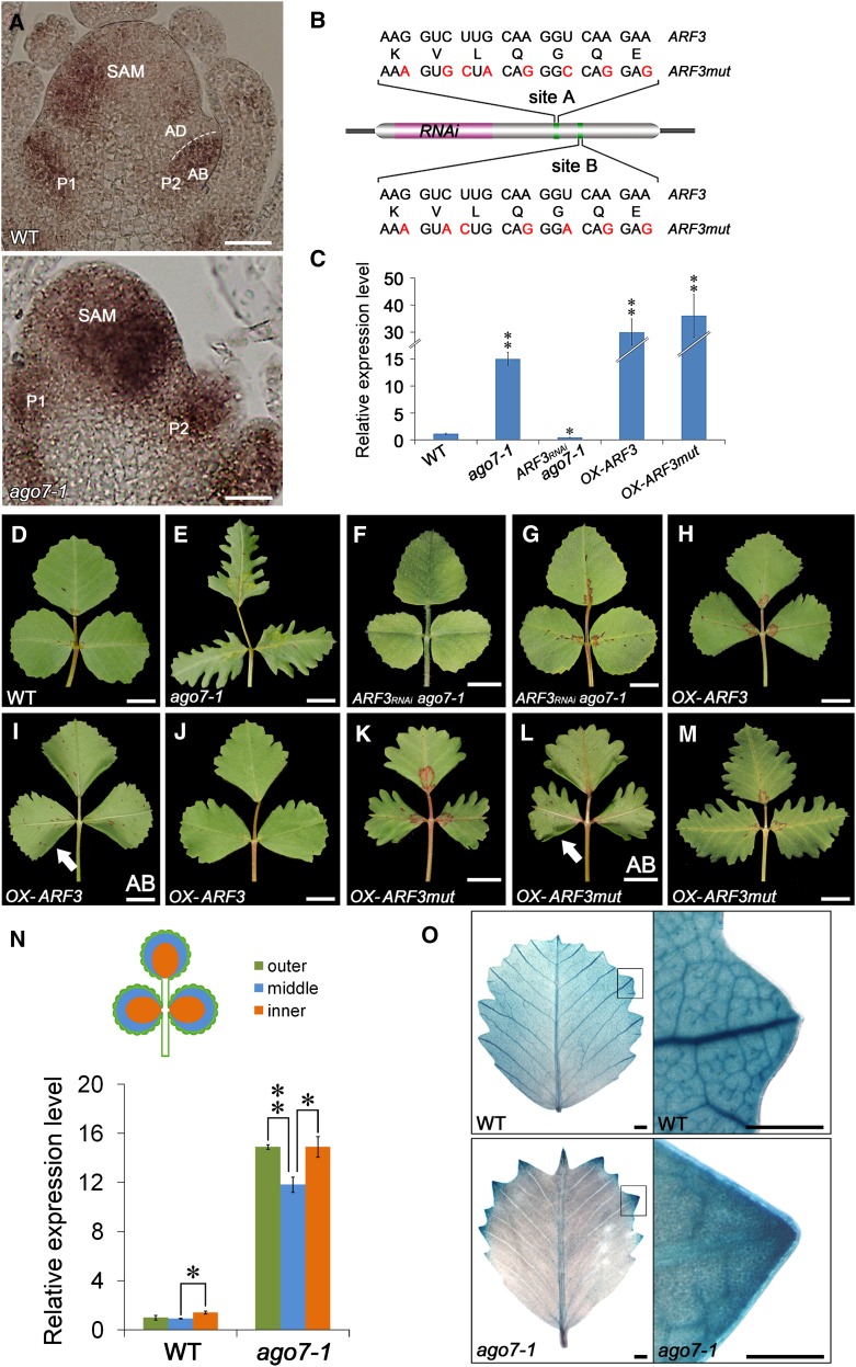
Leaf shape elaboration and organ separation are critical for plant morphogenesis. We characterized the developmental roles of lobed leaflet1 by analyzing a recessive mutant in the model legume Medicago truncatula. An ortholog of Arabidopsis thaliana argonaute7 (AGO7), Mt-AGO7/lobed leaflet1, is required for the biogenesis of a trans-acting short interfering RNA (ta-siRNA) to negatively regulate the expression of auxin response factors in M. truncatula. Loss of function in AGO7 results in pleiotropic phenotypes in different organs. The prominent phenotype of the ago7 mutant is lobed leaf margins and more widely spaced lateral organs, suggesting that the trans-acting siRNA3 (TAS3) pathway negatively regulates the formation of boundaries and the separation of lateral organs in M. truncatula. Genetic interaction analysis with the smooth leaf margin1 (slm1) mutant revealed that leaf margin formation is cooperatively regulated by the auxin/SLM1 (ortholog of Arabidopsis PIN-formed1) module, which influences the initiation of leaf margin teeth, and the TAS3 ta-siRNA pathway, which determines the degree of margin indentation. Further investigations showed that the TAS3 ta-siRNA pathway and no apical meristem (ortholog of Arabidopsis cup-shaped cotyledon) antagonistically regulate both leaf margin development and lateral organ separation, and the regulation is partially dependent on the auxin/SLM1 module.


Figures

Figure 1.
Figure 1.
lol1-1/ago7-1 Mutant of M. truncatula Shows Defects in Leaf Development. (A) Adult leaf of the wild type. (B) Adult leaf of lol1-1/ago7-1 mutant. (C) Four-week-old plant of the wild type. One juvenile leaf (first) and five adult leaves (second to sixth) are marked based on the order of leaf initiation. (D) to (G) Juvenile leaf of the wild type (D) and lol1-1/ago7-1 ([E] to [G]). The juvenile leaf of lol1-1/ago7-1 exhibits additional leaflets. The inset in (F) shows a close view of the rod-shaped leaflet indicated by an arrow. (H) to (K) Transverse sections of terminal leaflets in the wild type (H) and lol1-1/ago7-1 (I), and petioles in the wild type (J) and lol1-1/ago7-1 (K). The sectioning regions are shown in (A) and (B) by white lines, respectively. Arrows point to the vascular bundles on the adaxial side. Ab, abaxial side; Ad, adaxial side; Pa, palisade mesophyll cell; Ph, phloem; WT, wild type; X, xylem. Bar in (A) to (G) = 5 mm; bar in (H) to (K) = 200 μm.
Figure 2.
Figure 2.
lol1-1/ago7-1 Mutant of M. truncatula Shows Defects in Flower Development. (A) Flower phenotype in the wild type. Arrow points to the bottom of fused alae and keel. (B) to (D) Dissected floral organs of the wild type. The top view of vexillum (B), the top view (left) and bottom view (right) of fused alae and keel (C), and the top view of a dissected sepal (D). Arrows point to the bottom of fused alae and keel. (E) Flower phenotype in the lol1-1/ago7-1. Arrowhead points to the overseparated alae and keel. (F) to (H) Dissected floral organs of the lol1-1/ago7-1. The top view of vexillum (F), the separated alae and keel (G), and the top view of a sepal (H). Arrowheads point to the basal regions of overseparated alae and keel. (I) and (J) The side view of the central carpel in the wild type (I) and lol1-1/ago7-1 (J). The insets in (I) and (J) show the top view of central carpels. (K) and (L) Scanning electron microscopy analysis of the central carpel in the wild type (K) and lol1-1/ago7-1 (L). Arrow in (K) points to the closed central carpel. Arrowhead in (L) points to the opened central carpel and exposed ovules. (M) and (N) Scanning electron microscopy analysis of anthers in the wild type (M) and lol1-1/ago7-1 (N). Arrow in (M) points to the dehiscing anther. Arrowhead in (N) shows defect in anther dehiscence in lol1-1/ago7-1. (O) and (P) Pollen staining in the wild type (O) and lol1-1/ago7-1 (P). The size of pollen and anther sacs was uneven and pollen was partially viable in lol1-1/ago7-1. A, alae; AN, anther; C, carpel; FI, filament; K, keel; OV, ovule; WT, wild type. Bar in (A) to (J) = 2 mm; bar in (K) to (P) = 200 μm.
Figure 3.
Figure 3.
Molecular Cloning and Expression Pattern of AGO7 in M. truncatula. (A) Schematic representation of the gene structure of AGO7. Three exons (block) and two introns (line) are shown. Numbers indicate nucleotide positions of the site of mutations. (B) PCR amplification of AGO7 from the wild type and ago7 mutants. A single insertion (∼5.3 kb) was detected in each mutant line. (C) RT-PCR analysis of AGO7 transcripts in the wild type and ago7 mutants. Actin was used as the loading control. Three technical replicates were performed. (D) RT-PCR analysis of AGO7 expression in different plant organs. Actin was used as the loading control. Three technical replicates were performed. (E) to (J) In situ hybridization analysis of AGO7 mRNA in vegetative and reproductive apices of the wild type. Bar = 50 μm in (E) to (J). (E) to (G) Longitudinal sections of the SAM at stages 3 and 4 ([E] and [F], respectively). The sense probe was hybridized and used as the control (G). (H) to (J) Longitudinal sections of the floral apical meristem at stages 5 and 7 ([H] and [I]). The sense probe was hybridized and used as the control (J). AB, abaxial; AD, adaxial; AN, anther; C, carpel; P, petal; S, stage; WT, wild type.
Figure 4.
Figure 4.
Characterization of Putative TAS3 ta-siRNA and Target Genes in M. truncatula. (A) A diagram represents the biogenesis of predicted TAS3 ta-siRNAs from TAS3 transcript directed by miR390. Putative ta-siRNAs are shown alternately in yellow and green. (B) Alignment of TAS3 ta-siRNAs among Mt (M. truncatula), Lj (L. japonicus), At (Arabidopsis), and Os (rice). (C) RT-PCR analyses of TAS3 5′D7(+), TAS3 5′D8(+), and miR390 in the wild type (WT) and ago7-1. EF1α was used as a control. Three technical replicates were performed. (D) Diagram represents TAS3 ta-siRNA and the coding sequence of three putative target ARF genes. (E) Relative expression level of three ARF genes in the juvenile leaf (first foliage leaf) and second foliage leaf of the wild type and ago7-1. Values are the mean and sd of three biological replicates. *P < 0.05; **P < 0.01.
Figure 5.
Figure 5.
Involvement of AGO7 in Leaf Margin Development in M. truncatula. (A) and (B) Observation of margin cells in the wild type (WT) (A) and ago7-1 (B) by scanning electron microscopy. Arrows point to the margin cells. Arrowheads point to the epidermal cells. (C) and (D) Transverse sections of leaf margin in the wild type (C) and ago7-1 (D). Arrow points to the ridge-like structure on the surface of margin cells in the wild type. Arrowhead points to the smooth surface of margin cells in ago7-1. (E) to (H) Observation of margin cells at the teeth tips in the wild type ([E] and [G]) and ago7-1 ([F] and [H]). Margin cells harboring the auxin response marker DR5 (DR5rev:GFP) were observed by scanning electron microscopy ([E] and [F]) and confocal microscopy ([G] and [H]). Cells with the ridge-like structure on the surface are marked in green in (E) and (F). (I) to (L) Observation of margin cells in the leaf sinus in the wild type ([I] and [K]) and ago7-1 ([J] and [L]). The margin cells are marked in purple and epidermal cells are marked in yellow in (I) and (J). Arrows point to the elongated margin cells in the wild type. Arrowheads point to the small and unelongated margin cells in ago7-1. (M) and (N) A schematic illustration of the developmental difference of leaf margin cells in the wild type (M) and ago7-1 (N). Bar in (A) to (D) = 20 μm; bar in (E) to (L) = 100 μm.
Figure 6.
Figure 6.
ARF3 Expression Pattern and Auxin Distribution Is Altered in ago7-1. (A) In situ hybridization of ARF3 in leaf primordia of the wild type and ago7-1. (B) A diagram represents the coding sequence of ARF3. The fragment used for ARF3RNAi and the two mutated ta-siARF target sites are shown. (C) Transcript levels of ARF3 in wild-type, mutant and transgenic plants. Values are the means and sd of three biological replicates. *P < 0.05; **P < 0.01. (D) and (E) Adult leaves of the wild type (D) and ago7-1 (E). (F) and (G) Adult leaves of ago7-1 transgenic plants harboring the ARF3RNAi construct. The leaves at the vegetative stage (F) and reproductive stage (G) are shown. (H) to (M) Adult leaves of transgenic plants overexpressing ARF3 and ARF3mut. Leaves at the vegetative stage ([H], [I], [K], and [L]) and reproductive stage ([J] and [M]) are shown. Arrows point to the downward-curled leaf margin in (I) and (L). (N) Transcript levels of the ARF3 in the different regions (outer, middle, and inner) of leaflet in the wild type and ago7-1. Values are the means and sd of three biological replicates. *P < 0.05; **P < 0.01. (O) DR5:GUS expression in developing leaflet of the wild type and ago7-1. Close views (empty boxes) of margin serrations in the wild type and ago7-1 are shown on the right side. AB, abaxial; AD, adaxial; P: plastochron; WT, wild type. Bar in (A) = 50 μm; bar in (D) to (M) = 5 mm; bar in (O) = 1 mm.
Figure 7.
Figure 7.
Formation of Lobed Leaf Margin in ago7-1 Is Partially Dependent on SLM1 in M. truncatula. (A) Transcript levels of SLM1 in the wild type (WT) and ago7-1. Transcript levels were measured by qRT-PCR using leaves from 6-week-old plants. Values are the means and sd of three biological replicates. (B) and (C) Transcript levels of SLM1 in different regions of leaflets in the wild type (B) and ago7-1 (C). Values are the means and sd of three biological replicates. (D) to (G) SLM1 expression pattern in leaf buds ([D] and [E]) and fully expanded terminal leaflet ([F] and [G]) of the wild type and ago7-1, as determined by detecting the SLM1pro:GUS activity. (H) to (K) Adult leaves of the wild type (H), ago7-1 (I), slm1-1 (J), and ago7-1slm1-1 (K). (L) The outlines of terminal leaflets of the wild type (red), ago7-1 (blue), slm1-1 (khaki), and ago7-1 slm1-1 (green) are overlapped. Bar in (D) and (E) = 3 mm; bar in (F) to (K) = 5 mm.
Figure 8.
Figure 8.
Interaction Between the TAS3 ta-siRNA Pathway and NAM in Leaf Development in M. truncatula. (A) to (C) Leaf margin phenotype of the wild type (WT). Arrow points to the tip of leaf margin serration. (D) to (F) Leaf margin phenotype of nam-2. Arrows point to the fused leaflets in (D) and the relatively smooth leaf margin serration in (F). Arrowhead marks the clustered leaflets without rachis in (D). (G) and (H) Adult leaves (G) and leaf margin (H) of ago7-1. (I) and (J) Adult leaves (I) and leaf margin (J) of ago7-1 nam-2. Arrow indicates the fused leaflets. Arrowhead points to the clustered leaflets with partially recovered rachis. (K) The leaf series of the wild type, nam-2, ago7-1, and ago7-1nam-2. (L) Percentage of fused leaflets in the wild type and mutants (n = 50). (M) and (N) Length of petiole (M) and rachis (N) of the wild type and mutants. The data were measured on the first fully expanded trifoliate of 6-week-old plants. Means ± sd are shown (n = 40). Bar in (A), (D), (G),and (I) = 5 mm; bar in (C) and (F) = 150 μm.
Figure 9.
Figure 9.
Leaf Margin Development Regulated by NAM Is Dependent on SLM1. (A) to (C) Adult leaves of nam-2 (A), slm1-1 (B), and nam-2 slm1-1 (C). Arrow points to the clustered leaflets. Arrowheads indicate the fused leaflets. (D) Percentage of fused leaflets in the 6-week-old wild type and mutants (n = 50). (E) Transcript levels of NAM in the wild type (WT) and slm1-1. Transcript levels were measured by qRT-PCR using leaf buds from 6-week-old plants. Values are the means and sd of three biological replicates. (F) In situ hybridization and expression patterns of NAM in leaf primordia of the wild type and slm1-1. Empty boxes mark the leaf margin. Bar in (A) to (C) = 5 mm; bar in (F) = 50 μm.
Figure 10.
Figure 10.
A Proposed Model Illustrating the Functional Roles of the TAS3 ta-siRNA Pathway, NAM, and SLM1 in Leaf Margin Development and Lateral Organ Separation. SLM1 is an auxin efflux carrier that regulates auxin accumulation during leaf development. The auxin/SLM1 module is required for the initiation of leaf marginal teeth. NAM is a positive regulator that promotes the formation of leaf margin serrations and the separation of lateral organs in a SLM1-dependent manner. AGO7 is required for the production of ta-siARFs, which repress the expression of ARF3 and ARF4a,b by posttranscriptional cleavage in the TAS3 ta-siRNA pathway. The TAS3 ta-siRNA pathway plays crucial roles in the determination of leaf margin cell fate and the establishment of lateral organ polarity. It functions as a repressor in the formation of marginal indentation and separation of lateral organs.

Similar articles

Cited by

References

    1. Abdi H. (2007). Bonferroni and Sidak corrections for multiple comparisons. In Encyclopedia of Measurement and Statistics, N.J. Salkind, ed. (Thousand Oaks, California: Sage), pp. 103–107.
    1. Adenot X., Elmayan T., Lauressergues D., Boutet S., Bouché N., Gasciolli V., Vaucheret H. (2006). DRB4-dependent TAS3 trans-acting siRNAs control leaf morphology through AGO7. Curr. Biol. 16: 927–932 - PubMed
    1. Aida M., Tasaka M. (2006). Genetic control of shoot organ boundaries. Curr. Opin. Plant Biol. 9: 72–77 - PubMed
    1. Allen E., Xie Z., Gustafson A.M., Carrington J.C. (2005). microRNA-directed phasing during trans-acting siRNA biogenesis in plants. Cell 121: 207–221 - PubMed
    1. Barkoulas M., Hay A., Kougioumoutzi E., Tsiantis M. (2008). A developmental framework for dissected leaf formation in the Arabidopsis relative Cardamine hirsuta. Nat. Genet. 40: 1136–1141 - PubMed

Publication types

MeSH terms

LinkOut - more resources