Review
. 2013 Dec 23;203(6):875-81.
doi: 10.1083/jcb.201308087.

Phase transitions and size scaling of membrane-less organelles

Affiliations
Review

Phase transitions and size scaling of membrane-less organelles

Clifford P Brangwynne. J Cell Biol. .

Abstract

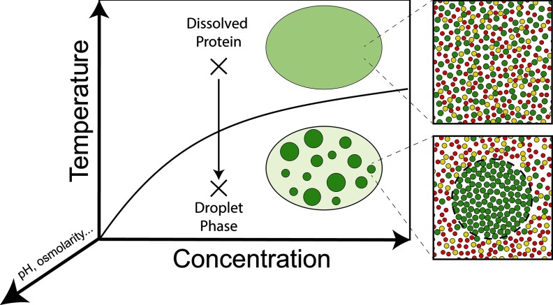
The coordinated growth of cells and their organelles is a fundamental and poorly understood problem, with implications for processes ranging from embryonic development to oncogenesis. Recent experiments have shed light on the cell size-dependent assembly of membrane-less cytoplasmic and nucleoplasmic structures, including ribonucleoprotein (RNP) granules and other intracellular bodies. Many of these structures behave as condensed liquid-like phases of the cytoplasm/nucleoplasm. The phase transitions that appear to govern their assembly exhibit an intrinsic dependence on cell size, and may explain the size scaling reported for a number of structures. This size scaling could, in turn, play a role in cell growth and size control.


Figures

Figure 1.
Figure 1.
RNA/protein droplets. (A) Nucleoli (and other RNP droplets) within the nucleus of an X. laevis oocyte. Adapted from Brangwynne et al. (2011). (B) In vitro droplets formed from myelin basic protein (MBP). Adapted from Aggarwal et al. (2013). (C) In vitro droplets of fluorescently labeled multi-domain SH34/PRM4 proteins. Adapted with permission from Macmillan Publishers Ltd.: Nature (Li et al., 2012).
Figure 2.
Figure 2.
Phase transitions and size scaling. (A) Growth of a cell without the production of droplet component(s) will lead to decreased total component concentration (conc.), shifting the location within the phase diagram. In this case, a single nucleated droplet would dissolve (no scaling). (B) In a situation where cell size changes either through growth with a constant total component concentration or where cell size changes through division, the location in the phase diagram will not change, and the size of a single nucleated droplet will scale with cell size.
Figure 3.
Figure 3.
Cell growth and RNP size scaling. In this image, dorsal root ganglia neurons of different size are stained with propidium iodide to visualize a single large nucleolus (white spot) within each cell nucleus. The size of nuclei and nucleoli increases with cell size. Adapted from Berciano et al. (2007) with permission from Elsevier.
Figure 4.
Figure 4.
Gravity and large droplets in large cells. (A and B) An intact X. laevis oocyte nucleus (GV) contains hundreds of RNP droplets, including nucleoli (red) and histone locus bodies (green), distributed through the GV (XY projection shown in top panels, XZ projection shown in bottom panels). A nuclear actin scaffold keeps these droplets from fusing. Bar, 50 μm. (C and D) Upon actin disruption, nucleoli undergo gravitational sedimentation to the bottom of the nucleus, where they undergo massive fusion events, ultimately resulting in a few large droplets (E). Panels B–E adapted from Feric and Brangwynne (2013), copyright Nature Publishing Group.
None

References

    1. Ader C., Frey S., Maas W., Schmidt H.B., Görlich D., Baldus M. 2010. Amyloid-like interactions within nucleoporin FG hydrogels. Proc. Natl. Acad. Sci. USA. 107:6281–6285 10.1073/pnas.0910163107 - DOI - PMC - PubMed
    1. Aggarwal S., Snaidero N., Pähler G., Frey S., Sánchez P., Zweckstetter M., Janshoff A., Schneider A., Weil M.-T., Schaap I.A.T., et al. 2013. Myelin membrane assembly is driven by a phase transition of myelin basic proteins into a cohesive protein meshwork. PLoS Biol. 11:e1001577 10.1371/journal.pbio.1001577 - DOI - PMC - PubMed
    1. An S., Kumar R., Sheets E.D., Benkovic S.J. 2008. Reversible compartmentalization of de novo purine biosynthetic complexes in living cells. Science. 320:103–106 10.1126/science.1152241 - DOI - PubMed
    1. Asherie N. 2004. Protein crystallization and phase diagrams. Methods. 34:266–272 10.1016/j.ymeth.2004.03.028 - DOI - PubMed
    1. Berciano M.T., Novell M., Villagra N.T., Casafont I., Bengoechea R., Val-Bernal J.F., Lafarga M. 2007. Cajal body number and nucleolar size correlate with the cell body mass in human sensory ganglia neurons. J. Struct. Biol. 158:410–420 10.1016/j.jsb.2006.12.008 - DOI - PubMed

Publication types

Substances