

doi: 10.1155/2013/197415.
Epub 2013 Dec 4.
Interhypothalamic adhesion in a 9-month-old male with cleft palate
Affiliations
- PMID: 24368961
- PMCID: PMC3867890
- DOI: 10.1155/2013/197415
Item in Clipboard
Interhypothalamic adhesion in a 9-month-old male with cleft palate
Case Rep Radiol.
2013.
Abstract
A 9-month-old male infant with multiple congenital anomalies including cleft lip and palate was referred to us for a brain MR to exclude additional intracranial abnormalities. Imaging revealed an interhypothalamic adhesion, which we present as a possible forme fruste of holoprosencephaly.
Figures

Sagittal FSPGR BRAVO T1WI (repetition time msec/echo time msec/inversion time msec, 10/4/450) showing a nodular parenchymal band in the third ventricle at the level of the hypothalamus between the anterior commissure and mammillary body (arrow). The pituitary infundibulum, adenohypophysis, and neurohypophysis are normal. Note metallic susceptibility artifact in the roof of the oral cavity from prior cleft palate repair.

Coronal FIESTA-C (repetition time msec/echo time, 10/4) showing a horizontal interhypothalamic parenchymal band crossing the third ventricle (arrow).

Postcontrast coronal FSPGR T1WI (repetition time msec/echo time msec/inversion time msec, 19/9/375) depicting a parenchymal band that is isointense to gray matter crossing the third ventricle and connecting the medial hypothalami (arrow).
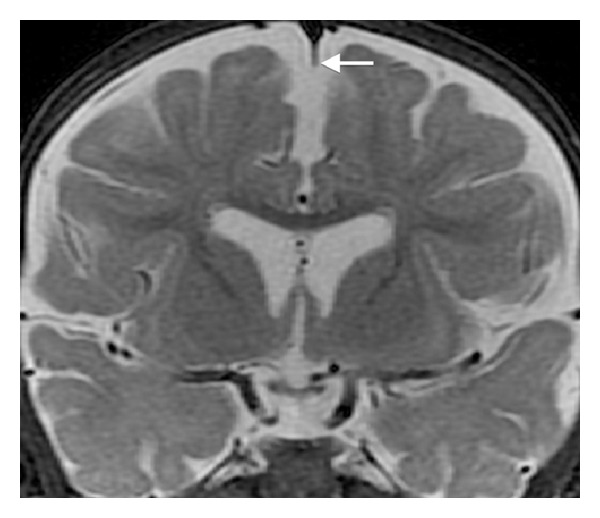
Coronal STIR (repetition time msec/echo time msec/inversion time msec, 4217/83/150) image demonstrating partial deficiency of the falx cerebri (arrow). Note the normal septum pellucidum.
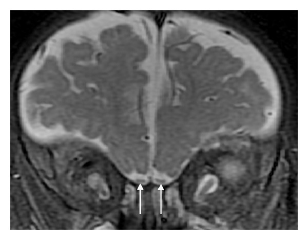
Coronal STIR (repetition time msec/echo time msec/inversion time msec, 4217/83/150) image demonstrating equivocal olfactory tract volume loss (arrows).

Coronal FIESTA-C (repetition time msec/echo time, 10/4) demonstrating malrotation of the hippocampal heads (arrows).
References
-
- Osborn AG. Osborn’s Brain: Imaging, Pathology, and Anatomy. Philadelphia, Pa, USA: Wolters Kluwer-Lippincott Williams & Wilkins; 2012.
-
- Swaiman K, Ashwal S, Ferriero DM, Ferriero D. Swaiman's Pediatric Neurology: Principles and Practice. 5th edition. Elsevier Saunders; 2012.
-
- Tortori-Donati P, Rossi A, Biancheri R. Brain malformations. In: Tortori-Donati P, Rossi A, editors. Pediatric Neuroradiology: Brain, Head, Neck and Spine. 1st edition. Berlin, Germany: Springer; 2009. pp. 86–94.
-
- Dias M, Partington M. Normal and abnormal embryology of the brain. In: Winn H, editor. Youmans Neurological Surgery. 6th edition. Philadelphia, Pa, USA: Elsevier Saunders; 2011. pp. 1883–1897.
LinkOut - more resources
Full Text Sources
Other Literature Sources
