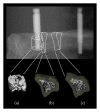
External mechanical microstimuli modulate the osseointegration of titanium implants in rat tibiae
- PMID: 24369009
- PMCID: PMC3866820
- DOI: 10.1155/2013/234093
External mechanical microstimuli modulate the osseointegration of titanium implants in rat tibiae
Abstract
Purpose: To assess the effect of external mechanical microstimuli of controlled magnitude on the microarchitecture of the peri-implant bone beds in rat tibiae.
Materials and methods: Tibiae of forty rats were fitted with two transcutaneous titanium cylinders. After healing, the implants were loaded to 1 to 3 N, five days/week for four weeks. These force levels translated into intraosseous strains of 700 ± 200 με, 1400 ± 400 με, and 2100 ± 600 με. After sacrifice, the implants' pullout strength was assessed. Second, the bone's microarchitecture was analyzed by microcomputed tomography (μCT) in three discrete regions of interest (ROIs). Third, the effect of loading on bone material properties was determined by nanoindentation.
Results: The trabecular BV/TV significantly increased in an ROI of 0.98 mm away from the test implant in the 1 N versus the 3 N group with an opposite trend for cortical thickness. Pull-out strength significantly increased in the 2 N relatively to the nonstimulated group. Higher values of E-modulus and hardness were observed in the trabecular bone of the 2 N group.
Conclusion: The in vivo mechanical loading of implants induces load-dependent modifications in bone microarchitecture and bone material properties in rat tibiae. In pull-out strength measurements, implant osseointegration was maximized at 2 N (1400 ± 400 με).
Figures


Similar articles
-
Peri-implant bone adaptations to overloading in rat tibiae: experimental investigations and numerical predictions.Clin Oral Implants Res. 2016 Nov;27(11):1444-1453. doi: 10.1111/clr.12760. Epub 2016 Feb 11. Clin Oral Implants Res. 2016. PMID: 26864329
-
Implementation of the "loaded implant" model in the rat using a miniaturized setup--description of the method and first results.Clin Oral Implants Res. 2012 Dec;23(12):1352-9. doi: 10.1111/j.1600-0501.2011.02349.x. Epub 2011 Dec 6. Clin Oral Implants Res. 2012. PMID: 22145779
-
Low protein intake is associated with impaired titanium implant osseointegration.J Bone Miner Res. 2006 Feb;21(2):258-64. doi: 10.1359/JBMR.051009. Epub 2005 Oct 18. J Bone Miner Res. 2006. PMID: 16418781
-
The role of TiO2 nanotube surface on osseointegration of titanium implants: Biomechanical and histological study in rats.Microsc Res Tech. 2020 Jul;83(7):817-823. doi: 10.1002/jemt.23473. Epub 2020 Mar 30. Microsc Res Tech. 2020. PMID: 32227674
-
PTH improves titanium implant fixation more than pamidronate or renutrition in osteopenic rats chronically fed a low protein diet.Osteoporos Int. 2010 Jun;21(6):957-67. doi: 10.1007/s00198-009-1031-x. Epub 2009 Oct 27. Osteoporos Int. 2010. PMID: 19859647
Cited by
-
Enhanced Bone Remodeling Effects of Low-Modulus Ti-5Zr-3Sn-5Mo-25Nb Alloy Implanted in the Mandible of Beagle Dogs under Delayed Loading.ACS Omega. 2019 Nov 1;4(20):18653-18662. doi: 10.1021/acsomega.9b02580. eCollection 2019 Nov 12. ACS Omega. 2019. PMID: 31737825 Free PMC article.
-
Improved Bone Micro Architecture Healing Time after Implant Surgery in an Ovariectomized Rat.J Hard Tissue Biol. 2016;25(3):257-262. doi: 10.2485/jhtb.25.257. J Hard Tissue Biol. 2016. PMID: 28133434 Free PMC article.
-
Preclinical mouse models for assessing axial compression of long bones during exercise.Bonekey Rep. 2015 Dec 23;4:768. doi: 10.1038/bonekey.2015.138. eCollection 2015. Bonekey Rep. 2015. PMID: 26788286 Free PMC article.
-
Effect of a dietary supplement on peri-implant bone strength in a rat model of osteoporosis.J Prosthodont Res. 2016 Apr;60(2):131-7. doi: 10.1016/j.jpor.2015.12.006. Epub 2016 Jan 16. J Prosthodont Res. 2016. PMID: 26787534 Free PMC article.
References
-
- Dayer R, Rizzoli R, Kaelin A, Ammann P. Low protein intake is associated with impaired titanium implant osseointegration. Journal of Bone and Mineral Research. 2006;21(2):258–264. - PubMed
-
- Maïmoun L, Brennan TC, Badoud I, Dubois-Ferriere V, Rizzoli R, Ammann P. Strontium ranelate improves implant osseointegration. Bone. 2010;46(5):1436–1441. - PubMed
-
- Dayer R, Brennan TC, Rizzoli R, Ammann P. PTH improves titanium implant fixation more than pamidronate or renutrition in osteopenic rats chronically fed a low protein diet. Osteoporosis International. 2010;21(6):957–967. - PubMed
-
- Lee KCL, Maxwell A, Lanyon LE. Validation of a technique for studying functional adaptation of the mouse ulna in response to mechanical loading. Bone. 2002;31(3):407–412. - PubMed
-
- Lambers FM, Schulte FA, Kuhn G, Webster DJ, Müller R. Mouse tail vertebrae adapt to cyclic mechanical loading by increasing bone formation rate and decreasing bone resorption rate as shown by time-lapsed in vivo imaging of dynamic bone morphometry. Bone. 2011;49(6):1340–1350. - PubMed
Publication types
MeSH terms
Substances
LinkOut - more resources
Full Text Sources
Other Literature Sources

