Review
. 2013:2013:543803.
doi: 10.1155/2013/543803. Epub 2013 Nov 28.

Synaptic dysfunction in prion diseases: a trafficking problem?

Affiliations
Review

Synaptic dysfunction in prion diseases: a trafficking problem?

Assunta Senatore et al. Int J Cell Biol. 2013.

Abstract

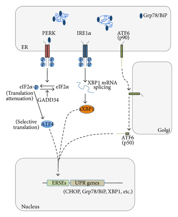
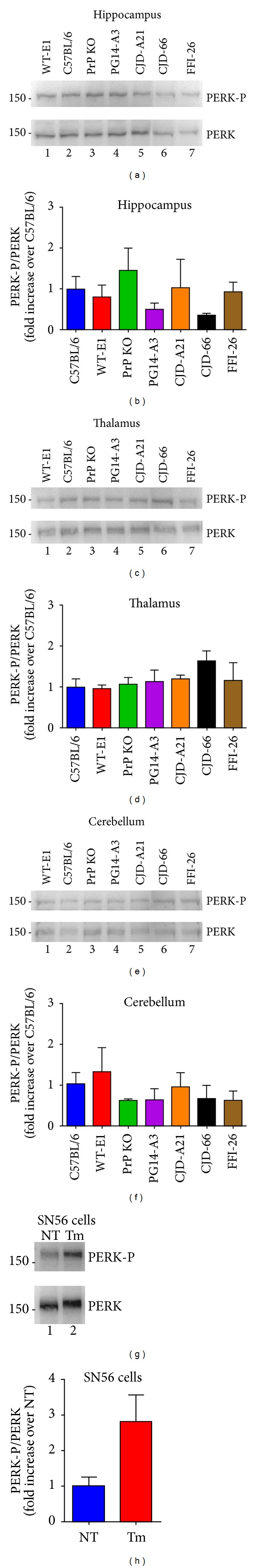
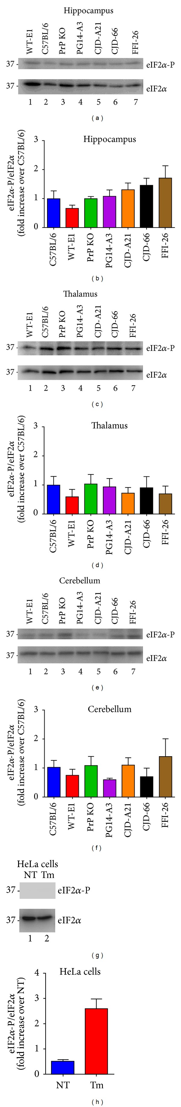
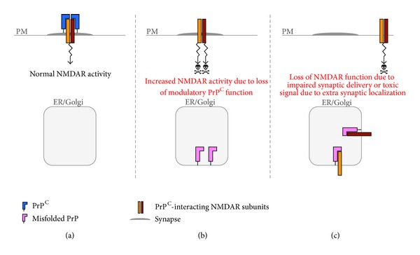
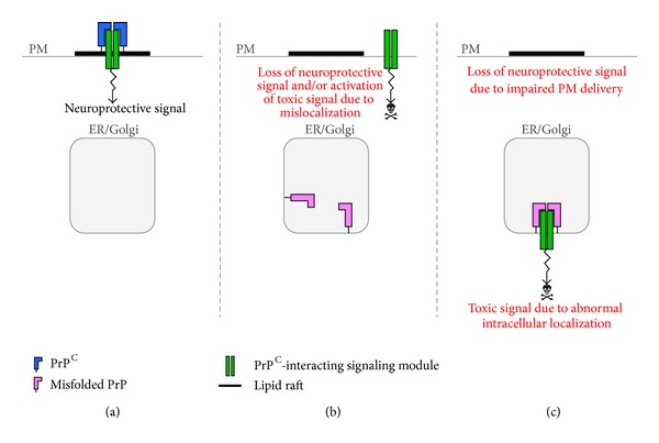
Synaptic dysfunction is an important cause of neurological symptoms in prion diseases, a class of clinically heterogeneous neurodegenerative disorders caused by misfolding of the cellular prion protein (PrP(C)). Experimental data suggest that accumulation of misfolded PrP(C) in the endoplasmic reticulum (ER) may be crucial in synaptic failure, possibly because of the activation of the translational repression pathway of the unfolded protein response. Here, we report that this pathway is not operative in mouse models of genetic prion disease, consistent with our previous observation that ER stress is not involved. Building on our recent finding that ER retention of mutant PrP(C) impairs the secretory trafficking of calcium channels essential for synaptic function, we propose a model of pathogenicity in which intracellular retention of misfolded PrP(C) results in loss of function or gain of toxicity of PrP(C)-interacting proteins. This neurotoxic modality may also explain the phenotypic heterogeneity of prion diseases.


Figures

Figure 1
Figure 1
UPR signaling pathways in mammalian cells. The UPR is mediated by three ER-resident transmembrane proteins that sense ER stress through Grp78/BiP binding/release to their luminal domains and/or through direct interaction with unfolded proteins. The kinase PERK (double-stranded RNA-activated protein kinase-like ER kinase) is activated by dimerization and phosphorylation. Once activated, it phosphorylates eIF2α (eukaryotic translation initiation factor 2). This inhibits protein translation, reducing the overload of misfolded proteins. This pathway also selectively enhances translation of ATF4 (activating transcription factor 4) that induces the expression of CHOP. In ER-stressed cells, IRE1α (inositol-requiring transmembrane kinase and endonuclease) multimerizes and autophosphorylates, setting in motion its RNAse activity. Activated IRE1α initiates the unconventional splicing of the mRNA encoding the transcriptional factor XBP1 (X-box-binding protein 1) to produce sXBP1, a more stable form of XBP1 with a potent transactivator domain that enhances transcription of genes involved in protein folding, secretion, and ER-associated degradation. Another ER stress sensor is ATF6 (activating transcription factor 6). This is a type II ER transmembrane protein whose cytosolic domain contains a bZIP transcriptional factor. ATF6 is transported to the Golgi where it is processed within the transmembrane domain to release the cytosolic domain, which translocates to the nucleus and induces expression of the ER chaperone Grp78/BiP and XBP1. GADD34, a protein phosphatase upregulated by the PERK pathway, dephosphorylates eIF2α to restore global protein synthesis. ERSEs: ER stress responsive elements.
Figure 2
Figure 2
Phosphorylation of PERK is not increased in the brains of mutant PrP mice. Phosphorylation of PERK was evaluated in brain extracts of the following mice: C57/BL6J (PrP level 1X), PrP KO (C57BL/6J/Prnp 0/0, European Mouse Mutant Archive, Rome, Italy; EM: 01723), Tg(WT-E1+/+) overexpressing 3F4-tagged wild-type PrP at ~4X, Tg(PG14-A3+/−) expressing 3F4-tagged PG14 PrP at ~1X, Tg(CJD-A21+/−) expressing 3F4-tagged D177N/V128 PrP at ~1X, Tg(CJD-66+/−) expressing untagged D177N/V128 PrP at ~2.5X, and Tg(FFI-26+/−) mice expressing untagged D177N/M128 PrP at ~2.5X. These mice were originally generated on a C57BL/6J X CBA hybrid and then bred with C57BL/6J/Prnp 0/0 mice ([92, 93] and manuscript in preparation). Proteins were extracted from the hippocampus, thalamus, and cerebellum of mice of the indicated strains/genotype ((a)–(f)) or from SN56 cells ((g) and (h)), using a lysis buffer containing 50 mM Tris, 150 mM NaCl, 2 mM EDTA, 1 mM MgCl2, 100 mM NaF, 10% glycerol, 1% Triton X-100, 1% Na deoxycholate, 0.1% SDS, and 125 mM sucrose, supplemented with Phos-STOP and protease inhibitors (Roche) [80]. Protein extracts (50 μg) were analyzed by Western blot with anti-PERK-P and antitotal PERK antibodies (1 : 1000; Cell Signaling) ((a), (c), (e), and (g)). Molecular mass markers are in kilodaltons. Phosphorylation levels were quantified by densitometric analysis of Western blots and expressed as the -fold increase over the level in C57BL/6 mice ((b), (d), (f), and (h)). Tunicamycin (Tm) treated HeLa cells were analyzed at 2 hours as control for UPR activation. Each value is the mean ± SEM of three animals of 300–350 days of age or from three independent cell preparations.
Figure 3
Figure 3
Phosphorylation of eIF2α is not increased in brains of mutant PrP mice. The same brain protein extracts (20 μg) as in Figure 2 ((a)–(f)) or lysates of HeLa cells ((g) and (h)) were analyzed by Western blot with anti-eIF2α-P and antitotal eIF2α antibodies (1 : 1000; Cell Signaling). Molecular mass markers are in kilodaltons. Phosphorylation levels were quantified by densitometric analysis of Western blots and expressed as the -fold increase over the level in C57BL/6 mice ((b), (d), (f), and (h)). Tunicamycin (Tm) treated HeLa cells were analyzed at 2 hours as control for UPR activation. Each value is the mean ± SEM of three animals of 300–350 days of age or from three independent cell preparations.
Figure 4
Figure 4
A role for intracellular PrP retention in NMDAR dysfunction. (a) PrPC on the plasma membrane (PM) attenuates NMDAR activity by associating with the NR2D subunit. (b) Owing to PrPC misfolding in transport organelles (ER/Golgi), PrPC is retained intracellularly. This results in increased NMDAR activation, potentially triggering neurotoxicity. (c) Intracellular retention of misfolded PrPC with NR2D and NR1 subunits results in impaired delivery of NMDARs to the cell surface or their abnormal targeting to extrasynaptic sites, leading to loss of NMDAR function and/or activation of neurotoxic stimuli.
Figure 5
Figure 5
Theoretical model for how intracellular retention could perturb PrPC-dependent signaling. (a) PrPC acts as scaffold molecules that keep a prosurvival signaling complex in lipid rafts of the plasma membrane (PM). The lipid raft localization would be essential to activate neuroprotective signaling. (b) Owing to retention in transport organelles (ER/Golgi), PrPC function is lost and the signaling complex localizes in nonraft regions of the PM, losing its neuroprotective activity and potentially eliciting a neurotoxic signal. (c) Misfolded PrP sequesters the signaling module in intracellular compartments, leading to loss of neuroprotective function on the cell membrane. Intracellular retention might also cause the complex to function abnormally and generate a toxic signal.

References

    1. Collinge J. Prion diseases of humans and animals: their causes and molecular basis. Annual Review of Neuroscience. 2001;24:519–550. - PubMed
    1. Watts JC, Balachandran A, Westaway D. The expanding universe of prion diseases. PLOS Pathogens. 2006;2(3):p. e26. - PMC - PubMed
    1. Knight RS, Will RG. Prion diseases. Journal of Neurology, Neurosurgery & Psychiatry. 2004;75(supplement 1):i36–i42. - PMC - PubMed
    1. Brown P, Gibbs CJ, Jr., Rodgers-Johnson P, et al. Human spongiform encephalopathy: the National Institutes of Health series of 300 cases of experimentally transmitted disease. Annals of Neurology. 1994;35(5):513–529. - PubMed
    1. Liberski PP, Sikorska B, Brown P. Kuru: the first prion disease. Advances in Experimental Medicine and Biology. 2012;724:143–153. - PubMed

LinkOut - more resources