

Review
doi: 10.1021/cr4004488.
Epub 2013 Dec 26.
Nonredox nickel enzymes
Affiliations
- PMID: 24369791
- PMCID: PMC5675112
- DOI: 10.1021/cr4004488
Item in Clipboard
Review
Nonredox nickel enzymes
Chem Rev.
.
Abstract
Conflict of interest statement
The authors declare no competing financial interest.
Figures
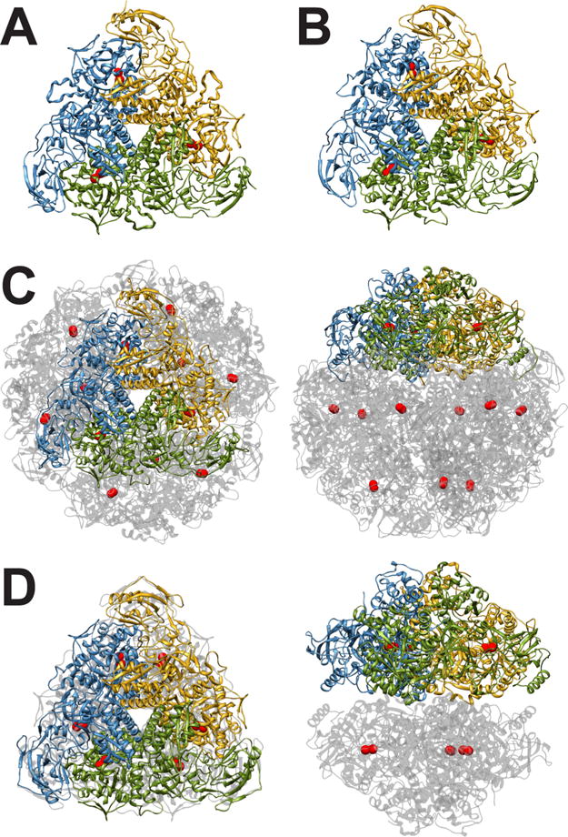
(A) Ribbon scheme of the functional oligomer (αβγ)3 of B. pasteurii urease. (B) Ribbon scheme of the functional oligomer (αβγ)3 of K. aerogenes urease. (C) Ribbon scheme of the functional oligomer [(αβ)3]4 of H. pylori urease seen through the ternary axis (left panel) and rotated by 90° along the horizontal axis (right panel). (D) Ribbon scheme of the functional oligomer [(α)3]2 of C. ensiformis urease seen through the ternary axis (left panel) and rotated by 90° along the horizontal axis (right panel).
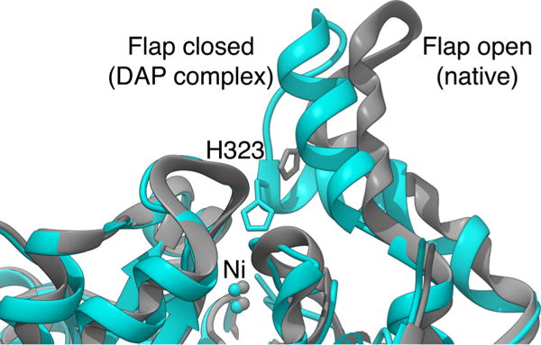
Ribbon scheme of the active site flap of B. pasteurii urease, highlighting the open and closed conformations observed in the native and the DAP-inhibited structures, respectively.
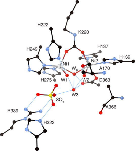
CrystalMaker drawing of the crystallographic structural models for the active site obtained for B. pasteurii urease (PDB code 2UBP) in the native state. The nickel ions are represented in gray, while CPK coloring is used for all other atoms. Hydrogen bonds are shown as thin blue lines. The BPU residue-numbering scheme (all residues belonging to the α subunit) is used. The residue indicated with the letter “K” is the carbamylated lysine.
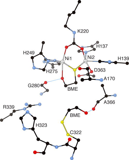
CrystalMaker drawing of the crystallographic structural model for the active site obtained for B. pasteurii urease complexed with β-mercaptoethanol (BME) (PDB code 1UBP). The nickel ions are represented in gray, while CPK coloring is used for all other atoms. Hydrogen bonds are shown as thin blue lines. The BPU residue-numbering scheme (all residues belonging to the α subunit) is used. The residue indicated with the letter “K” is the carbamylated lysine.
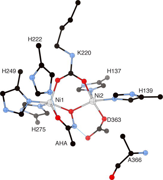
CrystalMaker drawing of the crystallographic structural model for the active site obtained for urease from B. pasteurii complexed with acetohydroxamic acid (AHA) (PDB code 4UBP). The nickel ions are represented in gray, while CPK coloring is used for all other atoms. Hydrogen bonds are shown as thin blue lines. The BPU residue-numbering scheme (all residues belonging to the α subunit) is used. The residue indicated with the letter “K” is the carbamylated lysine.
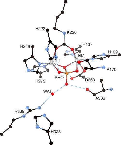
CrystalMaker drawing of the crystallographic structural model for the active site obtained for B. pasteurii urease complexed with phosphate (PHO) (PDB code 1IE7). The nickel ions are represented in gray and phosphorus is in orange, while CPK coloring is used for all other atoms. WAT = solvent molecule. Hydrogen bonds are shown as thin blue lines. The BPU residue-numbering scheme (all residues belonging to the α subunit) is used. The residue indicated with the letter “K” is the carbamylated lysine.
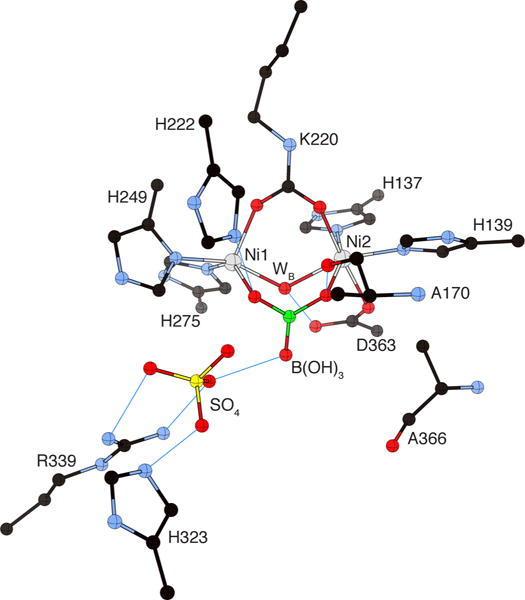
CrystalMaker drawing of the crystallographic structural model for the active site obtained for B. pasteurii urease complexed with boric acid B(OH)3 (PDB code 1S3T). The nickel ions are represented in gray and boron is in green, while CPK coloring is used for all other atoms. WB = nickel-bridging hydroxide. Hydrogen bonds are shown as thin blue lines. The BPU residue-numbering scheme (all residues belonging to the α subunit) is used. The residue indicated with the letter “K” is the carbamylated lysine.
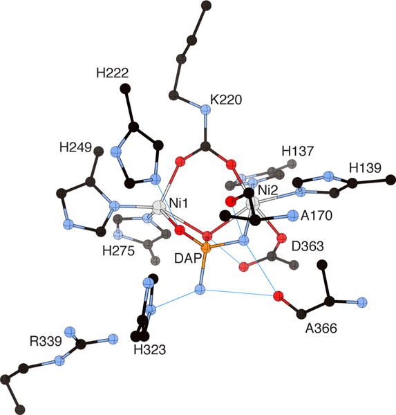
CrystalMaker drawing of the crystallographic structural model for the active site obtained for B. pasteurii urease complexed with diaminophosphate (DAP) (PDB code 3UBP). The nickel ions are represented in gray and phosphorus is in orange, while CPK coloring is used for all other atoms. Hydrogen bonds are shown as thin blue lines. The BPU residue-numbering scheme (all residues belonging to the α subunit) is used. The residue indicated with the letter “K” is the carbamylated lysine.
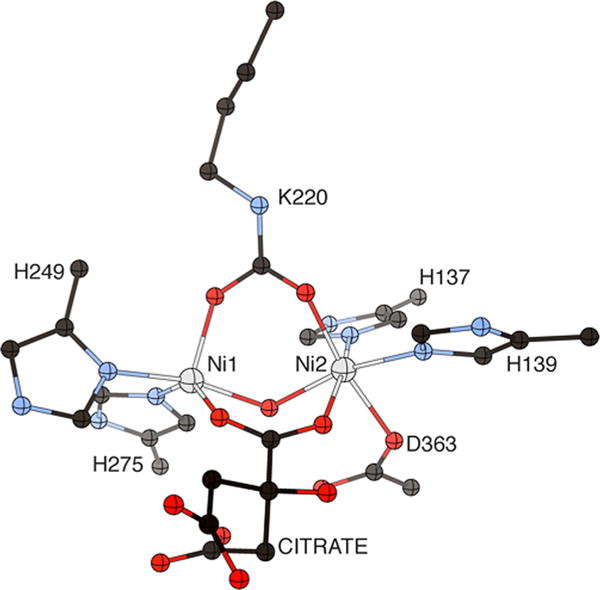
CrystalMaker drawing of the crystallograhic structural model for the active site obtained for B. pasteurii urease complexed with citrate (PDB code 4AC7). The nickel ions are represented in gray, while CPK coloring is used for all other atoms. The BPU residue-numbering scheme (all residues belonging to the alpha subunit) is used. The residue indicated with the letter “K” is the carbamylated lysine.

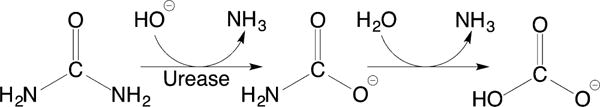
Structure-based urease catalytic mechanism of the enzymatic hydrolysis of urea. The BPU residue-numbering scheme is used.
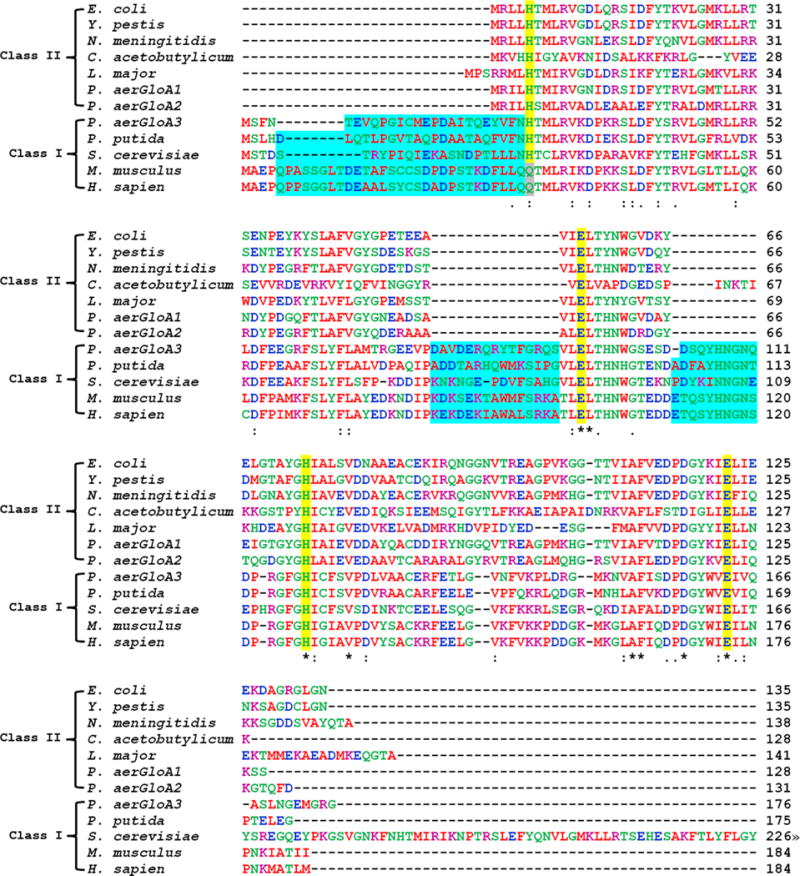
Sequence alignments of selected class I and class II glyoxalase I enzymes created using Clustal W2. Amino acids are colored by property (hydrophobic (red), acidic (blue), basic (purple), other (green)). Metal binding residues are highlighted in yellow. Residues marked with an asterisk (∗) are invariant; those marked by other symbols represent low (:) and moderate (.) variability. The N-terminal extension and additional loops found in class I enzymes are highlighted in blue. The S. cerevisiae sequence was truncated after 226 of 326 residues.
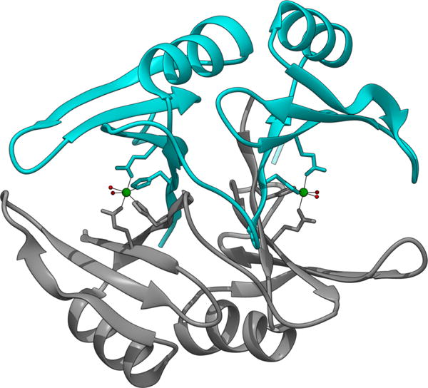
Ribbon diagram of the crystal structure of E. coli Glo I, (PDB code 1F9Z) showing the two subunits of the homo dimer in cyan and gray and the location of the two Ni sites (green spheres) at subunit interfaces.
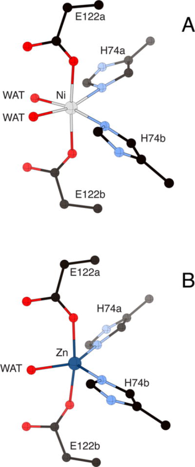
Comparison of the metal site structure of the Ni(II) complex (panel A, PDB code 1F9Z) and the Zn(II) complex (panel B, 1FA5) of E. coli Glo I showing the change in coordination number and geometry for the two metals. The nickel and zinc ions are represented in gray and dark blue, respectively, while CPK coloring is used for all other atoms. WAT = solvent molecules. Protein residues are distinguished by letters indicating the two different subunits of the enzyme.
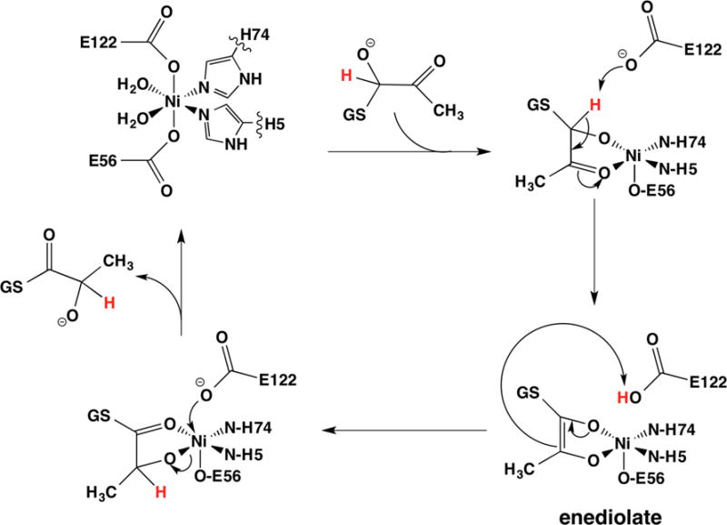
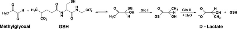
Putative reaction mechanism for the isomerization catalyzed by Glo I that involves coordination of the substrate.
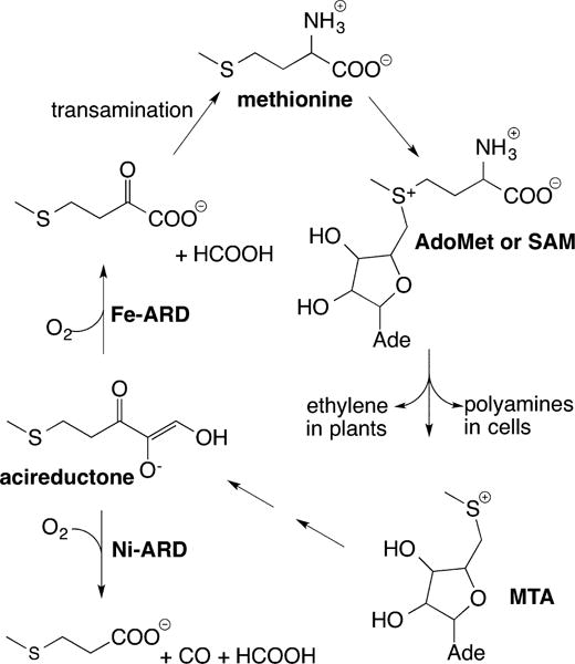
Simplified diagram of the methionine salvage pathway, illustrating key products, intermediates, and catalysis by Ni-ARD vs Fe-ARD.
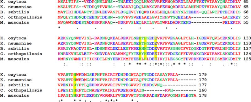
Sequence alignments of selected ARD enzymes created using Clustal W2. The sequences are numbered from Met0, since this residue is cleaved in the mature enzyme. Amino acids are colored by property (hydrophobic (red), acidic (blue), basic (purple), other (green)). Metal binding residues are highlighted in yellow. Residues marked with an asterisk (∗) are invariant; those marked by other symbols represent low (:) and moderate (.) variability.
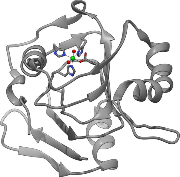
Ribbon diagram of the NMR structure of K. oxytoca Ni-ARD (PDB code 1ZRR) showing the cupin fold and the location of the metal ion (green sphere) with the ligand environment shown as sticks.
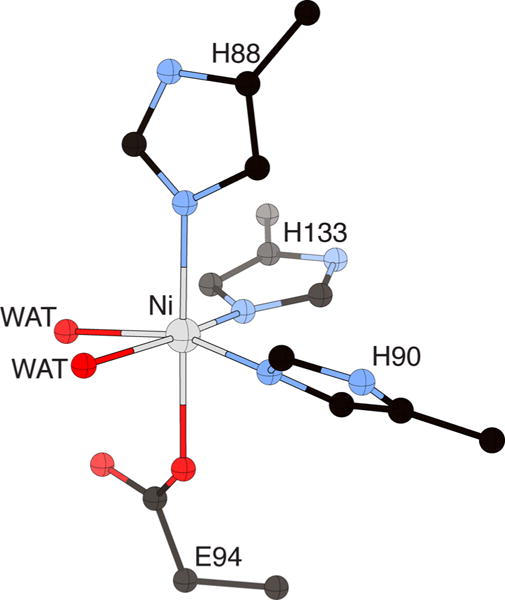
Metal site structure of K. oxytoca Ni-ARD (PDB code 1ZRR), showing the His3Glu coordination of the metal site and the two cis-aqua ligands in the positions used in binding substrate. The nickel ion is represented in gray, while CPK coloring is used for all other atoms. WAT = solvent molecules.
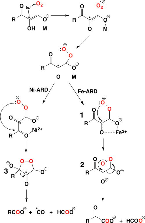
Proposed reaction mechanism illustrating the chelate hypothesis to explain the regioselectivity of the reactions catalyzed by Ni-ARD vs Fe-ARD. The results of incorporation of 18O and 14C labeling studies are indicated by the red O atoms and the asterisks (∗). (Adapted with permission from ref . Copyright 2007 John Wiley & Sons, Ltd.)
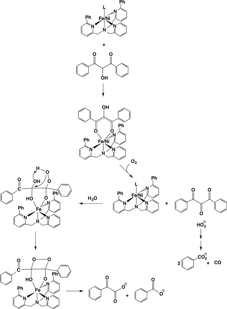
Model chemistry illustrating the role of substrate hydration in determining the regioselectivity of the reactions catalyzed by Ni-ARD vs Fe-ARD.
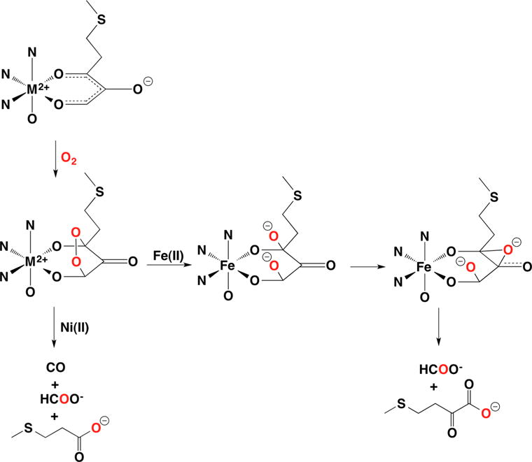
Mechanisms for Ni-ARD vs Fe-ARD catalysis from computational modeling indicate that the electronic structure of the metal ions leads to additional intermediates in the Fe-ARD reaction pathway.
References
-
- Sigel A, Sigel H, Sigel RKO, editors. Nickel and Its Surprising Impact on Nature. John Wiley & Sons, Ltd.; Chichester, England: 2007.
-
- Zambelli B, Ciurli S. Nickel and human health. In: Sigel A, Sigel H, Sigel RKO, editors. Interrelations between Essential Metal Ions and Human Diseases. Vol. 13 Springer Science and Business Media B.V.; Dordrecht, Germany: 2014.
Publication types
MeSH terms
Substances
Grants and funding
LinkOut - more resources
Full Text Sources
Other Literature Sources
