

Late kidney dysfunction in a kidney transplant recipient
- PMID: 24370771
- PMCID: PMC3944770
- DOI: 10.2215/CJN.07390713
Late kidney dysfunction in a kidney transplant recipient
Abstract
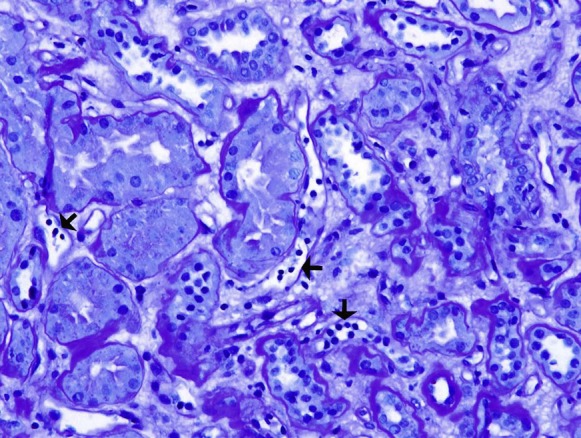
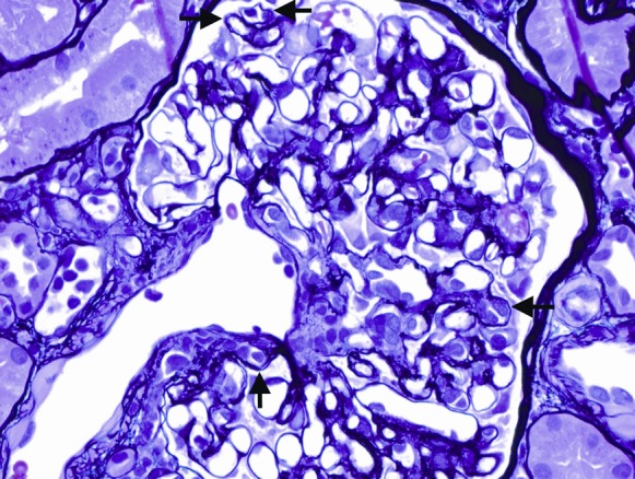
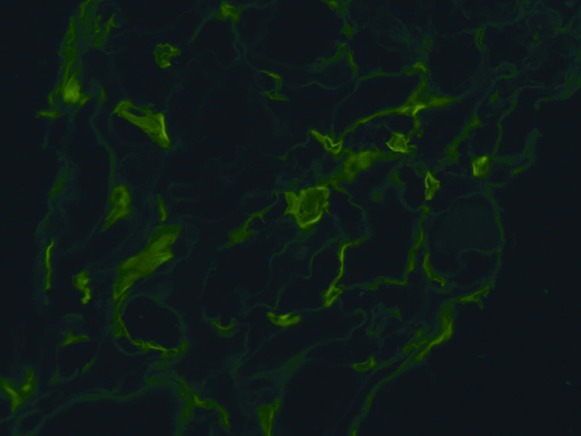
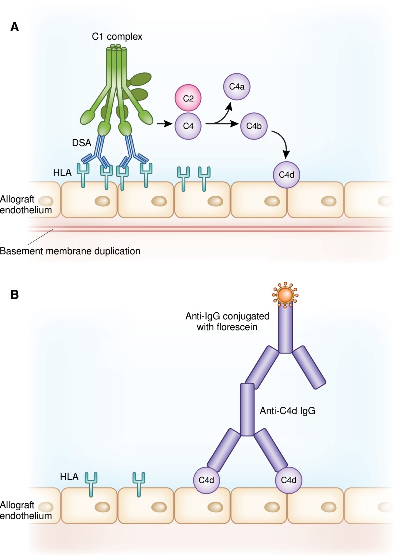
Late kidney transplant dysfunction may be a harbinger of graft failure. For many years, calcineurin inhibitor toxicity was felt to be the main cause for graft dysfunction with fibrosis and transplant loss. Recently this idea has come into question. With the observation that peritubular capillary C4d staining in kidney allografts may indicate antibody-mediated injury in conjunction with biopsy study findings, an appreciation for antibody-mediated rejection as a major cause of late graft dysfunction and loss has emerged. Twenty percent to 30% of patients develop de novo donor-specific antibodies after kidney transplantation. There are no US Food and Drug Administration-approved treatments for antibody-mediated rejection, nor have any randomized controlled trials assessed efficacy. Off-label treatment strategies include some combination of plasma exchange, intravenous immunoglobulin, and rituximab. Other approaches, including splenectomy, bortezomib, and eculizumab, have also been tried.
Figures
Similar articles
-
Probable C4d-negative accelerated acute antibody-mediated rejection due to non-HLA antibodies.Nephrology (Carlton). 2015 Jul;20 Suppl 2:75-8. doi: 10.1111/nep.12467. Nephrology (Carlton). 2015. PMID: 26031592 Review.
-
Rejection of the Renal Allograft in the Absence of Demonstrable Antibody and Complement.Transplantation. 2017 Feb;101(2):395-401. doi: 10.1097/TP.0000000000001118. Transplantation. 2017. PMID: 26901079
-
Antibody-mediated rejection due to anti-HLA-DQ antibody after pregnancy and delivery in a female kidney transplant recipient.Nephrology (Carlton). 2018 Jul;23 Suppl 2:81-84. doi: 10.1111/nep.13279. Nephrology (Carlton). 2018. PMID: 29968405
-
Clinicopathologic analysis of acute vascular rejection cases after renal transplantation.Transplant Proc. 2012 Jan;44(1):230-5. doi: 10.1016/j.transproceed.2011.11.002. Transplant Proc. 2012. PMID: 22310621
-
Pathology of chronic humoral rejection.Contrib Nephrol. 2009;162:75-86. doi: 10.1159/000170814. Epub 2008 Oct 31. Contrib Nephrol. 2009. PMID: 19001815 Review.
Cited by
-
Allograft rejection and tubulointerstitial fibrosis in human kidney allografts: interrogation by urinary cell mRNA profiling.Transplant Rev (Orlando). 2014 Jul;28(3):145-54. doi: 10.1016/j.trre.2014.05.003. Epub 2014 May 27. Transplant Rev (Orlando). 2014. PMID: 24929703 Free PMC article. Review.
-
Transplantology: Challenges for Today.Arch Immunol Ther Exp (Warsz). 2016 Dec;64(Suppl 1):37-45. doi: 10.1007/s00005-016-0439-1. Epub 2017 Jan 12. Arch Immunol Ther Exp (Warsz). 2016. PMID: 28083612 Free PMC article. Review.
-
Long-term unexpected consequence of two kidney transplants with full-match grafts: a report of two cases.Int J Clin Exp Med. 2015 Jun 15;8(6):10074-80. eCollection 2015. Int J Clin Exp Med. 2015. PMID: 26309702 Free PMC article.
-
Endothelial MMP-9 drives the inflammatory response in abdominal aortic aneurysm (AAA).Am J Transl Res. 2017 Dec 15;9(12):5485-5495. eCollection 2017. Am J Transl Res. 2017. PMID: 29312500 Free PMC article.
References
-
- Feucht HE, Schneeberger H, Hillebrand G, Burkhardt K, Weiss M, Riethmüller G, Land W, Albert E: Capillary deposition of C4d complement fragment and early renal graft loss. Kidney Int 43: 1333–1338, 1993 - PubMed
-
- Cornell LD: Renal allograft pathology in the sensitized patient. Curr Opin Organ Transplant 18: 327–336, 2013 - PubMed
-
- Racusen LC, Haas M: Antibody-mediated rejection in renal allografts: Lessons from pathology. Clin J Am Soc Nephrol 1: 415–420, 2006 - PubMed
-
- Racusen LC, Colvin RB, Solez K, Mihatsch MJ, Halloran PF, Campbell PM, Cecka MJ, Cosyns JP, Demetris AJ, Fishbein MC, Fogo A, Furness P, Gibson IW, Glotz D, Hayry P, Hunsickern L, Kashgarian M, Kerman R, Magil AJ, Montgomery R, Morozumi K, Nickeleit V, Randhawa P, Regele H, Seron D, Seshan S, Sund S, Trpkov K: Antibody-mediated rejection criteria — an addition to the Banff 97 classification of renal allograft rejection. Am J Transplant 3: 708–714, 2003 - PubMed
Publication types
MeSH terms
Substances
LinkOut - more resources
Full Text Sources
Other Literature Sources
Medical
