

Health of white sucker within the St. Louis River area of concern associated with habitat usage as assessed using stable isotopes
- PMID: 24370817
- PMCID: PMC3920021
- DOI: 10.1007/s10646-013-1167-5
Health of white sucker within the St. Louis River area of concern associated with habitat usage as assessed using stable isotopes
Abstract
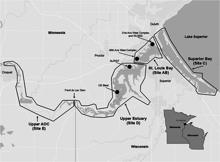
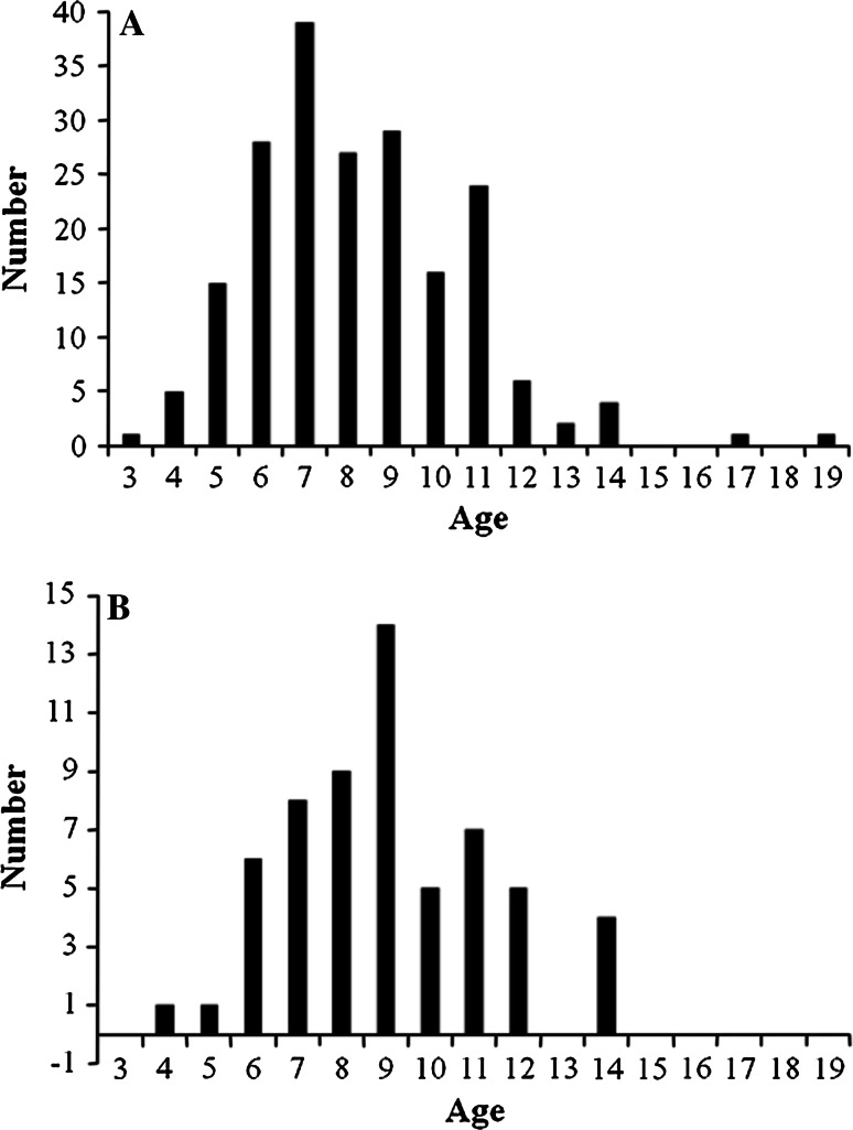
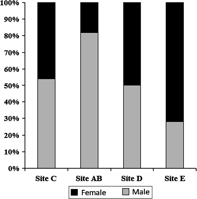
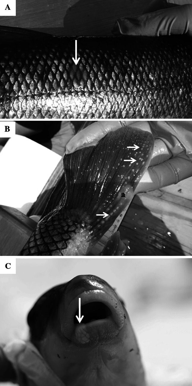
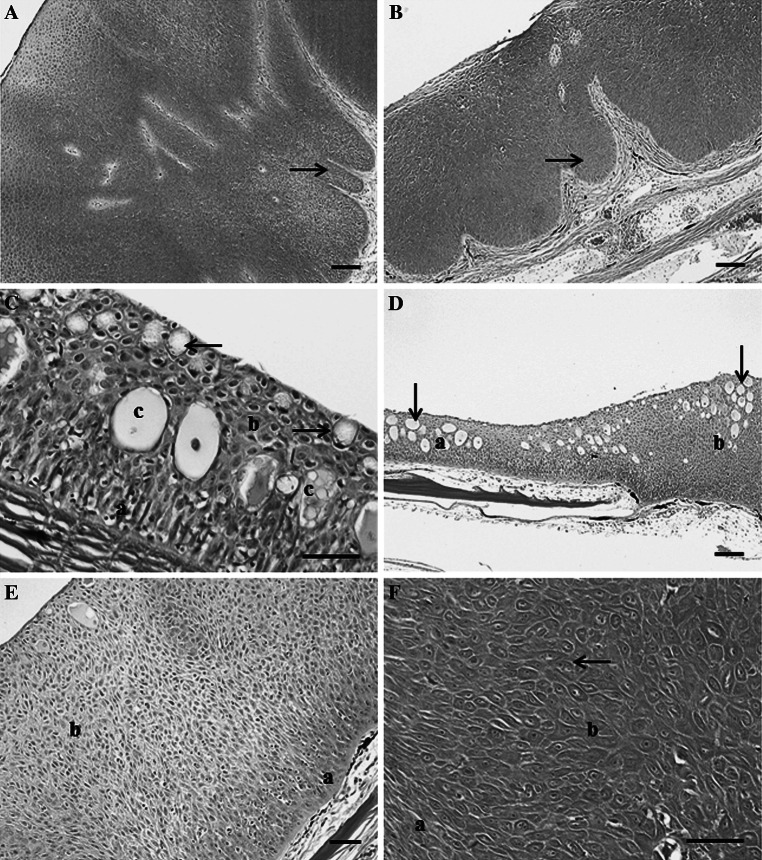
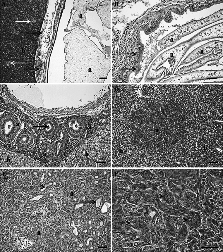
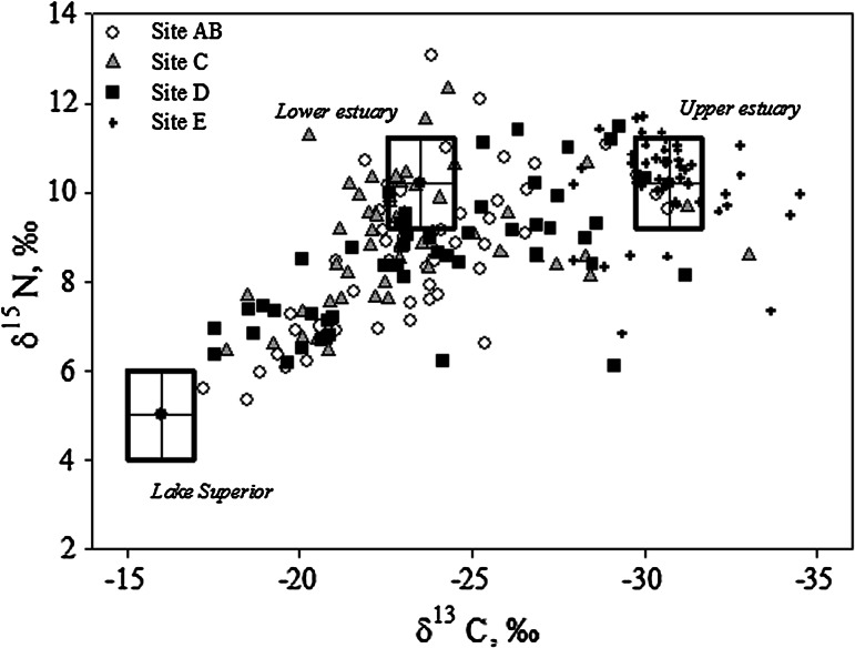
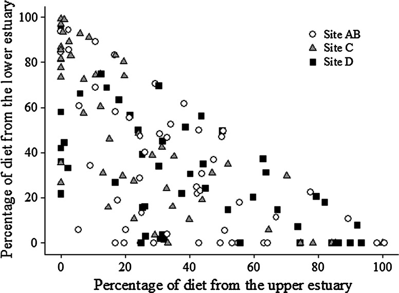
In Spring 2011, 200 adult white sucker were collected in four areas of the St. Louis River area of concern (AOC), located in Minnesota and Wisconsin, USA. The areas included the upper AOC as a reference area, the upper estuary, St. Louis Bay and Superior Bay. Grossly visible abnormalities were documented and preserved for microscopic analyses, as were five to eight representative pieces of liver tissue. A piece of dorsal muscle was preserved for stable isotope analyses and otoliths removed for age determination. The incidence of raised skin lesions (mucoid plaques) was high (31 %), however, microscopically only 4.5 % of the white suckers had neoplasia (papillomas). The remaining lesions were epidermal hyperplasia. Superior Bay had the lowest percentage of skin/lip lesions (10 %), while St. Louis Bay had the highest (44 %). St. Louis Bay also had the highest incidence of skin neoplasms (12 %). No hepatocellular neoplasms were documented, however bile duct tumors were observed in 4.5 % of the suckers. Foci of cellular alteration were observed in fish from all sites except the upper AOC. Stable isotope data indicated that most of the suckers relied on the St. Louis River AOC for the majority (>75 %) of their diet, indicating they were resident within the AOC and not in Lake Superior. The amount of diet obtained from the upper estuary was a significant predictor of skin lesion incidence. Hence, habitat use within the AOC appears to be an important risk factor for skin and possibly, liver lesions.
Figures
Similar articles
-
Influence of demographics, exposure, and habitat use in an urban, coastal river on tumor prevalence in a demersal fish.Sci Total Environ. 2020 Apr 10;712:136512. doi: 10.1016/j.scitotenv.2020.136512. Epub 2020 Jan 7. Sci Total Environ. 2020. PMID: 31945522 Free PMC article.
-
Tumours in white suckers from Lake Michigan tributaries: pathology and prevalence.J Fish Dis. 2017 Mar;40(3):377-393. doi: 10.1111/jfd.12520. Epub 2016 Aug 24. J Fish Dis. 2017. PMID: 27553424
-
Pathogenesis of skin and liver neoplasms in white suckers from industrially polluted areas in Lake Ontario.Sci Total Environ. 1990 May 1;94(1-2):105-23. doi: 10.1016/0048-9697(90)90367-4. Sci Total Environ. 1990. PMID: 2360036
-
Tumor prevalence and biomarkers of genotoxicity in brown bullhead (Ameiurus nebulosus) in Chesapeake Bay tributaries.Sci Total Environ. 2011 Dec 1;410-411:248-57. doi: 10.1016/j.scitotenv.2011.09.035. Epub 2011 Oct 11. Sci Total Environ. 2011. PMID: 21995877
-
Effects of harmful algal blooms and associated water-quality on endangered Lost River and shortnose suckers.Harmful Algae. 2020 Jul;97:101847. doi: 10.1016/j.hal.2020.101847. Epub 2020 Jun 20. Harmful Algae. 2020. PMID: 32732045 Review.
Cited by
-
Discovery and Genomic Characterization of a Novel Hepadnavirus from Asymptomatic Anadromous Alewife (Alosa pseudoharengus).Viruses. 2024 May 22;16(6):824. doi: 10.3390/v16060824. Viruses. 2024. PMID: 38932117 Free PMC article.
-
Indicators of exposure to estrogenic compounds at Great Lakes Areas of Concern: species and site comparisons.Environ Monit Assess. 2018 Sep 6;190(10):577. doi: 10.1007/s10661-018-6943-5. Environ Monit Assess. 2018. PMID: 30191322 Free PMC article.
-
Influence of demographics, exposure, and habitat use in an urban, coastal river on tumor prevalence in a demersal fish.Sci Total Environ. 2020 Apr 10;712:136512. doi: 10.1016/j.scitotenv.2020.136512. Epub 2020 Jan 7. Sci Total Environ. 2020. PMID: 31945522 Free PMC article.
-
Micronuclei and other erythrocyte nuclear abnormalities in fishes from the Great Lakes Basin, USA.Environ Mol Mutagen. 2017 Oct;58(8):570-581. doi: 10.1002/em.22123. Epub 2017 Sep 4. Environ Mol Mutagen. 2017. PMID: 28868735 Free PMC article.
-
Characterization of a Novel Hepadnavirus in the White Sucker (Catostomus commersonii) from the Great Lakes Region of the United States.J Virol. 2015 Dec;89(23):11801-11. doi: 10.1128/JVI.01278-15. Epub 2015 Sep 16. J Virol. 2015. PMID: 26378165 Free PMC article.
References
-
- Baumann PC. The use of tumors in wild populations of fish to assess ecosystem health. J Aquat Ecosyst Health. 1992;1:135–146. doi: 10.1007/BF00044045. - DOI
-
- Baumann PC, Smith IR, Metcalfe CD. Linkages between chemical contaminants and tumors in benthic Great Lakes fish. J Great Lakes Res. 1996;22:131–152. doi: 10.1016/S0380-1330(96)70946-2. - DOI
Publication types
MeSH terms
Substances
LinkOut - more resources
Full Text Sources
Other Literature Sources
Medical
