. 2014 Feb 3;55(2):706-15.
doi: 10.1167/iovs.13-13461.

Inhibition of ataxia telangiectasia mutated (ATM) kinase suppresses herpes simplex virus type 1 (HSV-1) keratitis

Affiliations

Inhibition of ataxia telangiectasia mutated (ATM) kinase suppresses herpes simplex virus type 1 (HSV-1) keratitis

Oleg Alekseev et al. Invest Ophthalmol Vis Sci. .

Abstract

Purpose: Herpes keratitis (HK) remains the leading cause of cornea-derived blindness in the developed world, despite the availability of effective antiviral drugs. Treatment toxicity and the emergence of drug resistance highlight the need for additional therapeutic approaches. This study examined ataxia telangiectasia mutated (ATM), an apical kinase in the host DNA damage response, as a potential new target for the treatment of HK.

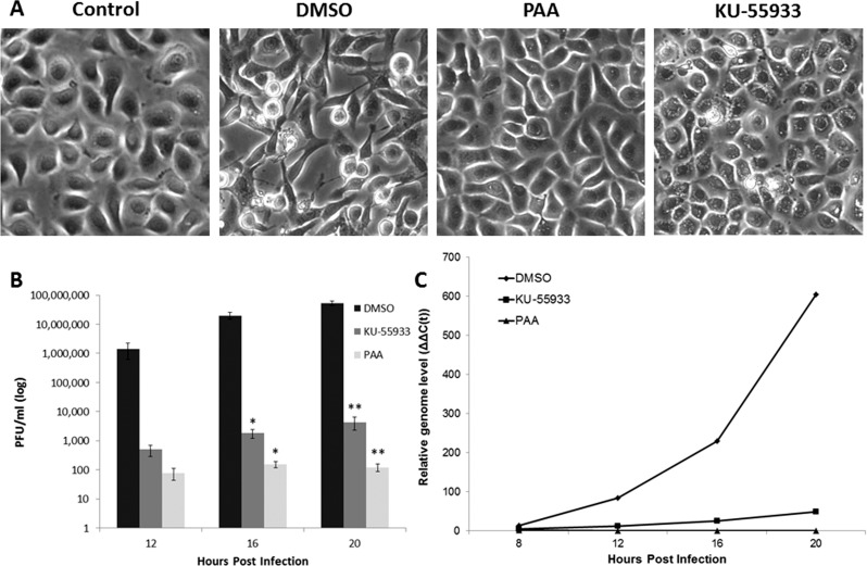
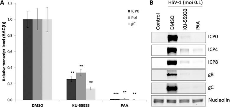
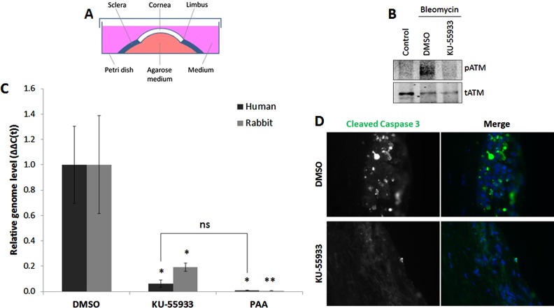
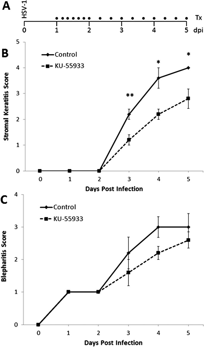
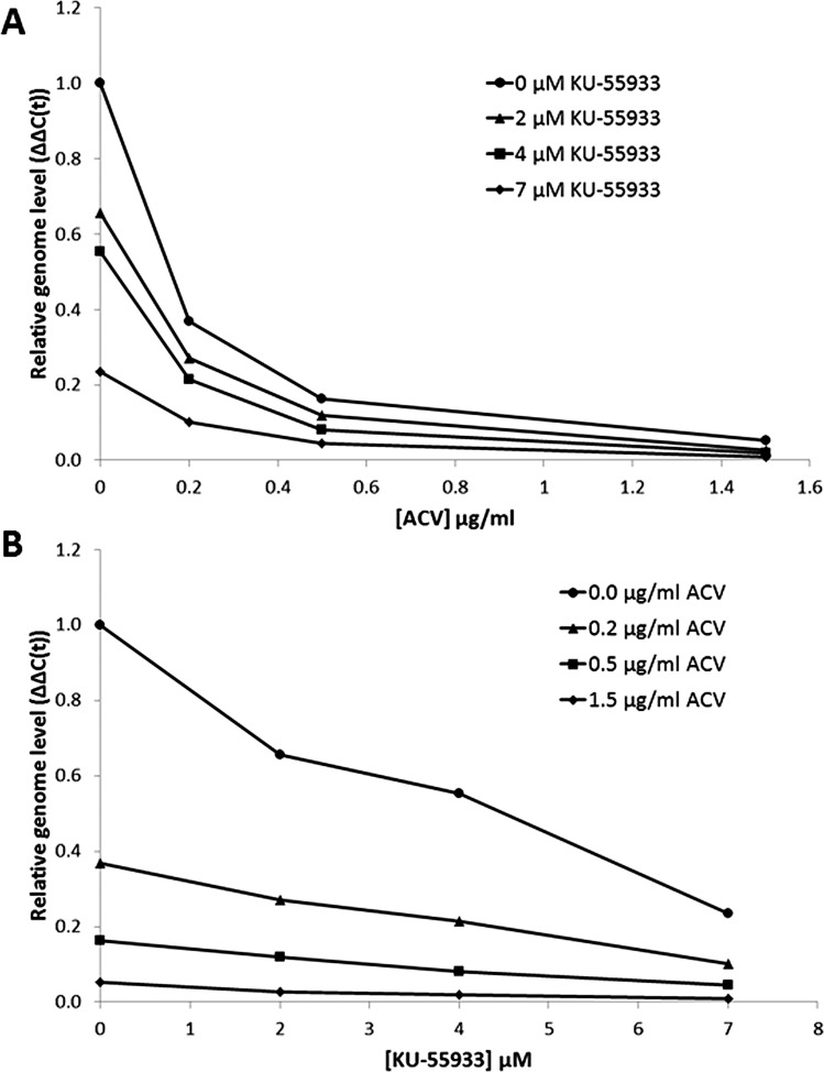
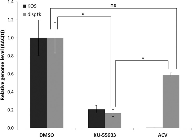
Methods: Small molecule inhibitor of ATM (KU-55933) was used to treat herpes simplex virus type 1 (HSV-1) infection in three experimental models: (1) in vitro--cultured human corneal epithelial cells, hTCEpi, (2) ex vivo--organotypically explanted human and rabbit corneas, and (3) in vivo--corneal infection in young C57BL/6J mice. Infection productivity was assayed by plaque assay, real-time PCR, Western blot, and disease scoring.

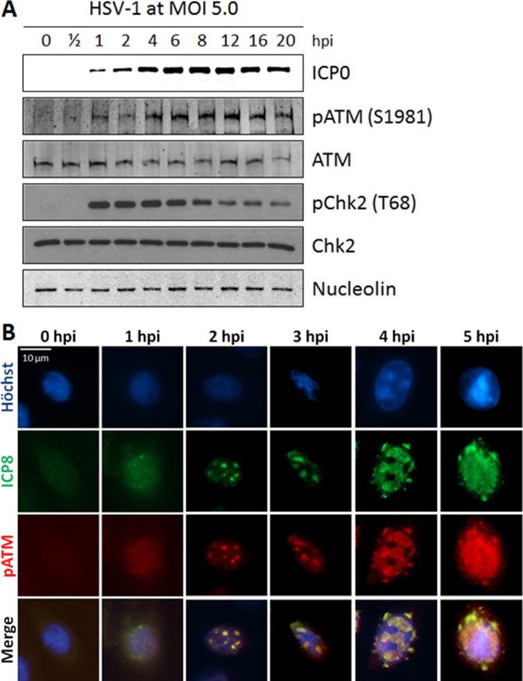
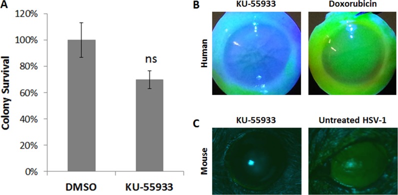
Results: Robust ATM activation was detected in HSV-1-infected human corneal epithelial cells. Inhibition of ATM greatly suppressed viral replication in cultured cells and in explanted human and rabbit corneas, and reduced the severity of stromal keratitis in mice. The antiviral effect of KU-55933 in combination with acyclovir was additive, and KU-55933 suppressed replication of a drug-resistant HSV-1 strain. KU-55933 caused minimal toxicity, as monitored by clonogenic survival assay and fluorescein staining.

Conclusions: This study identifies ATM as a potential target for the treatment of HK. ATM inhibition by KU-55933 reduces epithelial infection and stromal disease severity without producing appreciable toxicity. These findings warrant further investigations into the DNA damage response as an area for therapeutic intervention in herpetic ocular diseases.

Keywords: DNA damage response; HSV-1; ataxia telangiectasia mutated (ATM); cornea; herpes simplex keratitis.


Figures

Figure 1
Figure 1
HSV-1 activates ATM in human corneal epithelial cells. (A) hTCEpi cells were infected with HSV-1 at MOI 5.0. Lysates were collected at the indicated time points and analyzed by Western blot with antibodies specific to the indicated proteins. ICP0 staining was used to mark the progression of infection, and nucleolin is a loading control. Thr68 is an ATM-specific phosphorylation site on Chk2. (B) hTCEpi cells were infected with HSV-1 at MOI 5.0 and fixed at the indicated hpi. Cells were processed for indirect immunofluorescence with the indicated primary antibodies and counterstained with Hoechst 33258. ICP8 staining was used to visualize the viral replication compartments. Scale Bar: 10 μm. Data are representative of at least three independent experiments.
Figure 2
Figure 2
ATM inhibition suppresses HSV-1 replication in vitro. hTCEpi cells were infected at MOI 0.1 in the presence of ATM inhibitor (KU-55933, 10 μM). Control cells were neither infected nor treated. Mock treatment (DMSO) and viral replication inhibitor (PAA, 400 μg/mL) were used as negative and positive treatment controls, respectively. Under these experimental conditions, PAA contains activities that were found to inhibit several stages of HSV-1 gene expression. (A) Phase contrast images of hTCEpi cells were taken at 20 hpi. (B) Supernatants were collected at the indicated time points for analysis by plaque assay. Bars represent average viral titers ± SEM. (C) Total DNA was collected at the indicated time points for analysis by qPCR with primers for HSV-1 DNA polymerase and GAPDH. A representative experiment is shown. Bars represent relative ΔΔC(t) values ± SEM. n = 3 for all.
Figure 3
Figure 3
ATM inhibition reduces accumulation of viral transcripts and proteins in vitro. hTCEpi cells were infected at MOI 0.1 in the presence of ATM inhibitor (KU-55933, 10 μM). Mock treatment (DMSO) and viral replication inhibitor (PAA, 400 μg/mL) were used as negative and positive controls, respectively. Under these experimental conditions, PAA contains activities that were found to inhibit several stages of HSV-1 gene expression. Cells were collected for protein lysates or RNA isolation at 16 hpi. (A) Transcripts from all three HSV-1 gene families were detected with primers for ICP0 (immediate early), DNA polymerase (early), and glycoprotein C (true late). Bars represent relative ΔΔC(t) values ± SEM. (B) Viral protein accumulation was assayed by Western blot with antibodies against ICP0 and ICP4 (immediate early), ICP8 (early), glycoprotein B (leaky late), and glycoprotein C (true late). Control lysates were collected from cells that were neither infected nor treated. Nucleolin is a loading control. n = 2 for all.
Figure 4
Figure 4
ATM inhibition suppresses HSV-1 replication in explanted human and rabbit corneas. (A) Schematic representation of the ex vivo culture method of explanted corneoscleral buttons. Reprinted with permission from Alekseev O, Tran AH, Azizkhan-Clifford J. Ex vivo organotypic corneal model of acute epithelial herpes simplex virus type I infection. J Vis Exp. 2012;69:e3631, doi:10.3791/3631. (B) Ex vivo human corneas were pretreated for 1 hour with ATM inhibitor (KU-55933, 10 μM) or DMSO, followed by administration of bleomycin (200 μg/mL) for an additional hour. The epithelial layers were collected for protein lysates and analyzed by Western blot with antibodies against pATM (Ser1981) and total ATM. Each lysate was collected from three pooled corneas. (C, D) Human and rabbit corneas were infected with 1 × 10⁴ PFU/cornea. Treatments were applied at 1 hpi: ATM inhibitor (KU-55933, 10 μM) and mock treatment (DMSO). (C) PAA (400 μg/mL) was included as a positive control. Under these experimental conditions, PAA contains activities that were found to inhibit several stages of HSV-1 gene expression. DNA was isolated from the epithelial layers at 48 hpi and analyzed by qPCR with primers for HSV-1 DNA polymerase and GAPDH. Bars represent relative ΔΔC(t) values ± SEM. n = 6 for each treatment. (D) Alternatively, human corneas were processed for indirect immunofluorescence staining for cleaved caspase-3. Counterstain is Hoechst 33258.
Figure 5
Figure 5
KU-55933 reduces disease severity in the mouse model of herpes keratitis. (A) Corneas of 3-week-old C57BL/6J mice were infected with McKrae strain of HSV-1. Treatments with 200 μM KU-55933 (represented by black dots in the schematic) were initiated at 24 hpi and administered every 4 hours for 1 full day and then every 8 hours for the remainder of the experiment. dpi, days postinfection. Ocular disease severity was scored on a number scale (described in the Materials and Methods section) for stromal keratitis (B) and blepharitis (C). Data points represent average disease scores ± SEM. n = 5 mice per group.
Figure 6
Figure 6
KU-55933 exhibits low toxicity in corneal epithelium. (A) The toxicity of ATM inhibition in hTCEpi cells was assessed by colony survival assay after a 24-hour treatment with KU-55933 (10 μM). Bars represent average colony survival ± SEM. n = 3. (B) Ex vivo human corneas were treated with KU-55933 (10 μM) continually for 30 hours, and the epithelial toxicity was assessed by fluorescein staining. Toxic treatment with doxorubicin (100 μM) for 30 hours served as a positive control for detection of damage by staining. n = 2. (C) The eyes of uninfected healthy mice were treated with 200 μM KU-55933 administered at the same frequency and duration (4 days) as in the mouse ocular infection experiments (Fig. 5A). At the end of the experiment, the treated corneas were assessed for toxicity by fluorescein staining. A mouse cornea de-epithelialized as a consequence of untreated HSV-1 infection served as a positive staining control. n = 2.
Figure 7
Figure 7
ATM inhibition enhances the antiviral activity of acyclovir. hTCEpi cells were infected at MOI 0.1 in the presence of 16 different dose combinations of KU-55933 (0, 2, 4, and 7 μM) and acyclovir (0, 0.2, 0.5, and 1.5 μg/mL). Total DNA was collected at 16 hpi for analysis by qPCR with primers for HSV-1 polymerase and GAPDH. Viral genome replication was calculated using the ΔΔC(t) method. Data are representative of at least two independent experiments. The same data set was plotted in two different ways to highlight (A) the effect of KU-55933 on the acyclovir dose–response curve and (B) the effect of acyclovir on the KU-55933 dose–response curve.
Figure 8
Figure 8
ATM inhibition suppresses acyclovir-resistant HSV-1 infection. hTCEpi cells were infected at MOI 0.1 with wild-type or acyclovir-resistant HSV-1 (KOS strain and dlsptk strain, respectively) in the presence of ATM inhibitor (KU-55933, 10 μM). Mock treatment (DMSO) and viral polymerase inhibitor (acyclovir, 50 μg/mL) were used as negative and positive controls, respectively. Total DNA was collected at 16 hpi for analysis by qPCR with primers for HSV-1 polymerase and GAPDH. All values are normalized to the corresponding DMSO samples. Bars represent relative ΔΔC(t) values ± SEM. n = 2.

Similar articles

Cited by

References

    1. Rowe AM, St Leger AJ, Jeon S, Dhaliwal DK, Knickelbein JE, Hendricks RL. Herpes keratitis. Prog Retin Eye Res. 2013; 32: 88–101 - PMC - PubMed
    1. Toma HS, Murina AT, Areaux RG Jr, et al. Ocular HSV-1 latency, reactivation and recurrent disease. Semin Ophthalmol. 2008; 23: 249–273 - PubMed
    1. Farooq AV, Shukla D. Herpes simplex epithelial and stromal keratitis: an epidemiologic update. Surv Ophthalmol. 2012; 57: 448–462 - PMC - PubMed
    1. Young RC, Hodge DO, Liesegang TJ, Baratz KH. Incidence, recurrence, and outcomes of herpes simplex virus eye disease in Olmsted County, Minnesota, 1976-2007: the effect of oral antiviral prophylaxis. Arch Ophthalmol. 2010; 128: 1178–1183 - PMC - PubMed
    1. Wilhelmus KR. Antiviral treatment and other therapeutic interventions for herpes simplex virus epithelial keratitis. Cochrane Database Syst Rev. 2010; CD002898 - PMC - PubMed

Publication types

MeSH terms