Review
. 2014 Jan;127(1):71-90.
doi: 10.1007/s00401-013-1233-3. Epub 2013 Dec 27.

Human alcohol-related neuropathology

Affiliations
Review

Human alcohol-related neuropathology

Suzanne M de la Monte et al. Acta Neuropathol. 2014 Jan.

Abstract

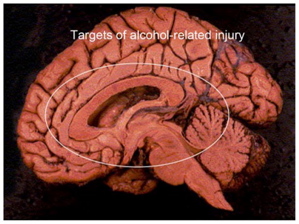
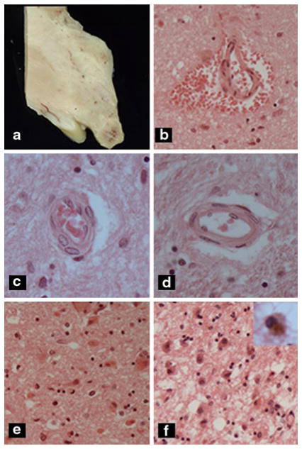
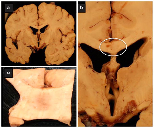
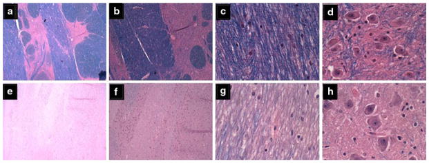
Alcohol-related diseases of the nervous system are caused by excessive exposures to alcohol, with or without co-existing nutritional or vitamin deficiencies. Toxic and metabolic effects of alcohol (ethanol) vary with brain region, age/developmental stage, dose, and duration of exposures. In the mature brain, heavy chronic or binge alcohol exposures can cause severe debilitating diseases of the central and peripheral nervous systems, and skeletal muscle. Most commonly, long-standing heavy alcohol abuse leads to disproportionate loss of cerebral white matter and impairments in executive function. The cerebellum (especially the vermis), cortical-limbic circuits, skeletal muscle, and peripheral nerves are also important targets of chronic alcohol-related metabolic injury and degeneration. Although all cell types within the nervous system are vulnerable to the toxic, metabolic, and degenerative effects of alcohol, astrocytes, oligodendrocytes, and synaptic terminals are major targets, accounting for the white matter atrophy, neural inflammation and toxicity, and impairments in synaptogenesis. Besides chronic degenerative neuropathology, alcoholics are predisposed to develop severe potentially life-threatening acute or subacute symmetrical hemorrhagic injury in the diencephalon and brainstem due to thiamine deficiency, which exerts toxic/metabolic effects on glia, myelin, and the microvasculature. Alcohol also has devastating neurotoxic and teratogenic effects on the developing brain in association with fetal alcohol spectrum disorder/fetal alcohol syndrome. Alcohol impairs function of neurons and glia, disrupting a broad array of functions including neuronal survival, cell migration, and glial cell (astrocytes and oligodendrocytes) differentiation. Further progress is needed to better understand the pathophysiology of this exposure-related constellation of nervous system diseases and better correlate the underlying pathology with in vivo imaging and biochemical lesions.


Figures

Fig. 1
Fig. 1
Mid-sagittal view of the brain with major targets of alcohol-mediated injury and degeneration circled. Structures included are the cingulate gyrus, corpus callosum, hypothalamus, periventricular thalamus, cerebellar vermis, basal ganglia, medial temporal structures, and periventricular white matter
Fig. 2
Fig. 2
Acute Wernicke’s encephalopathy. a Mammillary body with darkened area of hemorrhage. b Acute perivascular hemorrhage in hypothalamus. c Prominent reactive proliferation of vascular endothelial cells, d normal vessel, e, f hypothalamus with e acute or f chronic Wernicke’s encephaopathy. Note neuronal cytoplasmic eosinophilia and mild microvesicular as well a pericellular edema in e compared with gliosis, abundant hemosiderin-laden macrophages (see inset), and prominent spongiosis due to macrovesicular and microvesicular edema in f
Fig. 3
Fig. 3
Chronic alcoholic neurodegeneration. The brain was from a middle-aged alcoholic man with documented Wernicke–Korsakoff psychosis. a Coronal section through the cerebral hemispheres at the level of the hypothalamus and foramen of Munro demonstrating atrophy of the fornix and mammillary bodies. b Closer image of the atrophic hypothalamus, mammillary bodies, and fornix. The atrophy is extreme yet the typical rust discoloration associated with WKS was not observed. c Central demyelination in the mid-portion of the corpus callosum (region circled in panel b), with features suggestive of Marchiafava–Bignami
Fig. 4
Fig. 4
Central pontine myelinolysis. ad Control basis pontis showing ac intact myelin, a, b clear delineations between gray and white matter structures, and d abundant pontine neurons. eh Alcoholic basis pontis (same as Fig. 7a) showing eg almost complete loss of myelin, e, f virtually no delineation between gray and white mater structures, and b reduced abundance of glial cells, yet h preservation of neurons in pontine neurons (Luxol Fast Blue, Hematoxylin and Eosin stain)
Fig. 5
Fig. 5
Central pontine myelinolysis. Sections of pons from alcoholic patient in Fig. 4 were immunostained for a, b GFAP to detect reactive astrocytosis and gliosis, or c, d CD68 for macrophages and microglia. Examples of labeled cells are marked with arrows. Immunoreactivity was revealed with diaminobenzidine (brown) and sections were counterstained with hematoxylin (a, c, d 200× and b 400× original magnification)
Fig. 6
Fig. 6
Alcoholic white matter degeneration. a Periventricular frontal white matter from the anterior frontal region of a non-alcoholic middle-aged man. Other regions of brain showed similar degrees of myelin staining. bd Cerebral white matter degeneration is present in the brain of an alcoholic middle-aged man with dementia. White matter from the b anterior frontal, c periventricular frontal, and d periventricular region at the level of the hypothalamus with variable degrees of myelin pallor, vacuolation, and gliosis relative to normal
Fig. 7
Fig. 7
Alcoholic cerebellar degeneration. a Pronounced atrophy of cerebellar folia in the anterior vermis marked by widening of fissures and thinning of cortical and white matter structures. b Higher magnification of the cortex showing attenuation of cells in the inner granule cell (igc) layer, and proliferation of Bergmann’s glia (band-like; arrowheads) with loss of Purkinje cells. c A higher magnification image depicting severe degeneration of the cortex with subtotal depletion of neurons in the granule and Purkinje cell layers, and fiber loss in white matter cores (blue, center right). At the far right, the granule cell population is reduced but better preserved compared with the left side of the same folium
Fig. 8
Fig. 8
Alcoholic small fiber neuropathy detected by skin biopsy. Proximal (a, b) and distal (c, d) 8-mm punch biopsies of full-thickness skin were obtained from the medial thigh and lateral lower calf region from control (a, c) and alcoholic (b, d) patients. The tissues were fixed in 4 % paraformaldehyde, paraffin-embedded, and immunostained with antibodies to Ubiquitin 9.5. Immunoreactivity was detected with nickle-diaminobenzidine. Black linear staining corresponds to intra-epidermal nerve fibers Note the complexity of nerve fiber networks in the epidermis of control versus alcoholic specimens

Similar articles

Cited by

References

    1. Adachi J, Asano M, Ueno Y, et al. Alcoholic muscle disease and biomembrane perturbations (review) J Nutr Biochem. 2003;14(11):616–625. - PubMed
    1. Aggunlu L, Oner Y, Kocer B, Akpek S. The value of diffusion-weighted imaging in the diagnosis of Marchiafava–Bignami disease: apropos of a case. J Neuroimaging: Off J Am Soc Neuroimaging. 2008;18(2):188–190. doi: 10.1111/j.1552-6569.2007.00202.x. - DOI - PubMed
    1. Akai J, Akai K. Neuropathological study of the nucleus basalis of meynert in alcoholic dementia. Arukoru kenkyu to yakubutsu izon = Jpn J Alcohol Stud Drug Depend. 1989;24(2):80–88. - PubMed
    1. Albrecht J, Sidoryk-Wegrzynowicz M, Zielinska M, Aschner M. Roles of glutamine in neurotransmission. Neuron Glia Biol. 2010;6(4):263–276. doi: 10.1017/S1740925X11000093. - DOI - PubMed
    1. Altamirano J, Bataller R. Alcoholic liver disease: pathogenesis and new targets for therapy. Nat Rev Gastroenterol Hepatol. 2011;8(9):491–501. doi: 10.1038/nrgastro.2011.134. - DOI - PubMed

Publication types

MeSH terms

LinkOut - more resources