Comparative Study
. 2014 Apr 1;116(7):797-806.
doi: 10.1152/japplphysiol.00955.2013. Epub 2013 Dec 26.

Mature IGF-I excels in promoting functional muscle recovery from disuse atrophy compared with pro-IGF-IA

Affiliations
Comparative Study

Mature IGF-I excels in promoting functional muscle recovery from disuse atrophy compared with pro-IGF-IA

Soohyun Park et al. J Appl Physiol (1985). .

Abstract

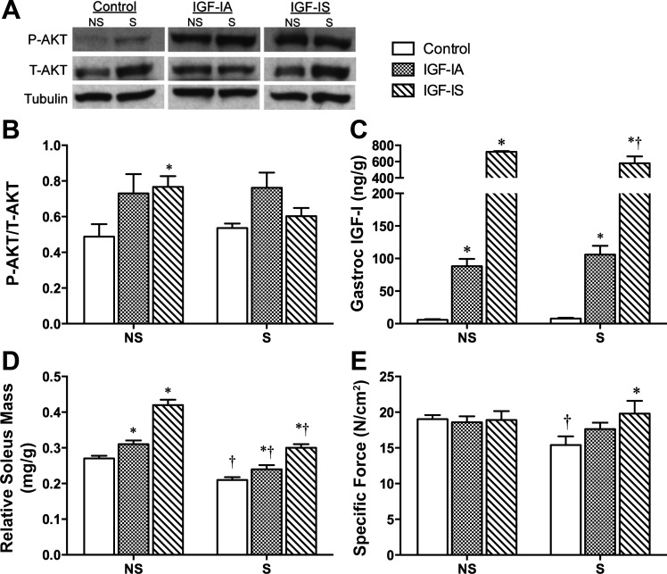
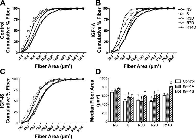
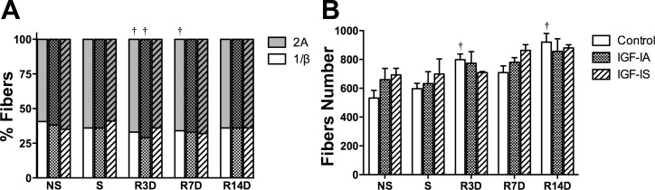
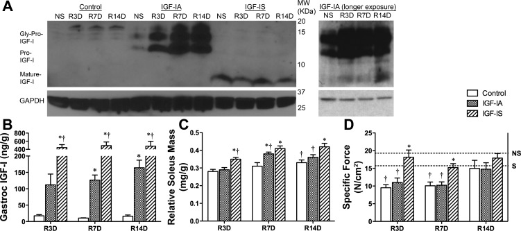
Prolonged disuse of skeletal muscle results in atrophy, and once physical activity is resumed, there is increased susceptibility to injury. Insulin-like growth factor-I (IGF-I) is considered a potential therapeutic target to attenuate atrophy during unloading and to enhance rehabilitation upon reloading of skeletal muscles, due to its multipronged actions on satellite cell proliferation, differentiation, and survival, as well as its actions on muscle fibers to boost protein synthesis and inhibit protein degradation. However, the form of IGF-I delivered may alter the success of treatment. Using the hindlimb suspension model of disuse atrophy, we compared the efficacy of two IGF-I forms in protection against atrophy and enhancement of recovery: mature IGF-I (IGF-IS) lacking the COOH-terminal extension, called the E-peptide, and IGF-IA, which is the predominant form retaining the E-peptide. Self-complementary adeno-associated virus harboring the murine Igf1 cDNA constructs were delivered to hindlimbs of adult female C57BL6 mice 3 days prior to hindlimb suspension. Hindlimb muscles were unloaded for 7 days and then reloaded for 3, 7, and 14 days. Loss of muscle mass following suspension was not prevented by either IGF-I construct. However, IGF-IS expression maintained soleus muscle force production. Further, IGF-IS treatment caused rapid recovery of muscle fiber morphology during reloading and maintained muscle strength. Analysis of gene expression revealed that IGF-IS expression accelerated the downregulation of atrophy-related genes compared with untreated or IGF-IA-treated samples. We conclude that mature-IGF-I may be a better option than pro-IGF-IA to promote skeletal muscle recovery following disuse atrophy.

Keywords: glycosylation; hindlimb suspension; skeletal muscle hypertrophy.


Figures

Fig. 1.
Fig. 1.
Viral expression of insulin-like growth factor-IS (IGF-IS) prevents muscle weakness associated with hindlimb suspension induced atrophy. A: representative immunoblots for P-AKT and T-AKT in tibialis anterior (TA) muscle lysates following viral injection and/or suspension. B: quantification of Western blot (A) revealed ∼40% increase in P-Akt by both viral constructs in muscles from nonsuspended mice, but no difference in Akt phosphorylation following hindlimb suspension. C: estimation of IGF-I content in gastrocnemius muscles measured by ELISA showed 10- to 60-fold increase in IGF-I content in viral injected muscles. D: soleus mass normalized to body mass demonstrated that both viral constructs caused a significant increase in muscle mass in nonsuspended mice, but that in all cases, suspension caused a significant loss of mass, which was at a similar proportion regardless of viral construct. E: specific force did not differ among treatment groups in soleus muscles from nonsuspended mice, and significantly decreased in untreated suspended muscles. However, only IGF-IS treatment significantly improved specific force following suspension compared with untreated muscles. NS, nonsuspended; S, suspended muscles. Bars represent means ± SE from 4–7 mice of each condition and time point. *P < 0.05 compared with control treatment at each time point; †P < 0.05 compared with the NS time point within the same treatment.
Fig. 2.
Fig. 2.
IGF-IS treatment accelerates recovery of fiber size in soleus muscles following reloading. A–C: cumulative fiber size distributions for control, IGF-IA, and IGF-IS treated soleus muscles from each time point determined by immunohistochemistry for laminin. A leftward shift of a line indicates a fiber area distribution with smaller fibers. Each time point is marked with different symbols; the thicker line represents NS, and the dotted line represents S. D: median fiber areas determined from the cumulative percent fiber distribution showed that IGF-IS treatment fiber size returned to nonsuspended values by 7 days reloading, whereas control and IGF-IA treatments achieved nonsuspended values by 14 days reloading. Data are means ± SE for n = 3–5 samples. *P < 0.05 compared with control treatment at each time point; †P < 0.05 compared with the NS time point within the same treatment.
Fig. 3.
Fig. 3.
Fiber type and fiber number analysis of soleus muscles via immunohistochemistry. A: percent fibers categorized into fast-twitch muscle (2A) and slow-twitch muscle (1/β) via immunohistochemistry. The normal fiber type ratio of 60:40 (2A:1/β) was retained in most muscles at all time points, but the control muscles showed significant increases in fast-twitch muscle composition during muscle reloading [for 3 days (R3D) and 7 days (R7D)], as did IGF-IA-treated muscles at R3D. Bars with no pattern, control muscles; stippled bars, IGF-IA treatment; striped bars, IGF-IS treatment. Overlay of no color or grey represents myosin heavy chain I/β and IIA, respectively. X-axis labels specify the time point. B: average fiber numbers of solei muscles showed that the viral injected muscles do not show a significant change in fiber number after suspension and during the muscle reloading while the control muscles showed increases in solei fiber numbers during the muscle reloading. Data are means ± SE for n = 3–5 samples. †P < 0.05 compared with the NS time point within the same treatment.
Fig. 4.
Fig. 4.
Diminished function caused by reloading is rescued by IGF-IS. A: immunoblotting of TA muscles for IGF-I before suspension and 3, 7, and 14 days after reloading shows that IGF-IA produces primarily pro- and glycosylated pro-IGF-I, whereas IGF-IS produces only mature IGF-I. Protein loading was 80 μg for control, 40 μg for IGF-IA, and 20 μg for IGF-IS. Increased protein loading (80 μg) of IGF-IA samples and longer exposure (right) reveals increased presence of mature IGF-I in the IGF-IA-treated muscles only during reloading. GAPDH was utilized as loading control for each blot. B: gastrocnemius IGF-I content measured by ELISA showed significant differences in IGF-I content in viral injected muscles throughout the reloading period after induced atrophy. C: the muscle-to-body mass ratios for soleus muscles demonstrated that muscles treated with IGF-IS had constantly higher ratios than those of control muscles during reloading. IGF-IA-treated muscles regained more mass by R7D than untreated controls. D: specific forces of soleus muscles following reloading showed that the control and IGF-IA-treated muscles were significantly weaker until R14D, but IGF-IS in treated muscles maintained the muscle strength during the entire reloading period. Dotted lines are average specific forces of nonsuspended (NS) and suspended (S) control soleus muscles from Fig. 1E. R3D, R7D, and R14D: reloading for 3, 7, and 14 days following hindlimb suspension. Data are means ± SE of 3–4 mice from each time point and treatment. *P < 0.05 compared with control treatment at each time point; †P < 0.05 compared with the NS time point within the same treatment.
Fig. 5.
Fig. 5.
Satellite cell activation in soleus muscles before and after hindlimb suspension and during muscle reloading following hindlimb suspension assessed via immunohistochemistry of Ki67 for proliferating cell marker, Pax7 for satellite cells, and DAPI for all nuclei. A: immunohistochemistry images of Ki67, Pax7, DAPI, and all three merged of IGF-IS-treated soleus at R3D. B: control and IGF-IS-treated muscles have elevated levels of Pax7-positive cells compared with nonsuspended muscle samples throughout reloading, yet IGF-IA-treated muscles have reduced satellite cells at 3 days of reloading, and increased Pax7-positive cells at 14 days of reloading compared with control muscles. C: the number of Ki67-positive cells normalized to 100 fibers showed that IGF-IS-treated solei had significantly higher numbers of proliferating cells in the early reloading process. D: the number of proliferating satellite cells was determined by colocalization of Ki67 and Pax7 immunostaining. IGF-IA-treated muscles did not display elevated proliferating satellite cells in the early recovery period, unlike IGF-IS and untreated samples. Data are means ± SE for n = 3–4 samples. *P < 0.05 compared with control treatment at each time point; †P < 0.05 compared with the NS time point within the same treatment; **P < 0.05 between IGF-IA and IGF-IS at the same time point.

Similar articles

Cited by

References

    1. Akima H, Ushiyama J, Kubo J, Tonosaki S, Itoh M, Kawakami Y, Fukuoka H, Kanehisa H, Fukunaga T. Resistance training during unweighting maintains muscle size and function in human calf. Med Sci Sports Exerc 35: 655–662, 2003 - PubMed
    1. Allen DL, Roy RR, Edgerton VR. Myonuclear domains in muscle adaptation and disease. Muscle Nerve 22: 1350–1360, 1999 - PubMed
    1. Alzghoul MB, Gerrard D, Watkins BA, Hannon K. Ectopic expression of IGF-I and Shh by skeletal muscle inhibits disuse-mediated skeletal muscle atrophy and bone osteopenia in vivo. FASEB J 18: 221–223, 2004 - PubMed
    1. Barton ER. Impact of sarcoglycan complex on mechanical signal transduction in murine skeletal muscle. Am J Physiol Cell Physiol 290: C411–C419, 2006 - PubMed
    1. Barton ER. The ABCs of IGF-I isoforms: impact on muscle hypertrophy and implications for repair. Appl Physiol Nutr Metab 31: 791–797, 2006 - PubMed

Publication types

MeSH terms

Substances

LinkOut - more resources