. 2014 Mar;88(6):3103-13.
doi: 10.1128/JVI.03202-13. Epub 2013 Dec 26.

Interferon-inducible protein IFI35 negatively regulates RIG-I antiviral signaling and supports vesicular stomatitis virus replication

Affiliations

Interferon-inducible protein IFI35 negatively regulates RIG-I antiviral signaling and supports vesicular stomatitis virus replication

Anshuman Das et al. J Virol. 2014 Mar.

Abstract

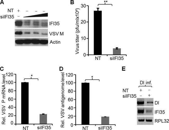
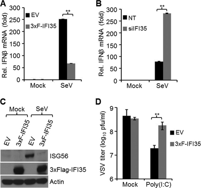
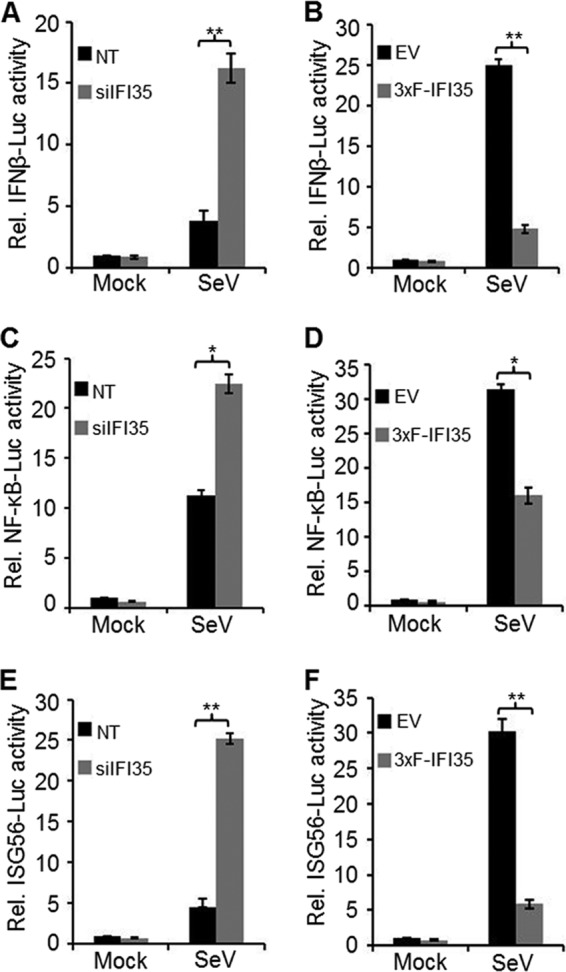
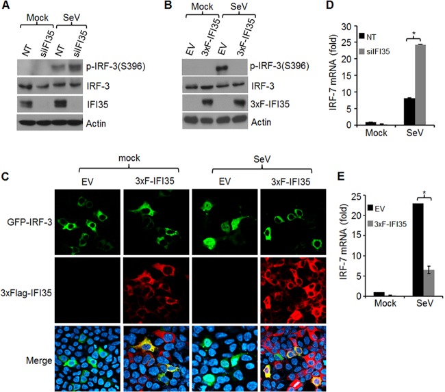
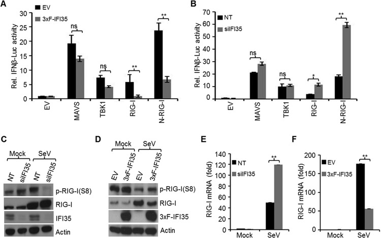
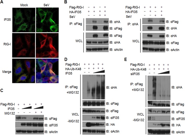
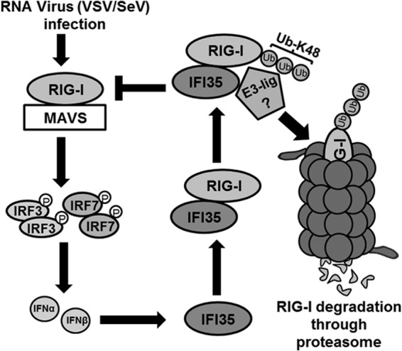
In a genome-wide small interfering RNA (siRNA) screen, we recently identified the interferon (IFN)-inducible protein 35 (IFI35; also known as IFP35) as a factor required for vesicular stomatitis virus (VSV) infection. Studies reported here were conducted to further understand the role and requirement of IFI35 in VSV infection. Consistent with the siRNA screening data, we found that depletion of IFI35 led to reduced VSV replication at the level of viral gene expression. Although no direct interaction of IFI35 with the viral replication machinery was observed, we found that IFI35 negatively regulated the host innate immune response and rescued poly(I·C)-induced inhibition of VSV replication. Promoter-driven reporter gene assays demonstrated that IFI35 overexpression suppressed the activation of IFN-β and ISG56 promoters, whereas its depletion had the opposite effect. Further investigation revealed that IFI35 specifically interacted with retinoic acid-inducible gene I (RIG-I) and negatively regulated its activation through mechanisms that included (i) suppression of dephosphorylation (activation) of RIG-I and (ii) proteasome-mediated degradation of RIG-I via K48-linked ubiquitination. Overall, the results presented here suggest a novel role for IFI35 in negative regulation of RIG-I-mediated antiviral signaling, which will have implications for diseases associated with excessive immune signaling.

Importance: Mammalian cells employ a variety of mechanisms, including production of interferons (IFNs), to counteract invading pathogens. In this study, we identified a novel role for a cellular protein, IFN-inducible protein 35 (IFP35/IFI35), in negatively regulating the host IFN response during vesicular stomatitis virus (VSV) infection. Specifically, we found that IFI35 inhibited activation of the RNA sensor, the retinoic acid-inducible gene I (RIG-I), leading to inhibition of IFN production and thus resulting in better replication of VSV. The identification of a cellular factor that attenuates the IFN response will have implications toward understanding inflammatory diseases in humans that have been found to be associated with defects in the regulation of host IFN production, such as systemic lupus erythematosus and psoriasis.


Figures

FIG 1
FIG 1
Depletion of IFI35 inhibits VSV replication. (A) HeLa cells were transfected with 15 nM NT siRNA or increasing amounts (5, 10, and 15 nM) of a combination of two different siRNAs targeting IFI35. At 60 h posttransfection, cells were infected with VSV (MOI = 1) for 4 h. Levels of IFI35 and VSV M were determined by immunoblotting with specific antibodies. Actin served as the loading control. (B) HeLa cells were transfected with 15 nM NT or IFI35 siRNA for 60 h and infected with VSV (MOI = 0.1) for 18 h. Supernatants were harvested, and virus titers were determined by plaque assay and expressed as PFU/ml. Average titers of viruses from NT- and siIFI35-treated cells were 2.7 × 10⁵ and 3.9 × 10⁴ PFU/ml, respectively. (C and D) IFI35 depletion inhibits VSV infection at the levels of virus transcription and replication. HeLa cells were transfected with 15 nM NT or IFI35 targeting siRNA for 60 h. Subsequently, the cells were infected with VSV (MOI = 0.1) for 12 h. VSV P mRNA (C) and VSV antigenomic RNA (D) levels in IFI35-depleted HeLa cells were determined by qRT-PCR. Values were normalized to the internal control β-actin level and expressed as relative changes over the NT sample value, which was set at 100. (E) IFI35 knockdown inhibits VSV DI RNA replication. NPeGFPL stable cells were transfected with 15 nM NT or IFI35 targeting siRNA for 60 h. The cells were then infected with DI particles (DI inf.) for 14 h, and the RNA replication products (both genomic and antigenomic) were quantitated by semiquantitative RT-PCR as described previously (41). IFI35 and RPL32 (internal control) mRNA levels were also examined. Data presented in panels B to D are from three independent experiments, and values represent means and standard deviations (SD) for duplicates. *, P < 0.05; **, P < 0.01.
FIG 2
FIG 2
IFI35 rescues VSV infection by negatively regulating host antiviral response. (A and B) IFI35 downregulates IFN-β mRNA synthesis. EV and 3xF-IFI35 stable cells (A) or HEK293 cells transfected with NT or IFI35 siRNA (B) were infected with SeV for 16 h. Total RNA was isolated, and IFN-β mRNA was quantified by qRT-PCR. Values were normalized to the internal control (glyceraldehyde-3-phosphate dehydrogenase [GAPDH]) and expressed as relative fold changes over the mock-infected NT or EV sample value, which was set at 1. Values represent means and SD for duplicate reactions from two independent experiments. **, P < 0.01. (C) IFI35 overexpression suppresses ISG56 induction. EV and 3xF-IFI35 stable cells were infected with SeV for 16 h. Cell lysates were used for immunoblotting using ISG56, Flag M2 (detects 3xF-IFI35), and actin antibodies. (D) IFI35 overexpression rescues poly(I·C)-induced suppression of VSV infection. EV and 3xF-IFI35 cells were transfected with 2 μg poly(I·C) for 16 h and infected with VSV (MOI = 0.1) for 18 h. The supernatants were harvested, and virus titers were determined by plaque assay on BHK-21 cells. Virus titers are expressed as log10 PFU/ml. Values represent means and SD for three independent experiments. **, P < 0.01.
FIG 3
FIG 3
IFI35 attenuates IFN-β activation and signaling. (A and B) Effect of IFI35 on IFN-β promoter activation. (A) HEK293 cells were transfected with 15 nM NT or IFI35 targeting siRNA for a total of 76 h. After the first 24 h, cells were transfected with 500 ng of IFN-β luciferase reporter plasmid (IFNβ-Luc) along with 50 ng of pRL-TK plasmid and were incubated for another 36 h. For the final 16 h prior to harvesting, cells were either mock infected or infected with SeV. Cell lysates were used for dual-luciferase assay. (B) EV and 3xF-IFI35 stable cells were transfected with 500 ng of IFN-β luciferase reporter plasmid along with 50 ng of pRL-TK plasmid for 32 h. Subsequently, cells were infected with SeV for 16 h, and cell lysates were used for dual-luciferase assay. (C and D) Effect of IFI35 on NF-κB promoter activation. Experimental conditions were similar to those for panels A and B, except that 500 ng of NF-κB luciferase construct (NF-κB-Luc) was transfected in place of IFNβ-Luc. (E and F) Effect of IFI35 on ISG56 promoter activation. Experimental conditions were similar to those for panels A and B, except that 500 ng of ISG56 luciferase construct (ISG56-Luc) was transfected in place of the IFNβ-Luc construct. Values presented in all the promoter reporter assays are normalized to NT or EV control cells and expressed as relative fold changes over the mock-infected NT or EV sample value, which was set at 1. Values represent means and SD for three independent experiments. *, P < 0.05; **, P < 0.01.
FIG 4
FIG 4
IFI35 downregulates activation of IRF-3 and IRF-7. (A and B) IFI35 downregulates IRF-3 phosphorylation. (A) HEK293 cells were transfected with 15 nM NT or IFI35 siRNA for 60 h and infected with SeV for another 16 h. Cell lysates were analyzed by immunoblotting using the indicated antibodies. (B) EV and 3xF-IFI35 stable cells were infected with SeV for 16 h, and cell lysates were analyzed by immunoblotting using the indicated antibodies. (C) IFI35 overexpression inhibits nuclear translocation of IRF-3. EV and 3xF-IFI35 stable cells were grown on coverslips, transfected with 1 μg GFP-IRF-3 plasmid for 32 h, and then infected with SeV for 16 h. 3xF-IFI35 was immunostained using anti-Flag antibody. Nuclei were stained with DAPI. (D and E) IFI35 downregulates IRF-7 induction. (D) HEK293 cells were transfected with 15 nM NT or IFI35 siRNA for 60 h and infected with SeV for another 16 h. Total RNA was isolated and subjected to qRT-PCR using IRF-7-specific primers. (E) IRF-7 mRNA was quantified as described for panel D, using EV and 3xF-IFI35 stable cells infected with SeV for 16 h. Values were normalized to the internal control GAPDH value and expressed as relative fold changes over the mock-infected EV or NT sample value, which was set at 1. Values represent means and SD for duplicate reactions from two independent experiments. *, P < 0.05.
FIG 5
FIG 5
IFI35 negatively regulates RIG-I pathway activation. (A) EV and 3xF-IFI35 stable cells were transfected with a plasmid encoding the RIG-I pathway components (0.5 μg each) MAVS, TBK1, RIG-I, or N-RIG-I, along with 0.25 μg IFNβ-Luc and 0.025 μg pRL-TK plasmid, for 48 h. The cells were lysed and used for dual-luciferase assays. (B) HEK293 cells were transfected with 15 nM NT or IFI35 siRNA for 24 h and then transfected for another 48 h with plasmids as described for panel A. Cells were lysed and used for dual-luciferase assays. Luciferase values were normalized to the EV or NT control cells and expressed as relative fold changes over the mock-infected EV or NT sample value, which was set at 1. Values represent means and SD for two independent experiments performed in duplicate. *, P < 0.05; **, P < 0.01; ns, not significant. (C and D) IFI35 negatively regulates RIG-I activation. (C) HEK293 cells were transfected with 15 nM NT or IFI35 siRNA for 60 h and infected with SeV for another 16 h. The cells were treated with 100 nM calyculin A for 30 min before harvesting. Cell lysates were analyzed by immunoblotting using the indicated antibodies. Actin served as a loading control. (D) EV and 3xF-IFI35 stable cells were infected with SeV for 16 h and processed as described for panel C. (E and F) Knockdown or overexpression of IFI35 enhances or suppresses RIG-I transcription, respectively. (E) HEK293 cells were transfected with 15 nM NT or IFI35 siRNA for 60 h and infected with SeV for another 16 h. Total RNA was isolated and used for quantification of RIG-I mRNA levels by qRT-PCR. (F) EV or 3xF-IFI35 stable cells were infected with SeV for 16 h and processed as described for panel E. Values were normalized to the internal control GAPDH level and expressed as relative fold changes over the mock-infected EV or NT sample value, which was set at 1. **, P < 0.01.
FIG 6
FIG 6
IFI35 interacts with and promotes degradation of RIG-I via K48-linked ubiquitination. (A) SeV infection enhances colocalization of IFI35 and RIG-I. HEK293 cells were grown on coverslips and mock infected or infected with SeV for 16 h. The cells were fixed and immunostained with the indicated antibodies. Nuclei were stained with DAPI, and images were collected at a magnification of ×60. (B) IFI35 interacts with RIG-I in transfected cells. HEK293 cells were transfected with 0.5 μg (each) of the indicated plasmids for 32 h. One set of cells was mock infected, while the other set was infected with SeV for another 16 h. Co-IP and immunoblotting (IB) were performed with the indicated antibodies. Expression of proteins from the transfected plasmids was analyzed in the whole-cell lysates (WCL) by using the indicated antibodies. (C) IFI35 promotes proteasomal degradation of RIG-I. Sets of HEK293 cells were transfected with 0.5 μg of Flag-RIG-I along with increasing amounts of IFI35 (0.5 and 1 μg) for 36 h. One set of cells was treated with 10 μM MG132 and the other set with DMSO for 12 h. Cell lysates were analyzed by immunoblotting with the indicated antibodies. (D) IFI35 overexpression enhances K48-linked ubiquitination of RIG-I. Sets of HEK293 cells were transfected with Flag-RIG-I and HA-Ub-K48 constructs (0.5 μg [each]) along with increasing amounts of plasmid expressing IFI35 (0.5, 1, and 2 μg) for 36 h. One set of cells was treated with MG132 (10 μM) for 12 h and subsequently lysed and used for co-IP and immunoblotting with the indicated antibodies. Another set of cells was treated with DMSO for 12 h, and the whole-cell lysates were analyzed by immunoblotting with the indicated antibodies. (E) IFI35 knockdown reduces K48-linked ubiquitination of RIG-I. Sets of HEK293 cells were transfected with NT siRNA or increasing amounts of IFI35 siRNA (5, 10, and 20 nM) for 24 h. The cells were transfected with Flag-RIG-I and HA-Ubiquitin-K48 constructs (0.5 μg [each]) for another 36 h. Cells were subsequently processed as described for panel D.
FIG 7
FIG 7
Proposed model depicting the role of IFI35 in negative regulation of RIG-I signaling. For the sake of simplicity, we show only the ubiquitin-mediated degradation pathway. Viruses such as VSV and SeV are recognized by RIG-I, which signals through downstream factors (IRF-3/IRF-7) leading to production of type I IFNs (IFN-α/β). IFN-α/β in turn induce the synthesis of ISGs, including IFI35, which interacts with RIG-I and inhibits its activation by promoting K48-linked ubiquitination and proteasomal degradation. The identity of the associated E3 ubiquitin ligase is unknown at this time. The negative-feedback loop mediated by IFI35 leads to downregulation of the host antiviral response.

References

    1. Gerlier D, Lyles DS. 2011. Interplay between innate immunity and negative-strand RNA viruses: towards a rational model. Microbiol. Mol. Biol. Rev. 75:468–490. 10.1128/MMBR.00007-11 - DOI - PMC - PubMed
    1. Rieder M, Conzelmann KK. 2009. Rhabdovirus evasion of the interferon system. J. Interferon Cytokine Res. 29:499–509. 10.1089/jir.2009.0068 - DOI - PubMed
    1. von Kobbe C, van Deursen JM, Rodrigues JP, Sitterlin D, Bachi A, Wu X, Wilm M, Carmo-Fonseca M, Izaurralde E. 2000. Vesicular stomatitis virus matrix protein inhibits host cell gene expression by targeting the nucleoporin Nup98. Mol. Cell 6:1243–1252. 10.1016/S1097-2765(00)00120-9 - DOI - PubMed
    1. Faria PA, Chakraborty P, Levay A, Barber GN, Ezelle HJ, Enninga J, Arana C, van Deursen J, Fontoura BM. 2005. VSV disrupts the Rae1/mrnp41 mRNA nuclear export pathway. Mol. Cell 17:93–102. 10.1016/j.molcel.2004.11.023 - DOI - PubMed
    1. Her LS, Lund E, Dahlberg JE. 1997. Inhibition of Ran guanosine triphosphatase-dependent nuclear transport by the matrix protein of vesicular stomatitis virus. Science 276:1845–1848. 10.1126/science.276.5320.1845 - DOI - PubMed

MeSH terms