. 2014 Feb 15;74(4):1166-78.
doi: 10.1158/0008-5472.CAN-13-1451. Epub 2013 Dec 26.

The niacin/butyrate receptor GPR109A suppresses mammary tumorigenesis by inhibiting cell survival

Affiliations

The niacin/butyrate receptor GPR109A suppresses mammary tumorigenesis by inhibiting cell survival

Selvakumar Elangovan et al. Cancer Res. .

Abstract

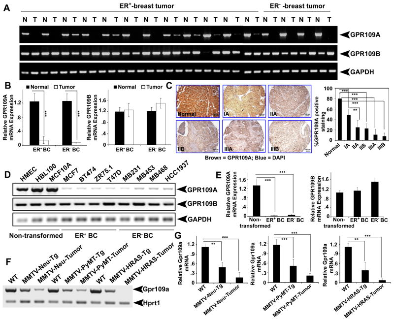
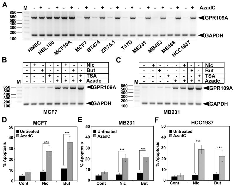
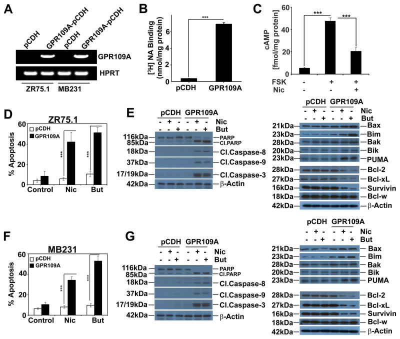
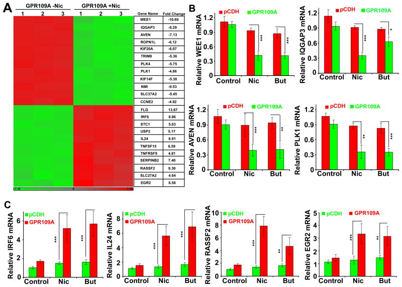
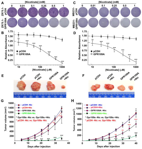
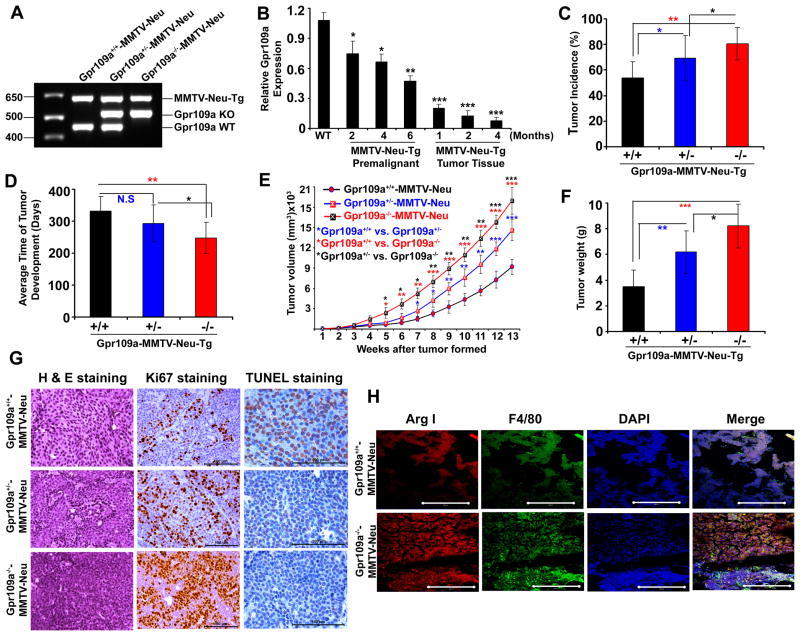
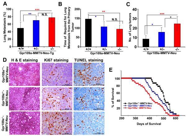
GPR109A, a G-protein-coupled receptor, is activated by niacin and butyrate. Upon activation in colonocytes, GPR109A potentiates anti-inflammatory pathways, induces apoptosis, and protects against inflammation-induced colon cancer. In contrast, GPR109A activation in keratinocytes induces flushing by activation of Cox-2-dependent inflammatory signaling, and the receptor expression is upregulated in human epidermoid carcinoma. Thus, depending on the cellular context and tissue, GPR109A functions either as a tumor suppressor or a tumor promoter. However, the expression status and the functional implications of this receptor in the mammary epithelium are not known. Here, we show that GPR109A is expressed in normal mammary tissue and, irrespective of the hormone receptor status, its expression is silenced in human primary breast tumor tissues, breast cancer cell lines, and in tumor tissues of three different murine mammary tumor models. Functional expression of this receptor in human breast cancer cell lines decreases cyclic AMP production, induces apoptosis, and blocks colony formation and mammary tumor growth. Transcriptome analysis revealed that GPR109A activation inhibits genes, which are involved in cell survival and antiapoptotic signaling, in human breast cancer cells. In addition, deletion of Gpr109a in mice increased tumor incidence and triggered early onset of mammary tumorigenesis with increased lung metastasis in MMTV-Neu mouse model of spontaneous breast cancer. These findings suggest that GPR109A is a tumor suppressor in mammary gland and that pharmacologic induction of this gene in tumor tissues followed by its activation with agonists could be an effective therapeutic strategy to treat breast cancer.


Conflict of interest statement

All authors declare no conflict of interest.

Figures

Figure 1
Figure 1
GPR109A expression is silenced in breast cancer. A and B, expression of GPR109A and GPR109B in ER-positive (ER+) and ER-negative (ER) breast tumor tissues and corresponding normal tissues analyzed by semi-quantitative and qPCR analyses, respectively. C. Representative images of GPR109A expression in normal and different breast tumor specimens. Images are shown at 10x magnification. D and E, GPR109A and GPR109B expressions in human immortalized normal and ER+ and ER breast cancer cell lines. F and G, Gpr109a expression in normal, pre-malignant and tumor tissues of MMTV-Neu-Tg, MMTV-PyMT-Tg and MMTV-HRAS-Tg mice. Data are means ± SEM. ***p<0.001; ***p<0.001 by t-test.
Figure 2
Figure 2
DNA methylation inhibitor re-activates GPR109A expression in human breast cancer cells. A, GPR109A expression was analyzed in human immortalized normal mammary epithelial cell lines and human breast cancer cell lines with and without 5′-aza-2′-deoxycytidine (AzadC). B, GPR109A expression was analyzed in two human breast cancer cell lines that were treated with HDAC inhibitors (butyrate and TSA) either alone or in combination with AzadC. Nicotinate, which does not inhibit HDACs, was used as a negative control. C, Three human breast cancer cell lines were first treated with 5′-AzadC and then treated with nicotinate or butyrate. Cells were harvested and cell cycle analysis was carried out using FACS. The apoptotic cell death was quantitated in sub G0/1 phase of the cell cycle. Values are expressed as means ± SEM. ***p<0.001 by t-test.
Figure 3
Figure 3
Stable expression of GPR109A in human breast cancer cell lines induces agonist-dependent apoptosis. A, ectopic expression of GPR109A in two human breast cancer cell lines. B GPR109A protein function in MB231-GPR109A cells was assessed by nicotinate binding assay. C, cAMP levels in MB231-GPR109A cells in the presence and absence of forskolin (10 μM) and nicotinate (1 mM). D and F, percent of apoptosis in control and GPR109A-expressing stable cell lines in the presence and absence of nicotinate and butyrate in ZR75.1 and MB231 cells. Data are means ± SEM. ***p<0.001 by t-test. E and G, expression of pro-apoptotic and anti-apoptotic proteins in control and GPR109A-expressing stable cell lines in the presence and absence of nicotinate or butyrate in ZR75.1 and MB231 cells.
Figure 4
Figure 4
GPR109A activation in breast cancer cells inhibits anti-apoptotic genes and induces pro-apoptotic genes. A, heat map generated for 12 genes that were up- and down-regulated in MB231-GPR109A cells in the presence and absence of nicotinate. B and C, qPCR confirmation of the changes in the expression of pro-survival and pro-apoptotic genes in control and GPR109A-expressing cells in the presence and absence of nicotinate or butyrate. Data are means ± SEM. *p<0.05; **p<0.01; ***p<0.001 by t-test.
Figure 5
Figure 5
GPR109A activation in breast cancer cells inhibits cell survival and mammary tumor growth. A–D, control and GPR109A-expressing ZR75.1 and MB231cells were subjected to colony-formation assay in the presence or absence of nicotinate at various concentrations for 2 weeks and the resulting colonies were stained with Giemsa dye (A and C) and the bound Giemsa dye were dissolved and quantified by spectrophotometer analysis (B and D). E–H, two days before tumor induction, female athymic Balb/c nude mice (6 mice per group) were treated with and without nicotiante (10 mM in drinking water). Two days after nicotinate treatment, ZR75.1-pCDH and ZR75.1-GPR109A as well as MB231-pCDH and MB231-GPR109A cells (1×10⁷ cells in 100 μl PBS) were injected subcutaneously in the mammary fat pad. Treatment with or without nicotinate continued throughout the experiment. Tumor volume was measured once in every 5 days for 30 days after cell injections (G and H). We compared tumor volume of pCDH-cells and GPR109A-expressing cells with and without nicotinate treatment and the statistical significance was calculated. At end of the experimental period, tumors were dissected and photographed. Representative images are shown (E and F). Data are means ± SEM. **p<0.01; ***p<0.001 by t-test.
Figure 6
Figure 6
Deletion of Gpr109a is associated with early onset of mammary tumor formation. A, Representative PCR image of genotype analysis of wild type and Gpr109a-knockout mice in MMTV-Neu-Tg background. B, Gpr109a expression was analyzed in wild type and MMTV-Neu-Tg mouse at different time of pre-malignant (2, 4 and 6 months) and different stages of tumor development (1, 2 and 4 months after the formation of the tumors. n=3 mice in each time point. Tumor incidence (C), time of tumor formation (D), tumor size (E), tumor weight (F), representative H & E histological sections (40x), Ki67 (40x) and TUNEL immunostaining (63x) (G) of Gpr109a+/+-, Gpr109a+/−- and Gpr109a−/− -MMTV-Neu-Tg mice. Data are means ± SEM (36 mice in each Gpr109a+/+-MMTV-Neu, Gpr109a+/−-MMTV-Neu and Gpr109a−/−-MMTV-Neu group). *p<0.05; **p<0.01; ***p<0.001 by t-test. H, Representative immunofluorescence (IF) images of tumor tissue sections from Gpr109a+/+- and Gpr109a−/− -MMTV-Neu-Tg mice probed for Arginase I (Arg I), F4/80 and DAPI. Images are shown at 40x magnification.
Figure 7
Figure 7
Gpr109a deletion increased lung metastasis and reduced survival. The average percent of lung metastasis (A), average time required for lung metastasis from the onset of primary tumor (B), and average number of metastatic nodules (C) were monitored in Gpr109a+/+-, Gpr109a+/−- and Gpr109a−/−-MMTV-Neu-Tg mice. To determine the morphological changes, we also monitored the H & E histological sections, Ki67 and TUNEL staining in these three groups of mice (D). Images are shown at 40x magnification. The mean survival time for these mice was also monitored (E). Data are means ± SEM (36 mice each in Gpr109a+/+-, Gpr109a+/− and Gpr109a−/− -MMTV-Neu mice). **p<0.01; ***p<0.001 by t-test.

References

    1. Offermanns S. The nicotinic acid receptor GPR109A (HM74A or PUMA-G) as a new therapeutic target. Trends Pharmacol Sci. 2006;27:384–90. - PubMed
    1. Schaub A, Futterer A, Pfeffer K. PUMA-G, an IFN-gamma-inducible gene in macrophages is a novel member of the seven transmembrane spanning receptor super family. Eur J Immunol. 2001;31:3714–25. - PubMed
    1. Tunaru S, Kero J, Schaub A, Wufka C, Blaukat A, Pfeffer K, Offermanns S. PUMA-G and HM74 are receptors for nicotinic acid and mediate its anti-lipolytic effect. Nat Med. 2003;9:352–55. - PubMed
    1. Wise A, Foord SM, Fraser NJ, Barness AA, Elshourbagy N, Eilert M, et al. Molecular identification of high and low affinity receptors for nicotinic acid. J Biol Chem. 2003;278:9869–74. - PubMed
    1. Soga T, Kamohara M, Takasaki J, Matsumoto S, Ohishi T, Hiyama H, et al. Molecular identification of nicotinic acid receptor. Biochem Biophys Res Commun. 2003;303:364–69. - PubMed

Publication types