. 2014 Feb 15;74(4):1261-1271.
doi: 10.1158/0008-5472.CAN-13-2062. Epub 2013 Dec 26.

Tumor suppressor alterations cooperate to drive aggressive mesotheliomas with enriched cancer stem cells via a p53-miR-34a-c-Met axis

Affiliations

Tumor suppressor alterations cooperate to drive aggressive mesotheliomas with enriched cancer stem cells via a p53-miR-34a-c-Met axis

Craig W Menges et al. Cancer Res. .

Abstract

Malignant mesothelioma is a highly aggressive, asbestos-related cancer frequently marked by mutations of both NF2 and CDKN2A. We demonstrate that germline knockout of one allele of each of these genes causes accelerated onset and progression of asbestos-induced malignant mesothelioma compared with asbestos-exposed Nf2(+/-) or wild-type mice. Ascites from some Nf2(+/-);Cdkn2a(+/-) mice exhibited large tumor spheroids, and tail vein injections of malignant mesothelioma cells established from these mice, but not from Nf2(+/-) or wild-type mice, produced numerous tumors in the lung, suggesting increased metastatic potential of tumor cells from Nf2(+/-);Cdkn2a(+/-) mice. Intraperitoneal injections of malignant mesothelioma cells derived from Nf2(+/-);Cdkn2a(+/-) mice into severe combined immunodeficient mice produced tumors that penetrated the diaphragm and pleural cavity and harbored increased cancer stem cells (CSC). Malignant mesothelioma cells from Nf2(+/-);Cdkn2a(+/-) mice stained positively for CSC markers and formed CSC spheroids in vitro more efficiently than counterparts from wild-type mice. Moreover, tumor cells from Nf2(+/-);Cdkn2a(+/-) mice showed elevated c-Met expression/activation, which was partly dependent on p53-mediated regulation of miR-34a and required for tumor migration/invasiveness and maintenance of the CSC population. Collectively, these studies demonstrate in vivo that inactivation of Nf2 and Cdkn2a cooperate to drive the development of highly aggressive malignant mesotheliomas characterized by enhanced tumor spreading capability and the presence of a CSC population associated with p53/miR-34a-dependent activation of c-Met. These findings suggest that cooperativity between losses of Nf2 and Cdkn2a plays a fundamental role in driving the highly aggressive tumorigenic phenotype considered to be a hallmark of malignant mesothelioma.


Figures

Figure 1
Figure 1
Kaplan-Meier survival curves depicting accelerated MM formation in Nf2+/−;p16/p19+/−mice vs. Nf2+/− and WT mice, following initial exposure to asbestos.
Figure 2
Figure 2
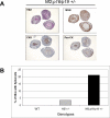
Ascites from asbestos-exposed Nf2+/−;p16/p19+/− mice harboring tumor spheroids. A) Representative spheroids from the ascites of Nf2+/−;p16/p19+/− mice stained with H&E (top left), mesothelin (MSN), cytokeratin 8 (CK8) and pan-cytokeratin (pan-CK). Ascitic spheroids were centrifuged, resuspended in 2% agarose/formalin, paraffin-embedded, and sectioned for histopathology. B) Graph depicting the frequency of tumor spheroids in ascites of asbestos-exposed Nf2+/−;p16/p19+/−, Nf2+/− and WT mice.
Figure 3
Figure 3
MM cells from Nf2+/−;p16/p19+/− mice show higher metastatic potential than those of Nf2+/− and WT mice, based on experimental metastasis assay using MM cell lines derived from asbestos-exposed Nf2+/−;p16/p19+/−, Nf2+/− and WT mice. A) Representative images of lung tumor development in recipient SCID mice 4 weeks after tail-vein injections of MM cells (3 cell lines per genotype, each injected in 3 mice), and ratios of recipient mice that developed lung tumors are indicated. H&E staining of representative lung sections (10X magnification) are shown in bottom panels (M, metastasis; L, lung). B) H&E sections of primary tumors, migrating/invading tumors and metastatic tumors in SCID mice injected i.p. with MM cells derived from Nf2+/−;p16/p19+/− mouse (D, diaphragm; I, intestines; L, lung; P, pancreas; S, spleen; T, tumor).
Figure 4
Figure 4
MM cells from Nf2+/−;p16/p19+/− mice harbor more CSCs than MM cells from WT mice. A) Equal numbers of MM cells from Nf2+/−;p16/p19+/− and WT mice were seeded on non-adherent plates in stem cell media, and colonies were photographed after 2 days. B) Flow cytometry analysis depicting CD24 surface staining of MM cells from Nf2+/−;p16/p19+/− and WT mice. C) Aldefluor assay showing percentage of CSCs in Nf2+/−;p16/p19+/−-derived MM cell line treated with increasing concentrations of pemetrexed.
Figure 5
Figure 5
c-Met is upregulated/activated in MM cells from Nf2+/−;p16/p19+/− mice. A) RT-PCR analysis of c-Met mRNA levels in MM cells from Nf2+/−;p16/p19+/− and WT mice. B) Immunoblot analysis of phospho-(P-)c-Met, total c-Met and actin levels in MM cells from WT and Nf2+/−;p16/p19+/− mice. C) IHC staining of P-c-Met and c-Met in MM tumor specimens from Nf2+/−;p16/p19+/− and WT mice.
Figure 6
Figure 6
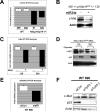
A p53-miR34a axis controls c-Met upregulation in MM cells from Nf2+/−;p16/p19+/−mice. A) RT-PCR analysis of RNA levels of miR34a in MM cells from Nf2+/−;p16/p19+/− and WT mice. B) Immunoblot analysis of c-Met protein levels in MM cells from Nf2+/−;p16/p19+/− mice 24 h post-nucleofection with miR34a or control miRNA. C) RT-PCR analysis of c-Met expression in MM cell lines 129 and 380, from Nf2+/−;p16/p19+/− mice, 24 h post-treatment with etoposide. D) Immunoblot analysis of c-Met and p53 protein levels 24 h post-treatment with etoposide in MM cell lines 129 and 380 from Nf2+/−;p16/p19+/− mice. E) RT- PCR analysis of miR34a levels in MM cell line 690, from WT mouse, 24 h post-nucleofection of control or Tp53 siRNA. F) Immunoblot analysis of c-Met and p53 in MM cell line 690, from WT mouse, 24 h post-nucleofection of control or Tp53 siRNAs.
Figure 7
Figure 7
c-Met activity is required for maintenance of CSCs in MM cells from Nf2+/−;p16/p19+/−mice. A) Cell viability assay of MM cell line 129 from Nf2+/−;p16/p19+/− mouse treated with increasing concentrations of c-Met inhibitor SGX523. B) Aldeflour assay showing the percentage of CSCs in MM line 129, from a Nf2+/−;p16/p19+/− mouse, following treatment with increasing concentrations of SGX523. C) Equal numbers of MM 129 cells from Nf2+/−;p16/p19+/−mouse were seeded on non-adherent plates in stem cell media, and colonies were photographed after 2-days in presence or absence of SU11274 or SGX523. D) Working model depicting role of c-Met upregulation in metastasis and CSC maintenance of MM cells from Nf2+/−;p16/p19+/− mice. Dotted line depicts the potential role of CSCs in metastatic potential.

References

    1. Finn RS, Brims FJ, Gandhi A, Olsen N, Musk AW, Maskell NA, et al. Postmortem findings of malignant pleural mesothelioma: a two-center study of 318 patients. Chest. 2012;142:1267–73. - PubMed
    1. Bianchi AB, Mitsunaga SI, Cheng JQ, Klein WM, Jhanwar SC, Seizinger B, et al. High frequency of inactivating mutations in the neurofibromatosis type 2 gene (NF2) in primary malignant mesotheliomas. Proc Natl Acad Sci U S A. 1995;92:10854–8. - PMC - PubMed
    1. Cheng JQ, Jhanwar SC, Klein WM, Bell DW, Lee WC, Altomare DA, et al. p16 alterations and deletion mapping of 9p21-p22 in malignant mesothelioma. Cancer Res. 1994;54:5547–51. - PubMed
    1. Ladanyi M, Zauderer MG, Krug LM, Ito T, McMillan R, Bott M, et al. New strategies in pleural mesothelioma: BAP1 and NF2 as novel targets for therapeutic development and risk assessment. Clin Cancer Res. 2012;18:4485–90. - PMC - PubMed
    1. Altomare DA, Vaslet CA, Skele KL, De Rienzo A, Devarajan K, Jhanwar SC, et al. A mouse model recapitulating molecular features of human mesothelioma. Cancer Res. 2005;65:8090–5. - PubMed

Publication types

MeSH terms