. 2014 Mar;42(5):3125-37.
doi: 10.1093/nar/gkt1347. Epub 2013 Dec 25.

Tracking the processing of damaged DNA double-strand break ends by ligation-mediated PCR: increased persistence of 3'-phosphoglycolate termini in SCAN1 cells

Affiliations

Tracking the processing of damaged DNA double-strand break ends by ligation-mediated PCR: increased persistence of 3'-phosphoglycolate termini in SCAN1 cells

Konstantin Akopiants et al. Nucleic Acids Res. 2014 Mar.

Abstract

To track the processing of damaged DNA double-strand break (DSB) ends in vivo, a method was devised for quantitative measurement of 3'-phosphoglycolate (PG) termini on DSBs induced by the non-protein chromophore of neocarzinostatin (NCS-C) in the human Alu repeat. Following exposure of cells to NCS-C, DNA was isolated, and labile lesions were chemically stabilized. All 3'-phosphate and 3'-hydroxyl ends were enzymatically capped with dideoxy termini, whereas 3'-PG ends were rendered ligatable, linked to an anchor, and quantified by real-time Taqman polymerase chain reaction. Using this assay and variations thereof, 3'-PG and 3'-phosphate termini on 1-base 3' overhangs of NCS-C-induced DSBs were readily detected in DNA from the treated lymphoblastoid cells, and both were largely eliminated from cellular DNA within 1 h. However, the 3'-PG termini were processed more slowly than 3'-phosphate termini, and were more persistent in tyrosyl-DNA phosphodiesterase 1-mutant SCAN1 than in normal cells, suggesting a significant role for tyrosyl-DNA phosphodiesterase 1 in removing 3'-PG blocking groups for DSB repair. DSBs with 3'-hydroxyl termini, which are not directly induced by NCS-C, were formed rapidly in cells, and largely eliminated by further processing within 1 h, both in Alu repeats and in heterochromatic α-satellite DNA. Moreover, absence of DNA-PK in M059J cells appeared to accelerate resolution of 3'-PG ends.


Figures

Figure 1.
Figure 1.
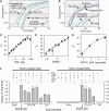
Principle of the Taqman PCR assay and verification of enzymatic manipulations. (A) Rationale. At AGT•ACT sites, NCS-C induces three types of bistranded lesions: (1) an abasic site (^) with a closely opposed break, (2) a DSB with a 3′-PG (•) terminus or (3) a DSB with a 3′-phosphate terminus. NaBH4 stabilizes the 5′-aldehyde, while putrescine converts abasic sites to strand breaks (dashed arrow). The resulting DSB ends are subjected to treatment with PNKP, TdT, TDP1 and CIP, such that 3′-phosphate DSBs are capped with unligatable tails, while 3′-PG DSBs are converted to 3′-hydroxyl DSBs, ligated to an anchor and amplified by PCR. (B) Quantitation by Taqman PCR. As PCR product accumulates, a fluorescent Taqman probe spanning the ligation site anneals to the joint sequence, and Taq polymerase releases the FAM fluorophore. (C) Verification of 3′-PG termini at the AGT sequence at Alu bp 224–226. An end-labeled fragment containing the BLUR8 Alu DNA clone was treated with NCS-C, and then treated with TDP1 and/or PNKP as indicated and analyzed on a denaturing sequencing gel. A substantial fraction of the band corresponding to a DSB at bp 226 required both TDP1 and PNKP to induce a shift to lower mobility, indicating the presence of DSBs with 3′-PG termini, and their conversion to 3-hydroxyl termini. (D) Verification that the enzymatic manipulation of DNA ends can be effected with near-quantitative efficiency. An internally labeled (asterisk) substrate containing a DSB with a one-base 3′ overhang and a 3′-PG or 3′-phosphate terminus (black circle) was treated successively with the indicated enzymes. A 12-base-labeled fragment was released from the 3′-end by TaqI and analyzed on a denaturing gel. The 3′-phosphate terminus is efficiently poly(dT)-tailed, while the 3′-PG terminus is converted to a 3′-hydroxyl and ligated to an oligomeric anchor duplex.
Figure 2.
Figure 2.
Detection and quantitation of 3′-PG, 3′-phosphate and 3′-hydroxyl DSB termini on NCS-C-induced DSBs. (A) Detection of DSBs in a cloned Alu sequence. A plasmid harboring the BLUR8 Alu sequence was treated with 100-nM NCS-C, then with NaBH4 and putrescine and finally with PNKP, TdT, TDP1 and CIP. TdT, TDP1 or NCS-C was omitted where indicated. DSB ends were then ligated to an anchor and quantitated by Taqman PCR. (B) Detection of DSB ends in DNA from cells treated with 5 -μM NCS-C. Chemical and enzymatic treatments and Taqman PCR, were the same as in (A), except that only 50 pg of ligated template DNA was added and a consensus Alu PCR primer was used. Various treatments were omitted as indicated. (C) Titration of the PCR assay for 3′-PG DSBs. Conditions are the same as for the Complete Rxn in (B), except that various dilutions of the ligated DNA were used as PCR template. The threshold cycle CT is plotted, (threshold level of 0.04 fluorescence units is indicated by red line in B). The slope of the best-fit line was exactly one PCR cycle per 2-fold dilution. (D) Same as (C), except that to mimic decreasing levels of 3′-PG DSBs in DNA from treated cells, the same ligated DNA was diluted in ligated DNA from untreated cells, keeping total template DNA constant at 50 pg. The best-fit slope was 1.28 PCR cycles per 2-fold dilution. (E) Dose response for formation of 3′-PG DSBs in SCAN1 lymphoblastoid cells by NCS-C. Cells were treated with NCS-C for 10 min at 37°C. (F) Detection and quantitation of 3′-PG, 3′-phosphate and 3′-hydroxyl DSB termini in DNA from NCS-C-treated cells (left), or in NCS-C-treated DNA (right). DNA was subjected to chemical and enzymatic manipulations as is in (B), with various treatments omitted as indicated. Ligatable DNA ends were then quantitated by LMPCR, and normalized to the sample where TdT treatment was omitted so that all 3′-PG, 3′-phosphate and 3′-hydroxyl DSB termini would be rendered ligatable. The types of ends that should be detected by each combination of treatments are listed below the graph. For simplicity, a change of one PCR cycle in CT was assumed to correspond to a factor of 2 in the number of ligatable ends. CT values from which these data were derived are shown in Supplementary Figure S3. In all panels, error bars show mean ± SEM for at least three PCR reactions.
Figure 3.
Figure 3.
Kinetics of 3′ processing of DSBs in lymphoblastoid cells. (A) Cells in PBS were treated with 5 -μM NCS-C for 5 min at 0°C, and then for the indicated times at 37°C, with complete medium added after 10 min. DNA was isolated and subjected to the complete enzyme reactions for detection of 3′-PGs, TdT and CIP only for detection of 3′-phosphates or no enzymes for detection of 3′-hydroxyls (OH), then subjected to LMPCR. Typical amplification profiles for 3′-PG lesions are shown. Values (mean ± SEM from three independent experiments, each with three to four PCR reactions per sample) are normalized to the total of all three lesions at 10 min. (B) DNA samples from the experiment shown in (A) were subjected to LMPCR with primers and a probe specific to α-satellite DNA, and similarly analyzed. Owing to the weaker LMPCR signal for the satellite sequence, PG and PO4 data at >30 min were indistinguishable from background. Profiles for 3′-hydroxyl lesions are shown. (C) Cells were incubated in PBS at 0°C for 5 min and then at 22°C for the indicated times or for 10 min at 37°C, and the same three types of DSB ends were determined. The abundance of each type of lesion, calculated from CT values, was normalized to the average level of total lesions at 30 and 60 min in each experiment. Error bars show mean ± SEM from four experiments.
Figure 4.
Figure 4.
Increased persistence of 3′-PG DSB ends in SCAN1 cells. (A) Cells were treated with 5 -μM NCS-C for 10 min at 37°C, and either harvested immediately or after incubation in complete medium for 20 or 60 min, and then 3′-PG DSB termini were detected by Taqman PCR. (B) Pooled real-time data (CT values, mean ± SEM) from four separate experiments, two with cells from patients 1646 (normal) and 1662 (SCAN1) and two with cells from patients 1668 (normal) and 1664 (SCAN1), with three to four PCR reactions for each sample. (C) Cell cycle profiles of normal (1646) and SCAN1 (1662) cells before and after treatment with 1-nM neocarzinostatin (NCS) for 1 h. G1, S and G2 fractions are indicated. Similar results were seen at other time points and in replicate experiments (Supplementary Figure S8).
Figure 5.
Figure 5.
Effect of DNA-PK inhibition or absence of DNA-PK on 3′-PG processing. (A) Normal lymphoblastoid cells (patient 1646) were grown in the presence 10 -μM KU-57788 (or 0.4% DMSO) for 2 h, treated with NCS-C as in Figure 4 and then diluted into complete medium with or without KU-57788 and harvested at the indicated times for determination of 3′-PG DSBs. (B) M059K or DNA-PK-deficient M059J glioma cells were trypsinized, treated with NCS-C as in (A), then harvested either immediately or after incubation in complete medium for 1 h. Levels of 3′-PG and 3′-hydroxyl DSBs were then determined by LMPCR. Error bars show mean ± SEM for four experiments. Differences between M059J and M059K in the ratio of PG- to hydroxyl-terminated DSB ends were significant (P < 0.01) at both time points.

References

    1. Pawelczak KS, Bennett SM, Turchi JJ. Coordination of DNA-PK activation and nuclease processing of DNA termini in NHEJ. Antioxid. Redox Signal. 2011;14:2531–2543. - PMC - PubMed
    1. Povirk LF. Processing of damaged DNA ends for double-strand break repair in mammalian cells. ISRN Mol. Biol. 2012;2012:345805. - PMC - PubMed
    1. Dedon PC, Goldberg IH. Free-radical mechanisms involved in the formation of sequence- dependent bistranded DNA lesions by the antitumor antibiotics bleomycin, neocarzinostatin, and calicheamicin. Chem. Res. Toxicol. 1992;5:311–332. - PubMed
    1. Povirk LF. DNA damage and mutagenesis by radiomimetic DNA-cleaving agents: bleomycin, neocarzinostatin and other enediynes. Mutat. Res. 1996;355:71–89. - PubMed
    1. Inamdar KV, Pouliot JJ, Zhou T, Lees-Miller SP, Rasouli-Nia A, Povirk LF. Conversion of phosphoglycolate to phosphate termini on 3′ overhangs of DNA double-strand breaks by the human tyrosyl-DNA phosphodiesterase hTdp1. J. Biol. Chem. 2002;276:24323–24330. - PubMed

Publication types

MeSH terms