. 2014 Apr;42(6):3982-97.
doi: 10.1093/nar/gkt1350. Epub 2013 Dec 26.

Converging pathways involving microRNA-206 and the RNA-binding protein KSRP control post-transcriptionally utrophin A expression in skeletal muscle

Affiliations

Converging pathways involving microRNA-206 and the RNA-binding protein KSRP control post-transcriptionally utrophin A expression in skeletal muscle

Adel Amirouche et al. Nucleic Acids Res. 2014 Apr.

Abstract

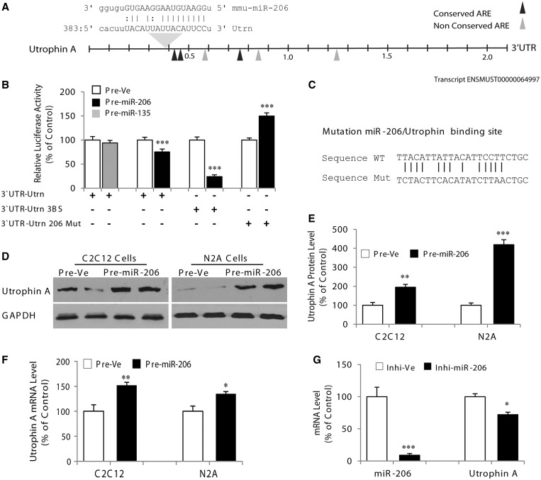
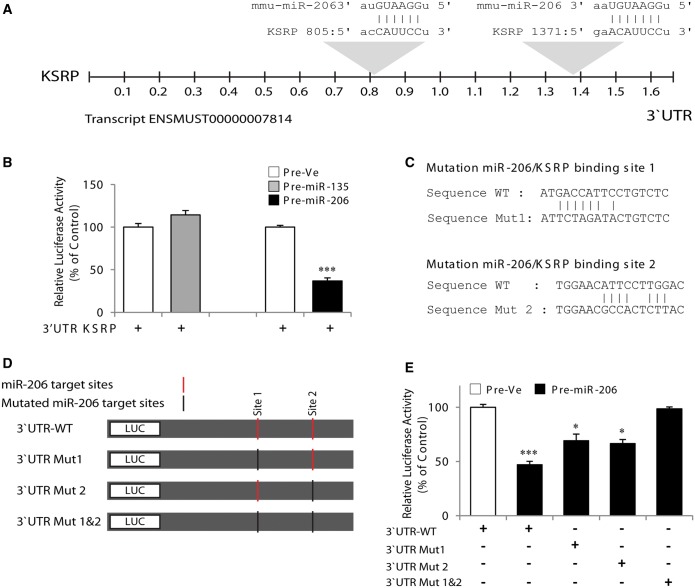
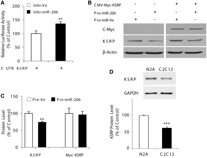
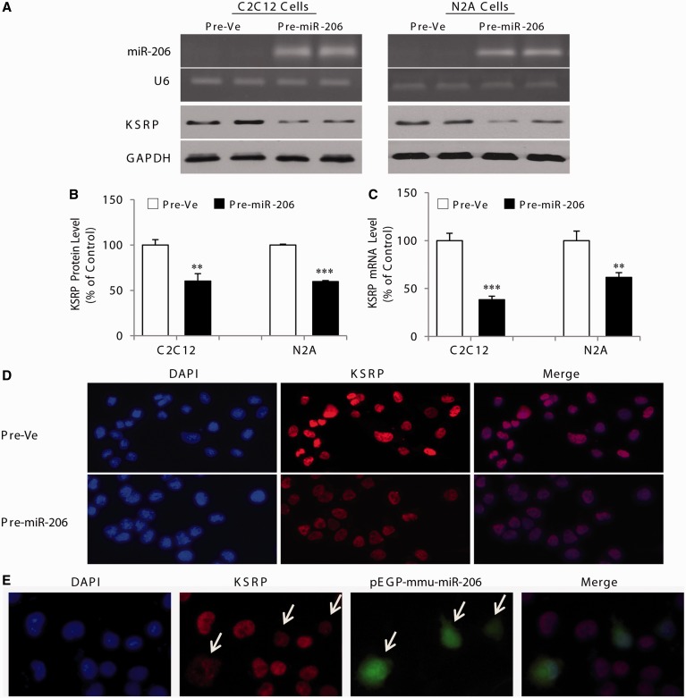
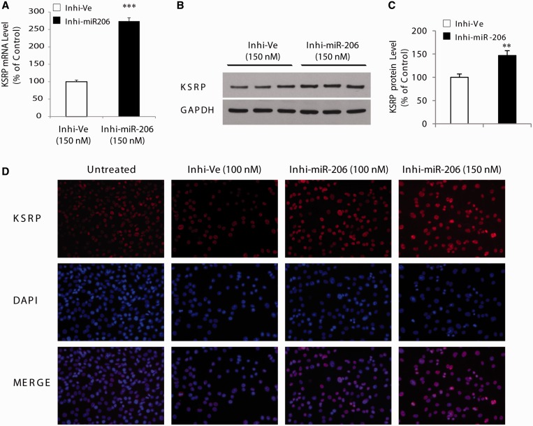
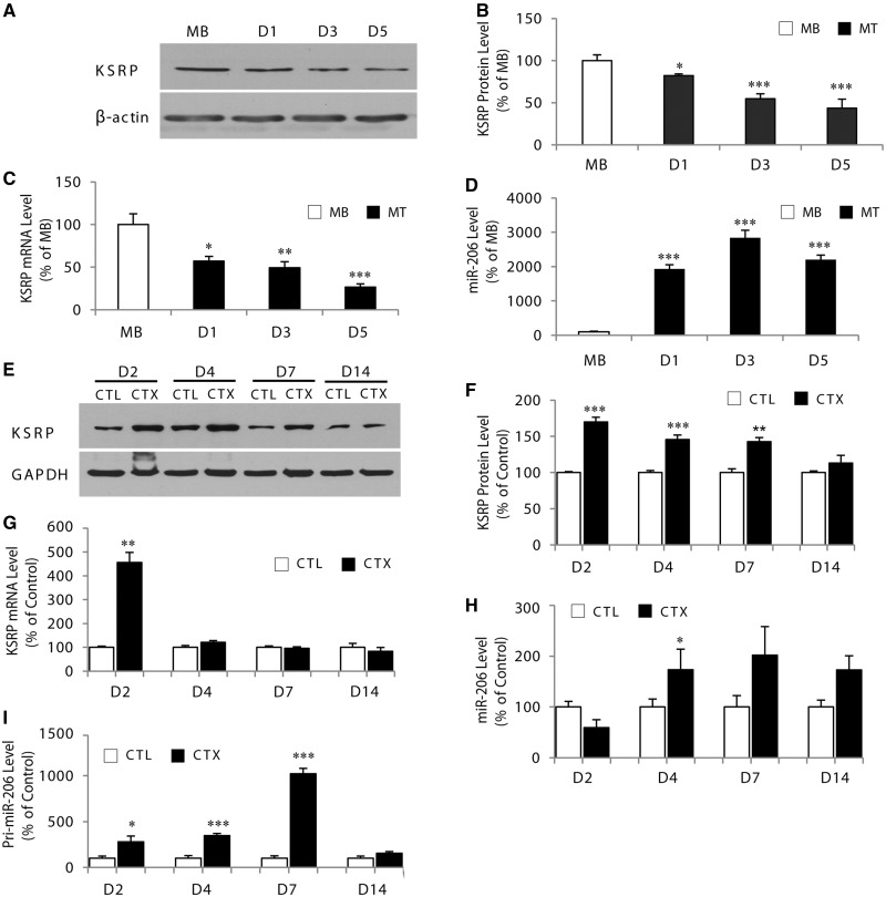
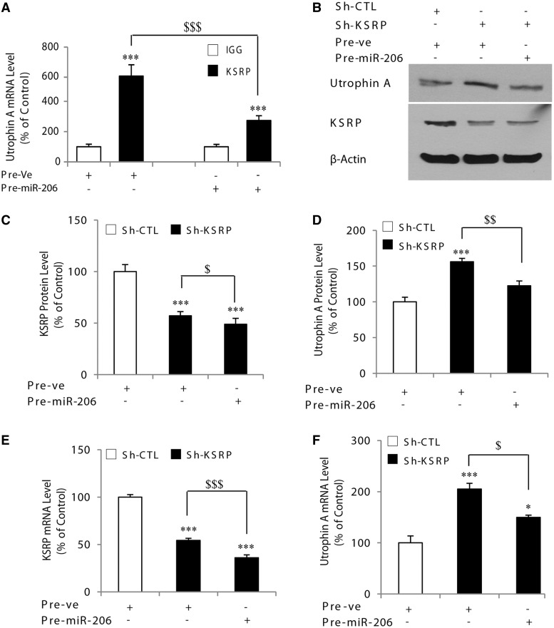
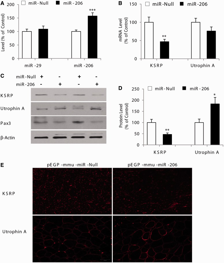
Several reports have previously highlighted the potential role of miR-206 in the post-transcriptional downregulation of utrophin A in cultured cells. Along those lines, we recently identified K-homology splicing regulator protein (KSRP) as an important negative regulator in the post-transcriptional control of utrophin A in skeletal muscle. We sought to determine whether these two pathways act together to downregulate utrophin A expression in skeletal muscle. Surprisingly, we discovered that miR-206 overexpression in cultured cells and dystrophic muscle fibers causes upregulation of endogenous utrophin A levels. We further show that this upregulation of utrophin A results from the binding of miR-206 to conserved sites located in the 3'-UTR (untranslated region) of KSRP, thus causing the subsequent inhibition of KSRP expression. This miR-206-mediated decrease in KSRP levels leads, in turn, to an increase in the expression of utrophin A due to a reduction in the activity of this destabilizing RNA-binding protein. Our work shows that miR-206 can oscillate between direct repression of utrophin A expression via its 3'-UTR and activation of its expression through decreased availability of KSRP and interactions with AU-rich elements located within the 3'-UTR of utrophin A. Our study thus reveals that two apparent negative post-transcriptional pathways can act distinctively as molecular switches causing repression or activation of utrophin A expression.


Figures

Figure 1.
Figure 1.
MicroRNA-206 enhances endogenous utrophin A expression. (A) Schematic representation of the utrophin A 3′-UTR showing the presence of a binding site of miR-206. (B) C2C12 myoblasts were cotransfected with reporter constructs containing the utrophin A full-length 3′-UTR, utrophin A full-length 3′-UTR with miR-206 seed sequence mutated or utrophin A full-length 3′-UTR with three binding sites (3BS) of miR-206, together with pre-miR-206 or pre-Ve. Pre-miR-135, a miRNA not predicted to target KSRP, was also used as control. (C) Shows the conserved binding site of miR-206 that was mutated within the utrophin A full-length 3′-UTR. (D) Representative immunoblots of utrophin A protein levels after pre-miR-206 or pre-Ve transfection in C2C12 cells and N2a cells. For the western blots, GAPDH was used to ensure equal loading. (E) Represents quantification of utrophin A protein levels in C2C12 cells and N2a cells transfected with pre-miR-206 or pre-Ve. (F) RT-qPCR analyses of utrophin A mRNA expression in C2C12 cells and N2a cells transfected with pre-miR-206 or pre-Ve. Note the increase in utrophin A protein and mRNA levels on pre-miR-206 overexpression. (G) Shows the results of RT-qPCR analyses for utrophin A mRNA in C2C12 cells transfected with the miR-206 inhibitor (150 nM). Values are means ± standard error (SE) (n = 3/group in triplicate). *P < 0.05; **P < 0.01; ***P < 0.001; relative to corresponding control (Figure 1B, E–G: unpaired t-tests).
Figure 2.
Figure 2.
MicroRNA-206 targets the KSRP 3′-UTR. (A) Schematic representation of the mouse KSRP 3′-UTR showing the presence of binding sites for miR-206. (B) C2C12 cells were cotransfected with a reporter construct containing the mouse 3′-UTR of KSRP, together with pre-miR-206 or pre-Ve. Pre-miR-135, a miRNA not predicted to target KSRP, was also used as control. (C) Base pairings of miR-206 with the 3′-UTR KSRP showing sites of mutations. (D) Schematic representation of reporter constructs containing the wild-type 3′-UTR of KSRP or mutated versions. Red vertical bars represent miR-206 target sites; black vertical bars represent mutated miR-206 target sites. (E) Shows luciferase activity observed with the different 3′-UTR constructs of KSRP on overexpression of pre-miR-206 in C2C12 cells. Values are means ± SE (n = 3/group in triplicate). *P < 0.05; ***P < 0.001 relative to corresponding control (Figure 2B: unpaired t-test; Figure 2E; One-way ANOVA and Bonferroni as post hoc test).
Figure 3.
Figure 3.
MicroRNA-206 specifically regulates KSRP expression via its 3′-UTR. (A) Represents the activity of the luciferase reporter construct containing the mouse KSRP 3′-UTR in C2C12 cells transfected with miR-206 inhibitor (100 nM) or negative control. (B) and (C) show representative western blots and their quantification, respectively, for KSRP and C-Myc with β-actin as a control. (D) Representative western blots and their quantification of KSRP protein levels in N2a and C2C12 cells. Values are means ± SE (n = 3/group in triplicate for luciferase experiments). **P < 0.01; ***P < 0.001; relative to corresponding control (Figure 3A–D: unpaired t-test).
Figure 4.
Figure 4.
MicroRNA-206 regulates expression of endogenous KSRP. (A) Top two panels: representative ethidium bromide-stained agarose gel showing mature miR-206 RT-PCR product amplified from C2C12 and N2a cells transfected with pre-miR-206. The U6 was used as control for loading. Bottom two panels: representative immunoblots of KSRP protein levels after pre-miR-206 transfection in C2C12 and N2a cells. (B) and (C) western blot quantification and qRT-PCR, respectively, for KSRP in C2C12 and N2a cells transfected with a pre-miR-206 or pre-Ve. (D) Shows immunofluorescence experiments for KSRP in control N2a cells (pre-Ve) or cells transfected with pre-miR-206. (E) Immunofluorescence for KSRP in N2a cells transfected with a mmu-miR-206 construct coupled with GFP (pEGP-mmu-miR-206).The reduction in KSRP levels was only seen in GFP-positive cells (arrows). As a control, no staining was observed in experiments with no primary antibody. Values are means ± SE (n = 3/group in triplicate). **P < 0.01; ***P < 0.001; relative to corresponding control (Figure 4B and C: unpaired t-test).
Figure 5.
Figure 5.
Inhibition of miR-206 causes derepression of KSRP. (A) Inhibition of miR-206 promotes increases in KSRP expression in C2C12 cells. Real-time qPCR analysis revealed that the endogenous mRNA levels of KSRP were increased with the miR-206 inhibitor at 150 nM. (B) Shows western blot analysis of KSRP with GAPDH as control. (C) Represents quantification of KSRP protein levels in C2C12 cells transfected with the miR-206 inhibitor. (D) Shows immunofluorescence experiments for KSRP in control N2a cells transfected with miR-206 inhibitor at 100 nM and 150 nM or negative control at 100 nM. Note the increase in KSRP in the presence of the inhibitor. As a control, no staining was observed in experiments with no primary antibody. Values are means ± SE (n = 3/group in triplicate). **P < 0.01; ***P < 0.001; relative to corresponding control (Figure 5A: one-way ANOVA and Bonferroni as post hoc test; Figure 5C: unpaired t-test).
Figure 6.
Figure 6.
Expression of KSRP and miR-206 is inversely correlated during skeletal muscle cell differentiation and after CTX-induced degeneration/regeneration. (A) Representative western blot showing KSRP level in myoblasts (MB) and myotubes at day 1, 3 and 5 of differentiation. (B) KSRP levels were quantified and are expressed as a percent of the levels seen in myoblasts. The ß-actin served as loading control. (C) RT-qPCR of KSRP mRNA levels during differentiation of C2C12 myoblasts. (D) RT-qPCR analysis of miR-206 expression normalized to U6 levels in C2C12 myoblasts and myotubes at day 1, 3 and 5. (E) Representative western blots showing KSRP levels in TA muscles from mdx mice injected with cardiotoxin (CTX) or saline (as a control: CTL) 2, 4, 7 and 14 days after injection. (F) Western blot analysis of KSRP protein levels after CTX-induced muscle degeneration and regeneration. (G) Represents quantification of KSRP mRNA levels in TA muscles from mdx mice after CTX-induced injury. (H) and (I) RT-qPCR revealed an increase in miR-206 and pri-miR-206 levels during CTX-induced injury. Values are means ± SE (n = 4/group). **P < 0.01; ***P < 0.001; relative to corresponding control (Figure 6B: unpaired t-test; Figure 6B–D: unpaired one-way ANOVA and Bonferroni as post hoc test; Figure 6F–I: paired one-way ANOVA and Bonferroni as post hoc test). At D14 in Figure 6H, P = 0.059.
Figure 7.
Figure 7.
Competition of the miR-206 and KSRP pathways on the regulation of utrophin A. (A) Represents the results of RIP assays in C2C12 cells transfected with either negative control or pre-miR-206. Samples were immunoprecipitated with either IgG as control or with an antibody against KSRP. GAPDH was used as a control. Real-time qPCR analysis revealed a decreased amount of utrophin A mRNA interacting with KSRP on miR-206 overexpression. (B) Representative western blots of utrophin A and KSRP in C2C12 cells transfected with pre-miR-206 or negative control and with sh-RNA against KSRP. The ß-actin served as a loading control. (C and D) Represents quantification of KSRP and utrophin A protein levels in C2C12 cells transfected with pre-miR-206 or negative control and with sh-RNA against KSRP. (E and F) Show the results of RT-qPCR analyses for KSRP and utrophin A mRNA, respectively, in C2C12 cells transfected with pre-miR-206 or negative control and with sh-RNA against KSRP. Levels of KSRP and utrophin A mRNA are standardized to 18S mRNA. Values are means ± SE (n = 4/group). *P < 0.05; **P < 0.01, ***P < 0.001; relative to corresponding control (Figure 7A, C–F: one-way ANOVA and Bonferroni as post hoc test). $ < 0.05; $$ < 0.01; $$$ < 0.001.
Figure 8.
Figure 8.
MicroRNA-206 represses KSRP expression causing upregulation of utrophin A in mdx mouse muscle fibers. The TA muscles from mdx mice were electroporated with pEGP-mmu-miR-206 or the corresponding control (pEGP-mmu-miR-Null in the contralateral TA muscle) and 10 days later, both TA muscles were excised and analyzed. (A) Shows the results of RT-qPCR analyses for miR-206 level in TA muscles electroporated with pEGP-mmu-miR-206 or pEGP-mmu-miR-Null. MiR-29C was used as a control. (B) Shows the results of RT-qPCR analyses for utrophin A and KSRP mRNAs in TA muscles electroporated with pEGP-mmu-miR-206 or pEGP-mmu-Null. (C) Shows representative western blot analysis of KSRP, utrophin A and Pax3 with β-actin as control for two different samples for each treatment. (D) Represents quantification of utrophin A and KSRP protein expression in TA muscles electroporated with miR-206 vector or corresponding control. (E) Are representative examples of muscle cross sections stained with an antibody against KSRP and utrophin A from mdx mouse muscle electroportated with pEGP-mmu-miR-206 or pEGP-mmu-miR-Null. As a control, no staining was observed in experiments with no primary antibody. Values are means ± SE (n = 6–8). *P < 0.05; **P < 0.01; ***P < 0.001; relative to corresponding control (Figure 1A, B and D: paired t-test).

Similar articles

Cited by

References

    1. Huntzinger E, Izaurralde E. Gene silencing by microRNAs: contributions of translational repression and mRNA decay. Nat. Rev. Genet. 2011;12:99–110. - PubMed
    1. Meister G. miRNAs get an early start on translational silencing. Cell. 2007;131:25–28. - PubMed
    1. Flynt AS, Lai EC. Biological principles of microRNA-mediated regulation: shared themes amid diversity. Nat. Rev. Genet. 2008;9:831–842. - PMC - PubMed
    1. Guller I, Russell AP. MicroRNAs in skeletal muscle: their role and regulation in development, disease and function. J. Physiol. 2010;588:4075–4087. - PMC - PubMed
    1. Luo W, Nie Q, Zhang X. MicroRNAs involved in skeletal muscle differentiation. J. Genet. Genomics. 2013;40:107–116. - PubMed

Publication types