

Staphylococcus aureus Colonization in Acute and Chronic Skin Lesions of Patients with Atopic Dermatitis
- PMID: 24371386
- PMCID: PMC3870207
- DOI: 10.5021/ad.2013.25.4.410
Staphylococcus aureus Colonization in Acute and Chronic Skin Lesions of Patients with Atopic Dermatitis
Abstract
Background: Staphylococcus aureus (SA) has peculiar abilities to colonize the skin in atopic dermatitis (AD) patients.
Objective: We sought to determine the colonization rates of SA in acute and chronic skin lesions of AD patients, to find any difference in colonization rates according to age and to find the influences of total immunoglobulin E (IgE) and eosinophil counts to the colonization of SA.
Methods: We evaluated the total IgE level and eosinophil counts, and cultured SA from the skin lesions of 687 AD patients (131 acute and 556 chronic skin lesions) and 247 control urticaria patients (July 2009 to November 2010; Samsung Medical Center Dermatology Clinic, Seoul, Korea).
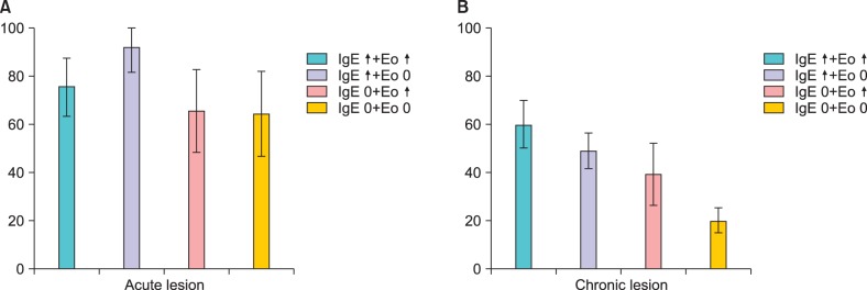
Results: The SA colonization rates were 74%, 38% and 3% in acute, chronic skin lesions and control skin, respectively, and they were increased with age in AD patients. The colonization rate in chronic skin lesions was higher in the high IgE/eosinophilia groups as compared to the normal IgE/eosinophil groups.
Conclusion: The SA colonization rate was higher in AD patients and especially in acute lesions, and had a tendency to increase with age. As the colonization rates were only higher in the high IgE/eosinophilia groups of chronic skin lesions, we suggested that SA may invade the skin through barrier defects in acute skin lesions, but the colonization in chronic lesions may be orchestrated through many different factors.
Keywords: Atopic dermatitis; Colonization rate; Eosinophil counts; Serum total IgE; Staphylococcus aureus.
Figures
References
-
- Breuer K, HAussler S, Kapp A, Werfel T. Staphylococcus aureus: colonizing features and influence of an antibacterial treatment in adults with atopic dermatitis. Br J Dermatol. 2002;147:55–61. - PubMed
-
- Gong JQ, Lin L, Lin T, Hao F, Zeng FQ, Bi ZG, et al. Skin colonization by Staphylococcus aureus in patients with eczema and atopic dermatitis and relevant combined topical therapy: a double-blind multicentre randomized controlled trial. Br J Dermatol. 2006;155:680–687. - PubMed
-
- Higaki S, Morohashi M, Yamagishi T, Hasegawa Y. Comparative study of staphylococci from the skin of atopic dermatitis patients and from healthy subjects. Int J Dermatol. 1999;38:265–269. - PubMed
-
- Lin YT, Wang CT, Chiang BL. Role of bacterial pathogens in atopic dermatitis. Clin Rev Allergy Immunol. 2007;33:167–177. - PubMed
-
- Guzik TJ, Bzowska M, Kasprowicz A, Czerniawska-Mysik G, Wójcik K, Szmyd D, et al. Persistent skin colonization with Staphylococcus aureus in atopic dermatitis: relationship to clinical and immunological parameters. Clin Exp Allergy. 2005;35:448–455. - PubMed
LinkOut - more resources
Full Text Sources
Other Literature Sources
Research Materials
