

doi: 10.1016/j.mmcr.2012.06.003.
eCollection 2012.
A case of onychomycosis caused by Aspergillus candidus
Affiliations
- PMID: 24371736
- PMCID: PMC3854621
- DOI: 10.1016/j.mmcr.2012.06.003
Item in Clipboard
A case of onychomycosis caused by Aspergillus candidus
Med Mycol Case Rep.
.
Abstract
Based on epidemiological studies, Aspergillus candidus has been demonstrated as an emerging fungal agent of toenail onychomycosis. Here we report a case of a toenail infection caused by A. candidus in a healthy 60-year-old woman. Based on macroscopic and microscopic characteristics of the culture as well as nucleotide sequencing of 28S region, the causative agent was identified as A. candidus.
Keywords: Aspergillus candidus; DNA sequencing; Onychomycosis.
Figures
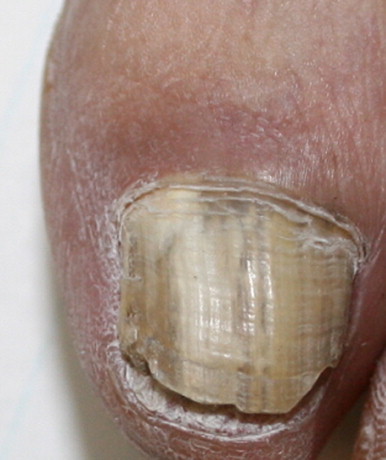
Clinical features of the toenail lesion caused by Aspergillus candidus. A total discolouration with a subungual hyperkeratosis is obviously observed.
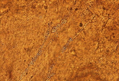
15% KOH slide preparation of the nail scraping showed rather broad septate and irregular hyphae (×400).
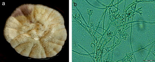
Colony morphology of the causing agent of onychomycosis on Czapek Yeast Agar (CYA) after incubating at 25° C for 6 days. (a) The picture depicts a pale yellow colony. And the microscopic view of filaments after performing slide culture: (b) the fungus possesses white, typically globose, conidial heads producing globose or subglobose, smooth, thin-walled conidia with spherical vesicles and metulae cover the entire surface of the vesicle. (Scale bar: 2 μm.) (For interpretation of the references to color in this figure legend, the reader is referred to the web version of this article.).
After 72 h of incubation at 35 °C, it was found that the isolate was resistant to Caspofungin with MIC>32 μg/mL and moderately susceptible to Fluconazole with MIC 2 μg/mL. However it was susceptible to Posaconazole with MICs 0.032 μg/mL.
References
-
- Welsh O., Vera-Cabrera L., Welsh E. Onychomycosis. Clinics in Dermatology. 2010;28(2):151–159. - PubMed
-
- Garcia-Martos P., Guarro J., Gené J., Mira J., Linares M., Ortoneda M. Onychomycosis caused by Aspergillus sclerotiorum. Journal de Mycologie Médicale. 2001;11(4):222–224.
-
- Chadeganipour M., Nilipour S., Ahmadi G. Study of onychomycosis in Isfahan, Iran. Mycoses. 2010;53(2):153–157. - PubMed
-
- Gupta A.K., Ryder J.E., Summerbell R.C. The diagnosis of nondermatophyte mold onychomycosis. International Journal of Dermatology. 2003;42(4):272–273. - PubMed
-
- Tosti A., Piraccini B.M., Lorenzi S. Onychomycosis caused by nondermatophytic molds: clinical features and response to treatment of 59 cases. Journal of the American Academy of Dermatology. 2000;42(2):217–224. - PubMed
LinkOut - more resources
Full Text Sources
