

Two-dimensional sum-frequency generation reveals structure and dynamics of a surface-bound peptide
- PMID: 24372101
- PMCID: PMC3956615
- DOI: 10.1021/ja408682s
Two-dimensional sum-frequency generation reveals structure and dynamics of a surface-bound peptide
Abstract
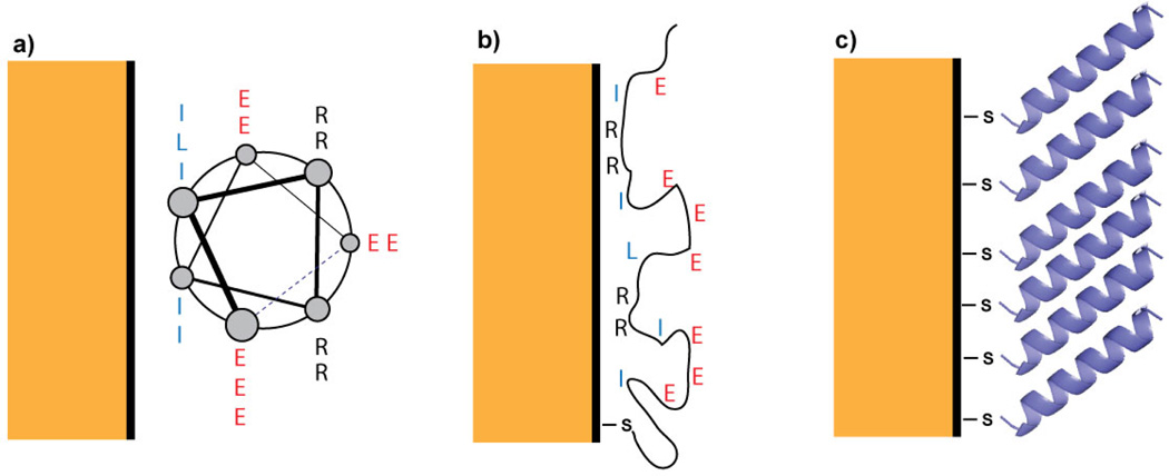
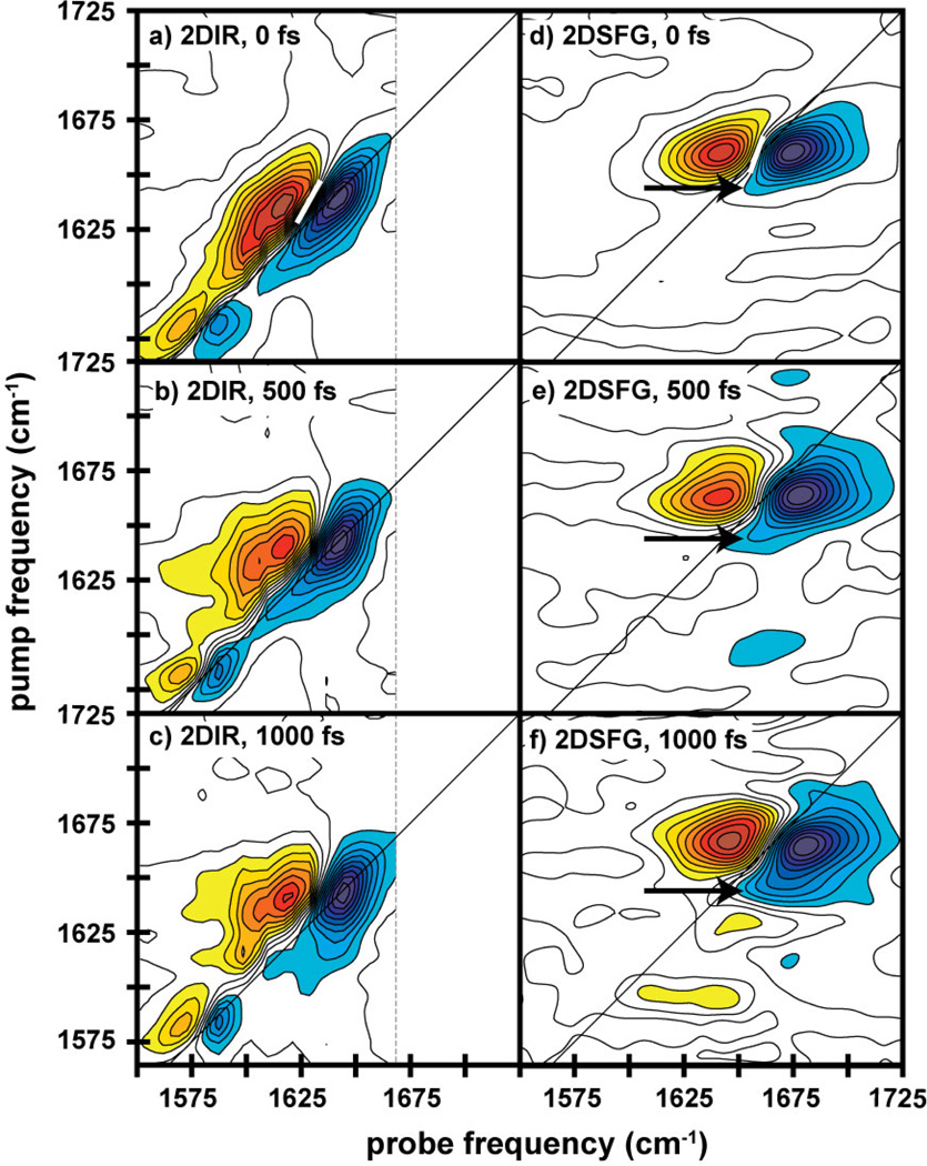
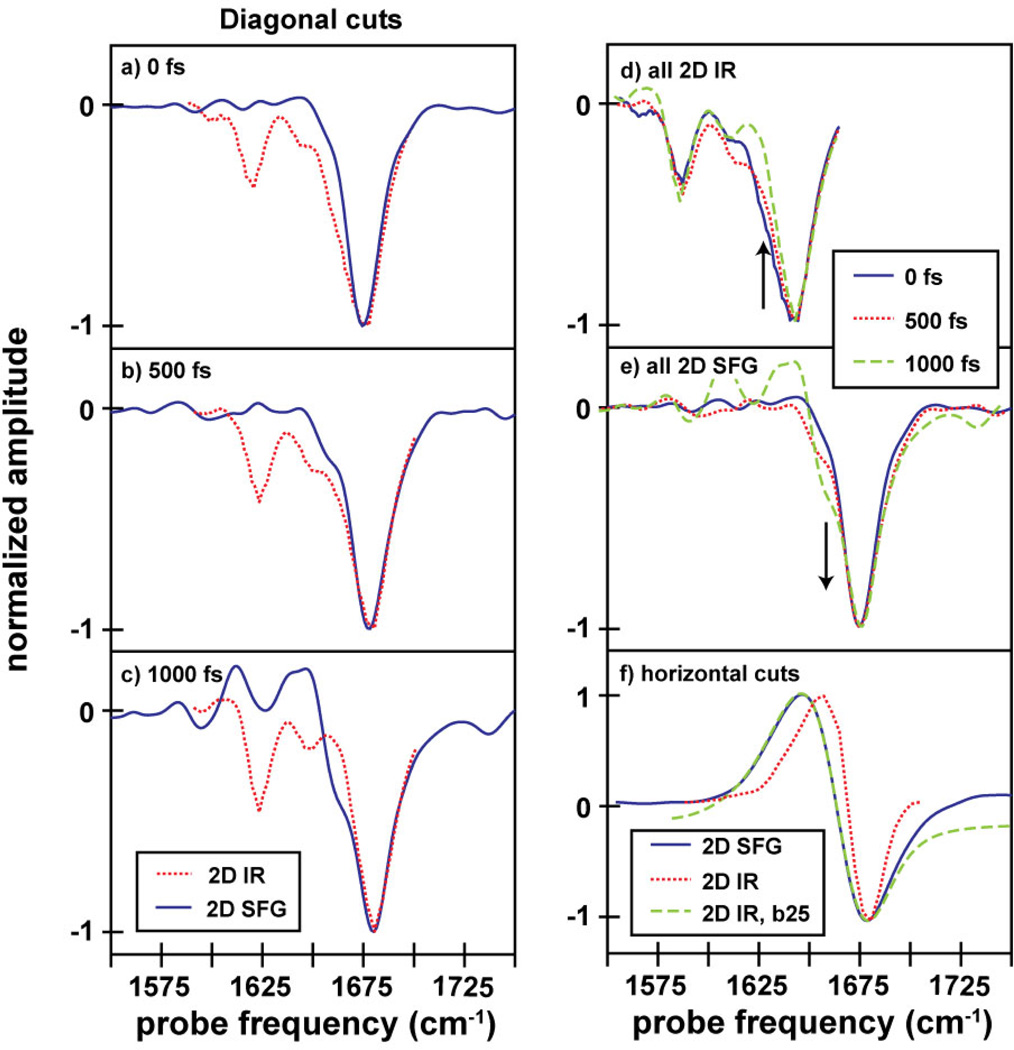
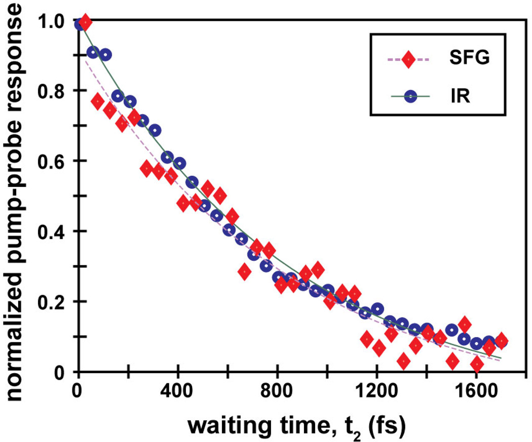
Surface-bound polypeptides and proteins are increasingly used to functionalize inorganic interfaces such as electrodes, but their structural characterization is exceedingly difficult with standard technologies. In this paper, we report the first two-dimensional sum-frequency generation (2D SFG) spectra of a peptide monolayer, which are collected by adding a mid-IR pulse shaper to a standard femtosecond SFG spectrometer. On a gold surface, standard FTIR spectroscopy is inconclusive about the peptide structure because of solvation-induced frequency shifts, but the 2D line shapes, anharmonic shifts, and lifetimes obtained from 2D SFG reveal that the peptide is largely α-helical and upright. Random coil residues are also observed, which do not themselves appear in SFG spectra due to their isotropic structural distribution, but which still absorb infrared light and so can be detected by cross-peaks in 2D SFG spectra. We discuss these results in the context of peptide design. Because of the similar way in which the spectra are collected, these 2D SFG spectra can be directly compared to 2D IR spectra, thereby enabling structural interpretations of surface-bound peptides and biomolecules based on the well-studied structure/2D IR spectra relationships established from soluble proteins.
Figures

Similar articles
-
Structural Characterization of Single-Stranded DNA Monolayers Using Two-Dimensional Sum Frequency Generation Spectroscopy.J Phys Chem B. 2015 Aug 20;119(33):10586-96. doi: 10.1021/acs.jpcb.5b07078. Epub 2015 Aug 10. J Phys Chem B. 2015. PMID: 26222775 Free PMC article.
-
Two-dimensional sum-frequency generation (2D SFG) spectroscopy: summary of principles and its application to amyloid fiber monolayers.Faraday Discuss. 2015;177:493-505. doi: 10.1039/c4fd00173g. Faraday Discuss. 2015. PMID: 25611039 Free PMC article.
-
Amide I two-dimensional infrared spectroscopy of proteins.Acc Chem Res. 2008 Mar;41(3):432-41. doi: 10.1021/ar700188n. Epub 2008 Feb 21. Acc Chem Res. 2008. PMID: 18288813 Review.
-
Adding a dimension to the infrared spectra of interfaces using heterodyne detected 2D sum-frequency generation (HD 2D SFG) spectroscopy.Proc Natl Acad Sci U S A. 2011 Dec 27;108(52):20902-7. doi: 10.1073/pnas.1115055108. Epub 2011 Dec 5. Proc Natl Acad Sci U S A. 2011. PMID: 22143772 Free PMC article.
-
Ultrafast nonlinear coherent vibrational sum-frequency spectroscopy methods to study thermal conductance of molecules at interfaces.Acc Chem Res. 2009 Sep 15;42(9):1343-51. doi: 10.1021/ar9000197. Acc Chem Res. 2009. PMID: 19388671 Review.
Cited by
-
Theoretical Sum Frequency Generation Spectroscopy of Peptides.J Phys Chem B. 2015 Jul 23;119(29):8969-83. doi: 10.1021/jp507861t. Epub 2014 Sep 24. J Phys Chem B. 2015. PMID: 25203677 Free PMC article.
-
Probing Site-Specific Structural Information of Peptides at Model Membrane Interface In Situ.J Am Chem Soc. 2015 Aug 19;137(32):10190-8. doi: 10.1021/jacs.5b04024. Epub 2015 Aug 11. J Am Chem Soc. 2015. PMID: 26241117 Free PMC article.
-
Two-dimensional infrared spectroscopy measures the structural dynamics of a self-assembled film only one molecule thick.Proc Natl Acad Sci U S A. 2016 May 3;113(18):4890-1. doi: 10.1073/pnas.1605263113. Epub 2016 Apr 19. Proc Natl Acad Sci U S A. 2016. PMID: 27095845 Free PMC article. No abstract available.
-
Structural Characterization of Single-Stranded DNA Monolayers Using Two-Dimensional Sum Frequency Generation Spectroscopy.J Phys Chem B. 2015 Aug 20;119(33):10586-96. doi: 10.1021/acs.jpcb.5b07078. Epub 2015 Aug 10. J Phys Chem B. 2015. PMID: 26222775 Free PMC article.
-
The photochemical reaction of phenol becomes ultrafast at the air-water interface.Nat Chem. 2021 Apr;13(4):306-311. doi: 10.1038/s41557-020-00619-5. Epub 2021 Feb 8. Nat Chem. 2021. PMID: 33558737
References
-
- Gooding JJ, Hibbert DB, Yang W. Sensors. 2001;1:75–90.
-
- Chaikoff EL, Wang HS, Wingert TM, Stephens S, Dluhy RA. MRS Proceedings. 1995;414:17.
-
- Bolduc OR, Correia-Ledo D, Masson JF. Langmuir. 2012;28:22–26. - PubMed
Publication types
MeSH terms
Substances
Grants and funding
LinkOut - more resources
Full Text Sources
Other Literature Sources
