. 2014 Apr 1;23(7):689-701.
doi: 10.1089/scd.2013.0362. Epub 2014 Feb 4.

Human and murine very small embryonic-like cells represent multipotent tissue progenitors, in vitro and in vivo

Affiliations

Human and murine very small embryonic-like cells represent multipotent tissue progenitors, in vitro and in vivo

Aaron M Havens et al. Stem Cells Dev. .

Abstract

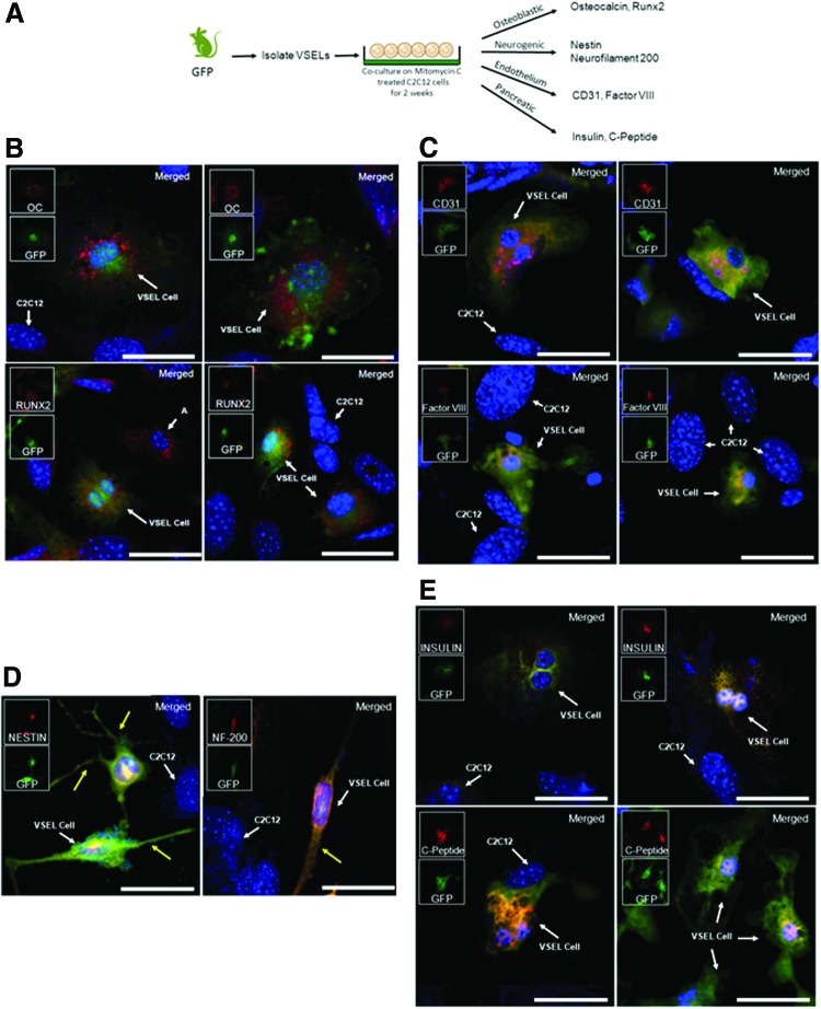
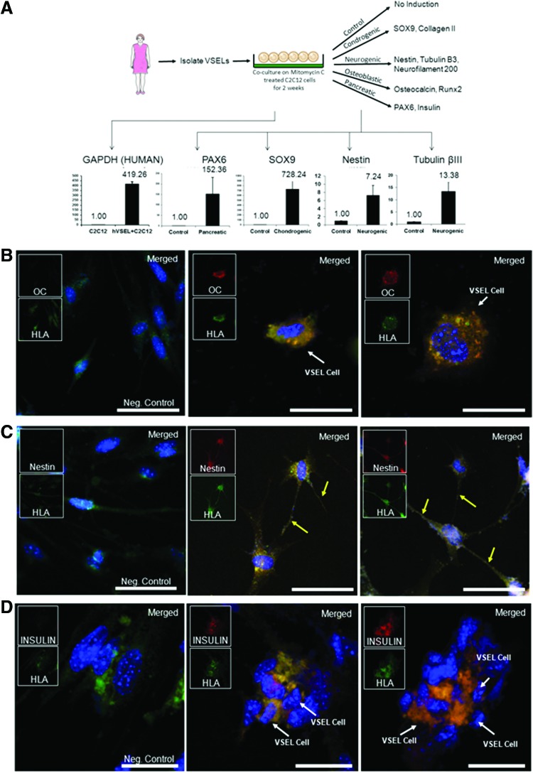
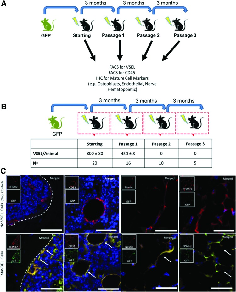
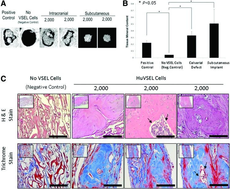
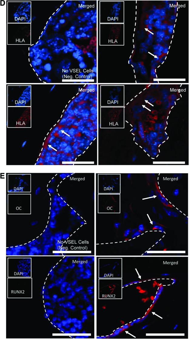
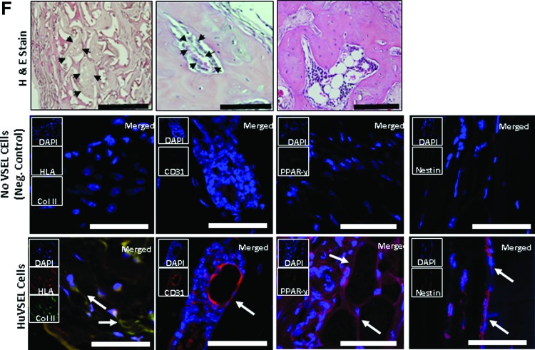
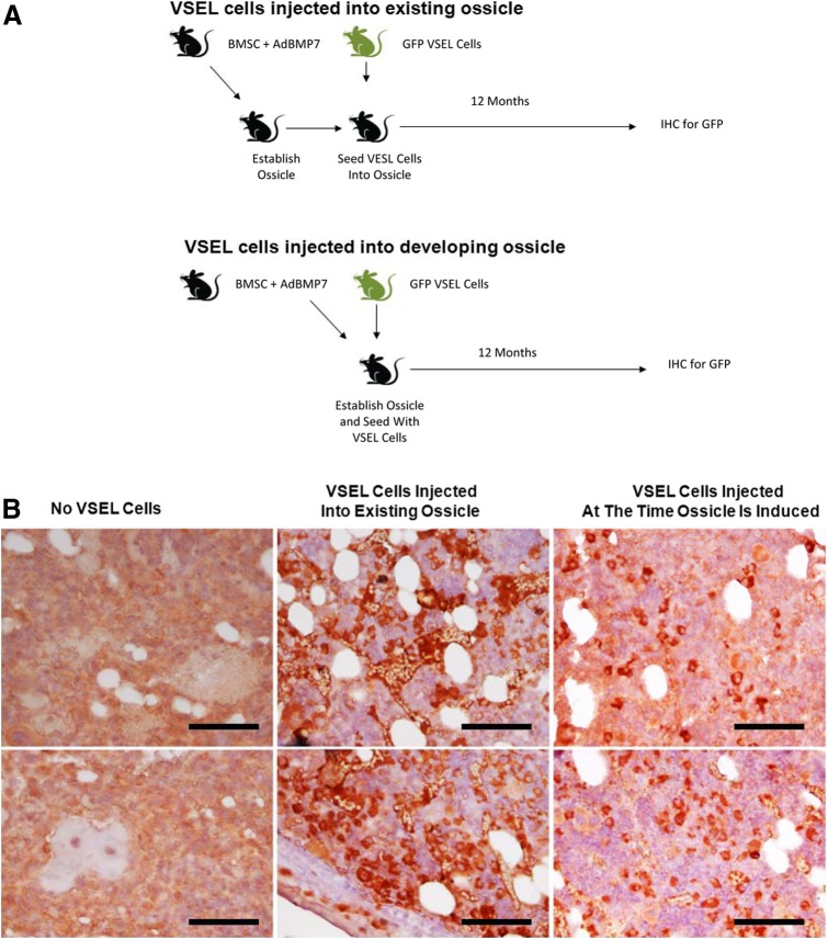
The purpose of this study was to determine the lineage progression of human and murine very small embryonic-like (HuVSEL or MuVSEL) cells in vitro and in vivo. In vitro, HuVSEL and MuVSEL cells differentiated into cells of all three embryonic germ layers. HuVSEL cells produced robust mineralized tissue of human origin compared with controls in calvarial defects. Immunohistochemistry demonstrated that the HuVSEL cells gave rise to neurons, adipocytes, chondrocytes, and osteoblasts within the calvarial defects. MuVSEL cells were also able to differentiate into similar lineages. First round serial transplants of MuVSEL cells into irradiated osseous sites demonstrated that ∼60% of the cells maintained their VSEL cell phenotype while other cells differentiated into multiple tissues at 3 months. Secondary transplants did not identify donor VSEL cells, suggesting limited self renewal but did demonstrate VSEL cell derivatives in situ for up to 1 year. At no point were teratomas identified. These studies show that VSEL cells produce multiple cellular structures in vivo and in vitro and lay the foundation for future cell-based regenerative therapies for osseous, neural, and connective tissue disorders.


Figures

<b>FIG. 1.</b>
FIG. 1.
In vitro MuVSEL cell differentiation into multiple germline tissues. (A) In primary culture, MuVSEL cells were freshly isolated from the bone marrow of GFP-labeled animals and cocultured on mitomycin-C-treated C2C12 cells for 2 weeks. Cells were placed in specific differentiation media conditions and were evaluated for expression for (B) osteoblastic [osteocalcin (OC) and Runx2], (C) endothelium (CD31, Factor VIII), (D) neural [nestin and neurofilament 200 (NF-200)]; yellow arrows indicate neuron extensions, and (E) insulin-producing endodermal (insulin and C-peptide)-specific markers. In each case, colocalization of the specific antibody (OC, Runx2, CD31, Factor VIII, nestin, neruofilament 20 each stained red) with GFP (stained green) was employed (merged staining appears yellow). Red and green inserts show each single color at 22% size of the merged images. The C2C12 cells did not express GFP, and therefore in combination with any of the aforementioned specific markers represent internal negative controls. (Note a few non-GFP expressing cells did stain for Runx 2 (labeled “A”) likely derived from C2C12 cells). Large panel immunohistochemistry images presented at 40×, scale bar=100 μm. GFP, green fluorescent protein; MuVSEL, murine very small embryonic like. Color images available online at www.liebertpub.com/scd
<b>FIG. 2.</b>
FIG. 2.
In vitro HuVSEL cell differentiation into multiple germline tissues. (A) In primary culture, HuVSEL cells were freshly isolated from peripheral blood and cocultured on mitomycin-C-treated C2C12 cells for 2 weeks. Expression of human GAPDH by QRT-PCR was over 400-fold higher in cocultures containing HuVSEL cells compared with cultures of C2C12 cells alone (lower left panel). After 2 weeks, cells were cultured in specific differentiation media for an additional 2 weeks. Gene expression changes compared with baseline and normalized to GAPDH were evaluated by QRT-PCR for early neural/pancreatic (PAX6), chondrogenic (SOX9), and early neurogenic (nestin and or βIII-tubulin) markers. Cells were also evaluated by immunofluorescence microscopy for expression of (B) osteoblastic (osteocalcin), (C) early neural (nestin, yellow arrows indicate neuron extensions), and (D) insulin-producing endodermal (insulin)-specific markers. In each case, colocalization with human HLA staining was assessed. The C2C12 cells did not stain for any of the aforementioned markers. Negative control (Neg. Control) indicates VSEL cells plated on C2C12 without specific tissue induction. Inserts show each single color at 22% size of the merged images. Large panel immunohistochemistry images presented at 40×, scale bar=100 μm. HuVSEL, human very small embryonic like; PAX6, paired box gene 6; QRT-PCR, real time reverse transcriptase polymerase chain reaction. Color images available online at www.liebertpub.com/scd
<b>FIG. 3.</b>
FIG. 3.
Serial transplantation of VSEL cells. (A) Serial transplant studies were performed by injecting GFP-labeled VSEL cells into the tibias of locally irradiated mice. Three months after each transplant, mice were sacrificed and the presence of GFP-derived VSEL cells and mature cellular lineages was evaluated by FACS and immunofluorescence microscopy of serially sectioned tibias. FACS (B) indicated that ∼60% of the number of GFP+ VSEL cells (small LinSca-1+CD45 cells) transplanted could be recovered after the first serial transplant from the injected tibia (Passage 1), but no GFP+ LinSca-1+CD45 cells could be recovered after subsequent transplants (Passage 2 and Passage 3). Second and third serial transplant data (mice were transplanted with small GFP+ CD45 Sca-1 cells) indicated that although GFP+ VSEL cells could not be isolated, abundant GFP+ cells could be identified by FACS remained at the site of injection. (C) Characteristic morphology and histologic locations of GFP-expressing osteoblasts (Runx2), endothelial cells (CD31), neural stem or progenitor cells (nestin), and adipocytes (PPAR-γ) were observed in the P1 mice suggesting that many of the donor MuVSEL cells had differentiated. Note: the brightness nestin-negative control insert panel was increased by 30% for visibility. Inserts show each single color at 22% size of the merged images. Arrows indicate dual staining cells. Large panel immunohistochemistry images presented at 40×, scale bar=100 μm. FACS, fluorescence activated cell sorting. Color images available online at www.liebertpub.com/scd
<b>FIG. 4.</b>
FIG. 4.
HuVSEL osseous repair of craniofacial defects. (A) Microcomputed tomography images of representative calvarial defects. HuVSEL cells together with collagen carrier matrix were implanted into either calvarial defects (center panels) or subcutaneously (right panels) at 2,000 HuVSEL cells/implant. Murine bone marrow stromal cells (BMSCs) transduced with AdCMVBMP-7 were implanted as a positive control (left panel). Collagen carrier alone was used as a negative control (second from left). When implanted both in calvarial defects and subcutaneously, 2,000 BMP-7 BMSCs and HuVSEL cells produced mineralized tissue, whereas collagen carrier alone did not. (B) Tissue mineral content for each implant was averaged for n=5–7 animals. Controls and implanted cell numbers were the same as in A. Implants using HuVSEL cells or BMP-7 BMSCs produced significantly more tissue mineral content than implants using collagen carrier alone (*P<0.05). (C) Representative sections of calvarial defects stained with H&E (top row) and Masson's trichrome (bottom row, which stains both collagen and bone blue). Histology is shown at 20× (inserts) and 40× magnification. Positive controls were implanted with murine BMSCs expressing BMP-7 (not shown). Negative controls (left panels) were implanted with collagen carrier alone (eg, no VSEL cells). Note the persistence of the collagen carrier matrix in the negative control group as well as the absence of an inflammatory cell infiltrate. Implants with 2,000 HuVSEL cells demonstrated woven bone containing marrow spaces (arrows). Scale bar=100 μm. (D) Immunostaining of implanted calvarial defects using a fluorescent antibody to human HLA. (E) Immunostaining for human-specific antibody to osteocalcin (OC) and Runx2 (both in red) merged with images of antinuclear stain (DAPI). Top left panel shows negative controls (Neg. Control: implanted with collagen vehicle only, but no VSEL cells) exhibiting no osteocalcin or Runx2 staining. In contrast, tissue sections of HuVSEL implants show significant osteocalcin and Runx2 staining along the bone margin as well as in the marrow cavity. Arrows indicate positive cells. Inserts show each single color at 22% size of the merged images. Large panel immunohistochemistry images presented at 40×, scale bar=100 μm. Dashed white line outlines the bone margins. (F) H&E staining of tissue sections (top row) show morphologically characteristic cartilage, endothelial, and adipose tissue within the calvarial defect. Immunostaining of defects implanted with HuVSEL cells (bottom row) shows robust staining for collagen type-II colocalized with human HLA (left panel, identifying chondrocytes) as well as staining surrounding the lumens of vascular structures detected with antibody to human CD31 (second from left). Human-specific antibody to PPAR-γ (second from right) identifies adipocytes within the calvarial defects. Staining for human nestin (right) identified cells with long processes between cell bodies, indicating early neuronal differentiation. In each case, no human-specific staining was present in the calvarial defects of cellular or scaffold control treated animals (center row). (Neg. Control: implanted with collagen vehicle only, but no VSEL cells) Histologic images presented at 40×, scale bar=100 μm. Arrows indicate positive staining. Color images available online at www.liebertpub.com/scd
<b>FIG. 5.</b>
FIG. 5.
Long-term subcutaneous ossicle implantation of MuVSEL cells. (A) Experimental Outline. Top: Murine BMSCs were infected with an adenovirus designed to overexpress BMP7 (AdBMP7), which were then implanted s.c. into SCID mice for up to 1 year. In some cases, the resulting ossicles were exposed and injected directly with MuVSEL cells isolated from GFP transgenic mice. Bottom: BMSCs infected with AdBMP7 were coinjected with MuVSEL cells isolated from GFP-expressing mice. At 12 months, all ossicles were recovered, decalcified, and stained with an antibody to GFP. (B) Staining for GFP expression in decalcified ossicles recovered BMSC/AdBMP7-implanted animals (left), from BMSC/AdBMP7-implanted animals, which after 1 month were exposed and injected with GFP-VSEL cells (middle), or ossicles established with BMSC/AdBMP7 that also contained GFP-VSEL cells at the time of implantation (right). Staining used anti-GFP HRP-AEC (red), counterstained with H&E (light blue). Little to no specific immunostaining for GFP was observed when the ossicles were not exposed to GFP-VSEL cells (left). Abundant staining of the bone marrow and bone structures were observed when the ossicles had been injected with GFP-VSEL cells. Positive staining for GFP appears in red. Scale bar=100 μm. Color images available online at www.liebertpub.com/scd

References

    1. Nakamura M. and Okano H. (2013). Cell transplantation therapies for spinal cord injury focusing on induced pluripotent stem cells. Cell Res 23:70–80 - PMC - PubMed
    1. Priori SG, Napolitano C, Di Pasquale E. and Condorelli G. (2013). Induced pluripotent stem cell-derived cardiomyocytes in studies of inherited arrhythmias. J Clin Invest 123:84–91 - PMC - PubMed
    1. Ye L, Swingen C. and Zhang J. (2013). Induced pluripotent stem cells and their potential for basic and clinical sciences. Curr Cardiol Rev 9:63–72 - PMC - PubMed
    1. Wang Z, Song J, Taichman RS. and Krebsbach PH. (2006). Ablation of proliferating marrow with 5-fluorouracil allows partial purification of mesenchymal stem cells. Stem Cells 24:1573–1582 - PubMed
    1. Taichman RS, Wang Z, Shiozawa Y, Jung Y, Song J, Balduino A, Wang J, Patel LR, Havens A, et al. (2010). Prospective identification and skeletal localization of cells capable of multi-lineage differentiation in vivo. Stem Cells Dev 19:1557–1570 - PMC - PubMed

Publication types

MeSH terms