Review
. 2013;14(8):743-52.
doi: 10.2174/1389201014666131226114611.

Ultrasound and microbubble guided drug delivery: mechanistic understanding and clinical implications

Affiliations
Review

Ultrasound and microbubble guided drug delivery: mechanistic understanding and clinical implications

Wang Tzu-Yin et al. Curr Pharm Biotechnol. 2013.

Abstract

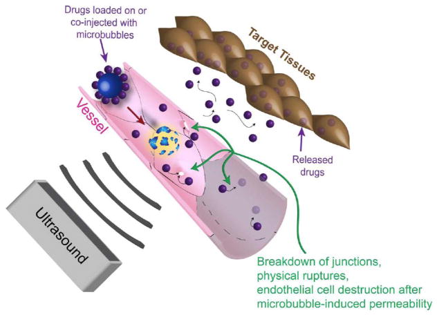
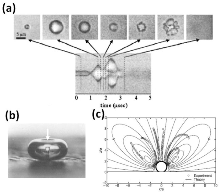
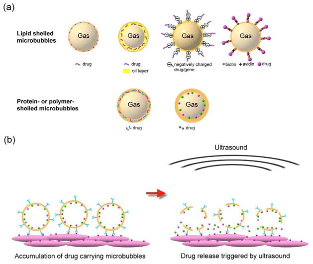
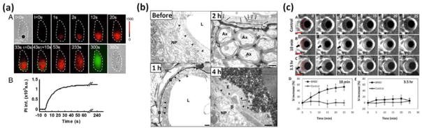
Ultrasound mediated drug delivery using microbubbles is a safe and noninvasive approach for spatially localized drug administration. This approach can create temporary and reversible openings on cellular membranes and vessel walls (a process called "sonoporation"), allowing for enhanced transport of therapeutic agents across these natural barriers. It is generally believed that the sonoporation process is highly associated with the energetic cavitation activities (volumetric expansion, contraction, fragmentation, and collapse) of the microbubble. However, a thorough understanding of the process was unavailable until recently. Important progress on the mechanistic understanding of sonoporation and the corresponding physiological responses in vitro and in vivo has been made. Specifically, recent research shed light on the cavitation process of microbubbles and fluid motion during insonation of ultrasound, on the spatio-temporal interactions between microbubbles and cells or vessel walls, as well as on the temporal course of the subsequent biological effects. These findings have significant clinical implications on the development of optimal treatment strategies for effective drug delivery. In this article, current progress in the mechanistic understanding of ultrasound and microbubble mediated drug delivery and its implications for clinical translation is discussed.


Conflict of interest statement

CONFLICT OF INTEREST

The authors confirm that this article content has no conflicts of interest.

Figures

Fig. 1
Fig. 1
Schematic drawing of ultrasound guided drug delivery. Ultrasound triggers microbubble cavitation at its focus, causing breakdown of tight junctions, opening on cellular membrane, and/or vessel disruption. These phenomena induce permeability change on the cells and vessels, allowing for drug delivery in a spatially localized region.
Fig. 2
Fig. 2
Ultrasound induced microbubble cavitation and fluid motions. (a) Microbubbles undergo volumetric expansion, contraction, fragmentation, and coalesce in response to the insonation ultrasound. They may continue to expand for several to several hundreds of microseconds and then collapse or dissolve after ultrasound is ceased. (b) During the violent collapse of microbubbles, fluid jets can form and impinge into a nearby surface. In this example, a fluid jet (white arrow) was formed in the center of a 2-mm bubble during the collapse and impinged into the surface where the bubble rested on. (c) The microbubble cavitation may induce local fluid motion called microstreaming. This example shows a computed fluid flow (solid lines) superimposed with measured flow (dots) near a microbubble undergoing volumetric oscillation. Figure panels (a), (b), and (c) are reprinted with permission from references [51, 46, 60], respectively.
Fig. 3
Fig. 3
(a) Examples of drug loading strategies. The drugs can be loaded on the shell of microbubbles, embedded in the shell matrix, or loaded in the internal void. For lipid-shelled microbubbles, the drug can be inserted in the lipid shell, in a thick oil layer underneath the lipid shell, on a positively charged shell through electrostatic force (for example, binding plasmid to cationic microbubble [86]), bonded to the shell via a biotin-avidin bridging system (for example, attaching liposomes to microbubbles using the bridging system [95]) or through covalent binding approaches (for example, adhering particles to microbubbles using a polymer [97]). For protein- or polymer-shelled microbubbles, the drug can be entrapped in the thick cross-linked protein or polymer matrix, or loaded in the internal void. (b) Surface modification of microbubbles allows for specific binding to cells at target sites, increasing local drug concentration. Application of ultrasound locally triggers the release of the accumulated drugs in the focal region.
Fig. 4
Fig. 4
Temporally resolved sonoporation process in (a) a cultured cell [66], (b) blood brain barrier disruption [77], and (c) blood-retinal barrier disruption [78]. (a) Increased red color indicates increased uptake of propidium iodide into the cell. Such uptake occurred most significantly within the first minute after ultrasound exposure, and gradually saturate at four minutes, indicating a resealing process. (b) Leakage of horseradish peroxidase molecules (black color) across the tight junctions was observed 1 h and 2 h after ultrasound and microbubble treatment, and was restored 4 h post treatment. (c) Enhanced contrast on MR images indicates perfusion of Magnevist across the barrier 10 minutes and 3.5 h after ultrasound and microbubble treatment. Overall, while a single bubble cavitation event induced by a short (8 μs) low intensity (0.17MPa) ultrasound can induce cellular uptake of propidium iodide for several minutes, exposure to long (10 ms) higher intensity (0.8–1 MPa) ultrasound pulses for a longer time (30 seconds) can induce permeability change across the natural barriers for up to 4 hours. Figure panels (a), (b), and (c) are reprinted with permission from references [66, 67], and [78], respectively.

Similar articles

Cited by

References

    1. Szabo TL. Diagnostic ultrasound imaging: inside out. 2004. Access Online via Elsevier.
    1. Dubinsky TJ, Cuevas C, Dighe MK, Kolokythas O, Joo HH. High-intensity focused ultrasound: Current potential and oncologic applications. Am J Roentgenology. 2008;190(1):191–199. - PubMed
    1. Kennedy JE. High-intensity focused ultrasound in the treatment of solid tumours. Nat Rev Cancer. 2005;5(4):321–327. - PubMed
    1. Rassweiler JJ, Knoll T, Kohrmann KU, McAteer JA, Lingeman JE, Cleveland RO, Bailey MR, Chaussy C. Shock wave technology and application: an update. Eur Urol. 2011;59(5):784–796. - PMC - PubMed
    1. Roberts WW, Hall TL, Ives K, Wolf JS, Jr, Fowlkes JB, Cain CA. Pulsed cavitational ultrasound: a noninvasive technology for controlled tissue ablation (histotripsy) in the rabbit kidney. J Urol. 2006;175(2):734–738. - PubMed

Publication types