. 2014 Jun;129(5):781-91.
doi: 10.1111/jnc.12645. Epub 2014 Jan 23.

Botulinum protease-cleaved SNARE fragments induce cytotoxicity in neuroblastoma cells

Affiliations
Free PMC article

Botulinum protease-cleaved SNARE fragments induce cytotoxicity in neuroblastoma cells

Jason Arsenault et al. J Neurochem. 2014 Jun.
Free PMC article

Abstract

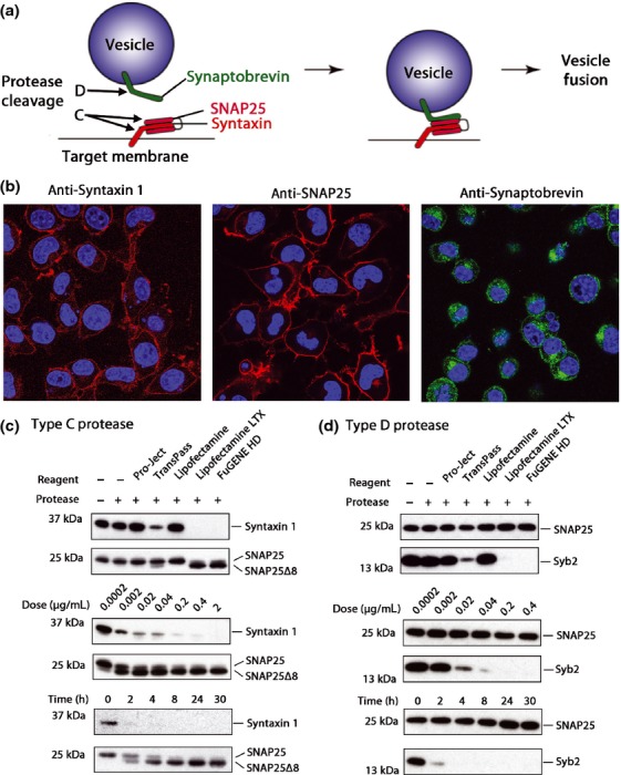
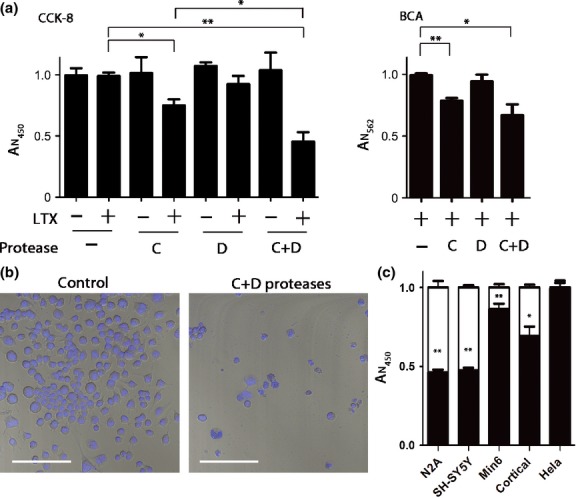
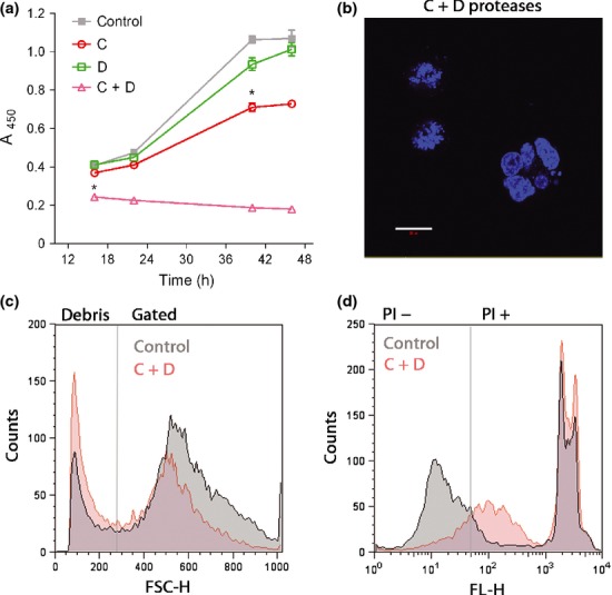
Soluble N-ethylmaleimide sensitive factor attachment protein receptors (SNAREs) are crucial for exocytosis, trafficking, and neurite outgrowth, where vesicular SNAREs are directed toward their partner target SNAREs: synaptosomal-associated protein of 25 kDa and syntaxin. SNARE proteins are normally membrane bound, but can be cleaved and released by botulinum neurotoxins. We found that botulinum proteases types C and D can easily be transduced into endocrine cells using DNA-transfection reagents. Following administration of the C and D proteases into normally refractory Neuro2A neuroblastoma cells, the SNARE proteins were cleaved with high efficiency within hours. Remarkably, botulinum protease exposures led to cytotoxicity evidenced by spectrophotometric assays and propidium iodide penetration into the nuclei. Direct delivery of SNARE fragments into the neuroblastoma cells reduced viability similar to botulinum proteases' application. We observed synergistic cytotoxic effects of the botulinum proteases, which may be explained by the release and interaction of soluble SNARE fragments. We show for the first time that previously observed cytotoxicity of botulinum neurotoxins/C in neurons could be achieved in cells of neuroendocrine origin with implications for medical uses of botulinum preparations. Ternary complex formation by synaptobrevin (green) and syntaxin/synaptosomal-associated protein of 25 kDa (red) is necessary for vesicle fusion, membrane trafficking, and cell homeostasis. Botulinum proteases cleave the three SNAREs proteins as indicated, resulting in a loss of cell viability. Lipofection reagents were used to deliver botulinum proteases or short SNARE peptides into neuroblastoma cells, revealing cytotoxic effects of SNARE fragments.

Keywords: SNARE; botulinum; cytotoxicity; neuro2A; syntaxin; transfection reagents.


Figures

Fig. 1
Fig. 1
Efficient transduction of botulinum type C and D proteases into neuroblastoma cells. (a) Schematic of the complex formation by synaptobrevin, v-soluble N-ethylmaleimide-sensitive factor attachment protein receptor (SNARE) (green) and syntaxin/SNAP25, t-SNAREs (red and fuchsia) necessary for vesicle fusion with the cell membrane. The substrates of the botulinum protease type C are syntaxin and SNAP25, whereas the type D protease cleaves only synaptobrevin. (b) Confocal image showing syntaxin 1 (red; left), SNAP25 (red; middle), and synaptobrevins 1-3 (green; right) in Neuro2A cells as revealed by immunocytochemistry. (c) Comparison of the efficiency of different reagents used to deliver botulinum protease type C (top panel). Cleavage of syntaxins evidenced by the disappearance of the immunoreactive band, whereas cleavage of SNAP25 is evidenced by the appearance of a proteolytic product SNAP25Δ8. SNAP25 immunoreactivity served as a loading control. Middle panel shows the dose dependence of SNARE cleavage following 20 h treatment. Bottom panel shows a time course using 0.2 μg/mL of the type C protease. (d) Comparison of different reagents used to deliver botulinum protease type D (top panel) and efficacy (lower panels) of synaptobrevin 2 (Syb2) cleavage evidenced by the disappearance of the immunoreactive band. SNAP25 was used as a loading control.
Fig. 2
Fig. 2
Cytotoxicity of the botulinum C and D proteases. (a) Cell counting assay (CCK-8) was used to monitor neuroblastoma N2A cell viability 40 h after application of the proteases in the presence or absence of Lipofectamine LTX. Botulinum protease type C significantly impaired viability compared with a control condition (*p < 0.01). Application of both proteases with Lipofectamine LTX further lowered cell survival (**p < 0.001). Absorbance at 450 nm (AN450) normalized to control values is proportional to cell count (left panel). The right panel shows the absorbance at 562 nm (AN562) proportional to total protein content in the wells as determined by the bicinchoninic acid (BCA) assay. Botulinum protease type C in the presence of Lipofectamine LTX significantly lowered viability compared with control condition (**p < 0.001) while the addition of the type D protease reduced survival further (*p < 0.01). Results are presented ± SD. (b) Confocal images showing a reduction in number of N2A cells stained with Hoechst 33342. Left panel shows cells treated with Lipofectamine LTX alone, whereas right panel shows cells treated with C and D proteases in the presence of Lipofectamine LTX. White bar: 100 μm. (c) CCK-8 assay was used to monitor viability of the indicated cells following the addition of Lipofectamine LTX with or without botulinum proteases. Control conditions where cells were treated with Lipofectamine LTX alone are shown as white columns. Results were normalized to control (LTX alone, ± SD). A significant (**p < 0.001) loss of viability compared with untreated controls was observed in N2A, SH-SY5Y, Min6, and rat brain cortical cells, which comprise both neurons and non-neuronal cells (*p < 0.01).
Fig. 3
Fig. 3
Time course of botulinum-triggered cytotoxic effects. (a) A late onset of toxicity observed following application of type C, type D, and C and D proteases. A significant (*p < 0.01) reduction in cell viability is observed at 16 h post-transduction for the combined C and D protease, whereas a significant (*p < 0.01) reduction in the type C protease-treated cells can be observed after 40 h. Results are presented ± SD. (b) Confocal image showing fragmented nuclei of N2A cells treated with the C and D proteases, indicating apoptosis. White bar: 20 μm. (c) Histogram of the forward scatter height (FSC-H) observed by flow cytometry. The gated region on the right contained morphologically normal (i.e., size and complexity) cells, whereas the left section shows the cell debris. Botulinum protease-treated cells (red) exhibit higher debris count compared with control (gray) (p < 0.05). (d) Histogram representation of FL-H (fluorescent height) signal of the propidium iodide (PI)-stained morphologically normal cells indicates a significant increase in cell death following application of botulinum proteases (p < 0.01).
Fig. 4
Fig. 4
The importance of syntaxin cleavage in botulinum-triggered cell demise. (a) Immunoblot showing that, in the presence of Lipofectamine LTX, the botulinum protease type C cleaves both syntaxin 1 and SNAP25 in N2A cells, whereas the type A protease cleaves SNAP25 only. Type C protease removes the last eight residues of SNAP25 (SNAP25Δ8), whereas type A removes the last nine (SNAP25Δ9). (b) The bar chart showing N2A cell demise following a 40 h treatment with the indicated proteases. Type A-induced cleavage of SNAP25, even in the presence of the type D protease, is insufficient to drive the cytotoxic effects. In contrast, cleavage of syntaxin by type C protease has a strong effect on cell survival (**p < 0.001). Results are normalized to control and presented ± SD.
Fig. 5
Fig. 5
Shorted soluble N-ethylmaleimide-sensitive factor attachment protein receptor (SNARE) fragments can interact and cause cytotoxicity upon transduction. (a) Western immunoblotting of N2A cells, treated with 0.002 μg of type C protease for up to 24 h, reveals cleaved syntaxin fragment 1-253 (syntaxinΔ35). In this instance immunoblotting was performed using the HPC-1 anti-syntaxin antibody. (b) Schematic of a SNARE pull-down reaction (upper panel). The lower panel showing cleavage of Glutathione S-transferase (GST)-synaptobrevin 2 (GST-Syb2) by type D protease and the ability of this product to bind syntaxin fragment (201-245) and SNAP25 as seen in the Coomassie-stained sodium dodecyl sulfate–polyacrylamide gel electrophoresis (SDS–PAGE) gel. Note SNAP25 is required for the syntaxin pull-down by GST-Syb2 and GST-Syb2D product highlighting the ternary nature of the SNARE interaction. (c) GST-SNAP25 pull-down experiment reveals interaction of the indicated shortened synaptobrevin and syntaxin fragments, strictly in a ternary manner. Syntaxin fragment 1-226 incorporating the regulatory syntaxin head with a truncated SNARE motif was revealed by Coomassie staining, whereas short SNARE peptides, carrying FITC, were imaged in the Bio-Rad XRS imaging station. (d) Bar chart showing cell survival measured using CCK-8 assay. Intracellular delivery of syntaxin fragment (201-245) and synaptobrevin (25-52) peptide using Lipofectamine LTX causes a significant reduction in cell viability, whereas transduction of control peptides, complexin (31-59) and penetratin, at the same concentration does not lead to cell death (*p < 0.05). Results are normalized to control and presented ± SD. (e) The short syntaxin fragment (201-245) applied to N2A cells causes cell death in a dose-dependent manner. Changes in cell viability are shown with and without Lipofectamine LTX. A significant loss of viability can be observed at doses as low as 5 μg/mL (*p < 0.01, ± SD).

Comment in

  • A role for SNAREs in neuronal survival?
    Harper CB, Bademosi AT, Coulson EJ, Meunier FA. Harper CB, et al. J Neurochem. 2014 Jun;129(5):753-5. doi: 10.1111/jnc.12699. Epub 2014 Apr 2. J Neurochem. 2014. PMID: 24697239 No abstract available.

Similar articles

Cited by

References

    1. Antonucci F, Rossi C, Gianfranceschi L, Rossetto O, Caleo M. Long-distance retrograde effects of botulinum neurotoxin A. J. Neurosci. 2008;28:3689–3696. - PMC - PubMed
    1. Arsenault J, Ferrari E, Niranjan D, Cuijpers SA, Gu C, Vallis Y, O'Brien J, Davletov B. Stapling of the botulinum type A protease to growth factors and neuropeptides allows selective targeting of neuroendocrine cells. J. Neurochem. 2013;126:223–233. - PMC - PubMed
    1. Bajohrs M, Darios F, Peak-Chew SY, Davletov B. Promiscuous interaction of SNAP-25 with all plasma membrane syntaxins in a neuroendocrine cell. Biochem. J. 2005;392:283–289. - PMC - PubMed
    1. Bangaru MLY, Woodliff J, Raff H, Kansra S. Growth suppression of mouse pituitary corticotroph tumor AtT20 cells by curcumin: a model for treating Cushing's disease. PLoS ONE. 2010;5:e9893. - PMC - PubMed
    1. Batcher E, Madaj P, Gianoukakis AG. Pancreatic neuroendocrine tumors. Endocr. Res. 2011;36:35–43. - PubMed

Publication types

MeSH terms