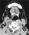
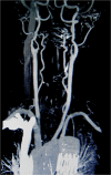
Airway obstruction by a retropharyngeal hematoma secondary to thoracic aortic aneurysm rupture
- PMID: 24373302
- PMCID: PMC3891989
- DOI: 10.1186/1749-8090-8-232
Airway obstruction by a retropharyngeal hematoma secondary to thoracic aortic aneurysm rupture
Abstract
Background: Retropharyngeal hematoma is a rare form of pharyngeal pathology and can present as acute airway obstruction. Among the many causes of retropharyngeal hematoma, thoracic aortic rupture is extremely rare.
Methods and results: A 78-year-old female with airway obstruction by a retropharyngeal hematoma secondary to thoracic aortic aneurysm rupture was successfully treated by total aortic arch replacement and open stent-graft insertion.
Conclusion: Rupture of the thoracic aorta should be considered as a rare but important cause of retropharyngeal hematoma and airway obstruction.
Figures


Similar articles
-
Acute Diastolic Heart Dysfunction Caused by Ruptured Descending Thoracic Aortic Aneurysm with Posterior Mediastinal Hematoma.J Card Surg. 2015 Jul;30(7):589-90. doi: 10.1111/jocs.12553. Epub 2015 May 18. J Card Surg. 2015. PMID: 25989223 No abstract available.
-
Should large mediastinal hematomas be drained after endovascular repair of ruptured descending thoracic aorta?J Thorac Cardiovasc Surg. 2007 Oct;134(4):1040-1. doi: 10.1016/j.jtcvs.2007.04.037. J Thorac Cardiovasc Surg. 2007. PMID: 17903526 No abstract available.
-
Airway obstruction secondary to thoracic aortic aneurysm leak. A case report.Eur Arch Otorhinolaryngol. 2006 Dec;263(12):1144-6. doi: 10.1007/s00405-006-0113-z. Epub 2006 Jul 20. Eur Arch Otorhinolaryngol. 2006. PMID: 16855827
-
Cardiac tamponade secondary to rupture of a distal aortic arch aneurysm.Jpn J Thorac Cardiovasc Surg. 2002 May;50(5):227-30. doi: 10.1007/BF03032293. Jpn J Thorac Cardiovasc Surg. 2002. PMID: 12048919 Review.
-
Ruptured right aortic arch aneurysm.Asian Cardiovasc Thorac Ann. 2004 Dec;12(4):360-2. doi: 10.1177/021849230401200417. Asian Cardiovasc Thorac Ann. 2004. PMID: 15585709 Review.
Cited by
-
Postintubation airway obstruction caused by a retrotracheal haematoma.BMJ Case Rep. 2016 May 25;2016:bcr2016215828. doi: 10.1136/bcr-2016-215828. BMJ Case Rep. 2016. PMID: 27226128 Free PMC article. No abstract available.
-
Vertebral artery ruptures manifesting as hoarseness.Braz J Otorhinolaryngol. 2020 Dec;86 Suppl 1(Suppl 1):11-13. doi: 10.1016/j.bjorl.2016.11.003. Epub 2016 Dec 13. Braz J Otorhinolaryngol. 2020. PMID: 28108273 Free PMC article. No abstract available.
-
Removal of infected pacemaker leads using an endoscopic minimally invasive cardiac surgical approach: a case report.Gen Thorac Cardiovasc Surg Cases. 2024 Jun 24;3(1):33. doi: 10.1186/s44215-024-00156-4. Gen Thorac Cardiovasc Surg Cases. 2024. PMID: 39517089 Free PMC article. No abstract available.
References
-
- Endo H, Kubota H, Tsuchiya H, Yoshimoto A, Takahashi Y, Inaba Y, Sudo K. Clinical efficacy of intermittent pressure augmented-retrograde cerebral perfusion. J Thorac Cardiovasc Surg. 2012. Epub ahead of print. - PubMed
-
- Jougon J, Zénnaro O. Acute cervico-mediastinal hematoma of parathyroid origin. Ann Chir. 1994;8:867–869. - PubMed
Publication types
MeSH terms
LinkOut - more resources
Full Text Sources
Other Literature Sources
Medical

