. 2014 Apr;133(4):1149-61.
doi: 10.1016/j.jaci.2013.10.051. Epub 2013 Dec 25.

Signaling lymphocytic activation molecule (SLAM)/SLAM-associated protein pathway regulates human B-cell tolerance

Affiliations

Signaling lymphocytic activation molecule (SLAM)/SLAM-associated protein pathway regulates human B-cell tolerance

Laurence Menard et al. J Allergy Clin Immunol. 2014 Apr.

Abstract

Background: Signaling lymphocytic activation molecule (SLAM)-associated protein (SAP) can mediate the function of SLAM molecules, which have been proposed to be involved in the development of autoimmunity in mice.

Objective: We sought to determine whether the SLAM/SAP pathway regulates the establishment of human B-cell tolerance and what mechanisms of B-cell tolerance could be affected by SAP deficiency.

Methods: We tested the reactivity of antibodies isolated from single B cells from SAP-deficient patients with X-linked lymphoproliferative disease (XLP). The expressions of SAP and SLAM family members were assessed in human bone marrow-developing B cells. We also analyzed regulatory T (Treg) cell function in patients with XLP and healthy control subjects.

Results: We found that new emigrant/transitional B cells from patients with XLP were enriched in autoreactive clones, revealing a defective central B-cell tolerance checkpoint in the absence of functional SAP. In agreement with a B cell-intrinsic regulation of central tolerance, we identified SAP expression in a discrete subset of bone marrow immature B cells. SAP colocalized with SLAMF6 only in association with clustered B-cell receptors likely recognizing self-antigens, suggesting that SLAM/SAP regulate B-cell receptor-mediated central tolerance. In addition, patients with XLP displayed defective peripheral B-cell tolerance, which is normally controlled by Treg cells. Treg cells in patients with XLP seem functional, but SAP-deficient T cells were resistant to Treg cell-mediated suppression. Indeed, SAP-deficient T cells were hyperresponsive to T-cell receptor stimulation, which resulted in increased secretion of IL-2, IFN-γ, and TNF-α.

Conclusions: SAP expression is required for the counterselection of developing autoreactive B cells and prevents their T cell-dependent accumulation in the periphery.

Keywords: B-cell tolerance; SLAM-associated protein; regulatory T cells; signaling lymphocytic activation molecule.


Figures

FIG 1
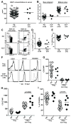
FIG 1. The central B-cell tolerance checkpoint is not functional in XLP patients
The frequencies of long IgH (15 or more a.a.) (A), long Igκ (B) and Igλ (C) (11 or more a.a.) in new emigrant/transitional B cells are represented for 11 healthy donors and 8 XLP patients. Each diamond represents an individual. The average is shown with a bar. (D) Antibodies from new emigrant/transitional B cells from XLP patients were tested by ELISA for reactivity against ssDNA, dsDNA, insulin and lipopolysaccharide (LPS) and the frequencies of polyreactive (E) and anti-nuclear clones are displayed in (E) and (F), respectively. Dotted line: ED38-positive control. Horizontal line: cutoff OD405 for positive reactivity. For each individual, the frequency of polyreactive (filled area) and non polyreactive (open area) clones is summarized in pie charts, with the total number of clones tested indicated in the centers.
FIG 2
FIG 2. SAP is expressed by some immature B cells
(A) PBMC depleted from CD20+ B cells or purified naive B cells were stained for CD3 or IgM (green), SAP or isotype control (red). (B) CD19+ adult bone marrow cells were stained for IgM, Igκ and Igλ, VpreB (green) and SAP or isotype control (red). The proportion of cells in SAP+ vs SAP cells that displayed a clustered (c) vs not clustered (n.c) IgM and Igκ/Igλ staining pattern is shown in (C). *:p-value<0.05; **: p-value<0.01. ***: p-value<0.001. (D) Dot plots show the proportion of CD3+ T cells in peripheral blood mononuclear cells from healthy donors (HD PBMC), CD20-enriched B cells using magnetic beads, sorted mature naive (MN) B cells and CD19+ fetal liver cells. SAP (E) and CD3E (F) gene expression was assessed by quantitative RT-PCR in MN B cells, CD19+ fetal liver and bone marrow cells and EBV cell lines. Jurkat and healthy donor (HD) CD4+ T cells were used as positive control for both SAP and CD3E expression.
FIG 3
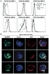
FIG 3. SAP and SLAMF6 co-localize in B cells with aggregated BCRs
(A) Overlays display the expression of SLAM family members by CD19+CD27CD10+IgM pre-B cells (dotted line) and CD19+CD27CD10+IgM+ immature B cells (bold line) from bone marrow and peripheral CD19+CD27CD10IgM+ mature naive B cells (thin line). (B) CD19+ enriched bone marrow B cells were stained for SLAMF6 (green), Igκ/Igλ (blue) and SAP (red). Representative cells without (top row) or with (middle and bottom row) co-staining of SLAMF6, Igκ/Igλ and SAP are shown. Clustered Igκ/Igλ, SLAMF6 and SAP appear white as the superposition of green, red and blue and is indicated by an arrow.
FIG 4
FIG 4. Defective peripheral B-cell tolerance checkpoint in XLP patients
(A) Antibodies from mature naive B cells from XLP patients were tested by ELISA for anti-HEp-2 cell reactivity and the frequencies of HEp-2 reactive, polyreactive, and antinuclear mature naive B cells are shown in (B), (C) and (D). Dotted line: ED38-positive control. Horizontal lines: cutoff OD405 for positive reactivity. For each individual, the frequency of HEp-2 reactive (filled area) and non HEp-2 reactive (open area) clones is summarized in pie charts, with the total number of clones tested indicated in the centers.
FIG 5
FIG 5. XLP patients display normal serum BAFF concentrations and Treg frequencies
(A). Serum BAFF concentrations (pg/ml) in healthy donors (n=73) and XLP patients (n=5). (B) Evaluation of the number of cell divisions undergone in vivo by KREC analysis on new emigrant and mature naive B cells of healthy donors (n=28 and n=32 respectively) and XLP patients (n=5). (C) Representative CD25 and CD127 (left) and Helios and FOXP3 staining (right) on CD3+CD4+ T cells from a healthy donor and an XLP patient. (D) CD3+CD4+CD25hiCD127loFOXP3+ Treg cell frequencies in 35 healthy donors and 6 XLP patients. (E) Frequencies of FOXP3+Helios+ cells in the CD3+CD4+CD25hiCD127loFOXP3+ population in 35 healthy donors and 6 XLP patients. (F) Representative staining of CD25, CD127 and CD45RO expression in an XLP patient (bold line) and HD (thin line) for FOXP3+Helios+ Treg cells and FOXP3Helios CD4+ T cells. MFI of CD25 (G), CD127 (H) and frequency of CD45RO+ cells (I) for Helios+FOXP3+ Treg and HeliosFOXP3 CD4+ non-Treg T cells from healthy donors and XLP patients.
FIG 6
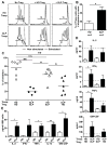
FIG 6. Increased resistance of SAP-deficient responder CD4+ T cells to suppression by Tregs
(A) Representative histograms of Treg mediated suppression of autologous and heterologous CFSE labeled Tresp cells on day 4.5 from a XLP patient compared to a healthy donor (HD). Dashed line displays non-stimulated Tresp. (B) Increased percentage of Tresp that divided 4 times or more in XLP patients compared to HD in the ‘Tresp only’ culture after 4.5 days. The autologous and heterologous suppressive activities of Tregs from HD and XLP patients are summarized in (C). (D) Increased concentrations of IL-2, IFNγ, TNFα and GM-CSF in supernatants of XLP ‘Tresp+Treg’ cocultures. The doted line represents the level of each cytokine in the supernatant of the stimulated ‘HD Tresp only’ well. (E) displays concentrations of the indicated cytokines in stimulated HD or XLP CD4+ T cells monocultures after 4 days. *:p-value<0.05; **: p-value<0.01.
FIG 7
FIG 7. Elevated Th2/Tfh cytokine concentrations in the serum of XLP patients
(A) IL-2, IFNγ, TNFα, IL-7, IL-4, IL-6, IL-10, IL-21 cytokine concentrations in the serum of 5 XLP patients and 29 healthy donors were measured by Luminex or ELISA. *:p-value<0.05; **: p-value<0.01. ****: p-value<0.0001. (B) Representative histograms of Treg mediated suppression of autologous CFSE labeled Tresp cells on day 4.5 from a healthy donor in the presence of the indicated cytokines. Dashed line displays non-stimulated Tresp and the suppressive activities of Tregs in the presence of diverse cytokines are summarized in (C). The XLP cytokine mix includes IL-4, IL-6, IL-7, IL-10, IL-21 and TNFα found elevated in XLP patients.

Similar articles

Cited by

References

    1. Nichols KE, Harkin DP, Levitz S, Krainer M, Kolquist KA, Genovese C, et al. Inactivating mutations in an SH2 domain-encoding gene in X-linked lymphoproliferative syndrome. Proc Natl Acad Sci U S A. 1998 Nov 10;95(23):13765–70. [Research Support, U.S. Gov’t, P.H.S.] - PMC - PubMed
    1. Coffey AJ, Brooksbank RA, Brandau O, Oohashi T, Howell GR, Bye JM, et al. Host response to EBV infection in X-linked lymphoproliferative disease results from mutations in an SH2-domain encoding gene. Nat Genet. 1998 Oct;20(2):129–35. [Research Support, Non-U.S. Gov’t Research Support, U.S. Gov’t, P.H.S.] - PubMed
    1. Sayos J, Wu C, Morra M, Wang N, Zhang X, Allen D, et al. The X-linked lymphoproliferative-disease gene product SAP regulates signals induced through the co-receptor SLAM. Nature. 1998 Oct 1;395(6701):462–9. [Research Support, Non-U.S. Gov’t Research Support, U.S. Gov’t, P.H.S.] - PubMed
    1. Cannons JL, Tangye SG, Schwartzberg PL. SLAM family receptors and SAP adaptors in immunity. Annu Rev Immunol. 2011;29:665–705. [Review] - PubMed
    1. Wandstrat AE, Nguyen C, Limaye N, Chan AY, Subramanian S, Tian XH, et al. Association of extensive polymorphisms in the SLAM/CD2 gene cluster with murine lupus. Immunity. 2004 Dec;21(6):769–80. [Research Support, Non-U.S. Gov’t Research Support, U.S. Gov’t, P.H.S.] - PubMed

Publication types

MeSH terms

Substances