

A sensory neuron-expressed IL-31 receptor mediates T helper cell-dependent itch: Involvement of TRPV1 and TRPA1
- PMID: 24373353
- PMCID: PMC3960328
- DOI: 10.1016/j.jaci.2013.10.048
A sensory neuron-expressed IL-31 receptor mediates T helper cell-dependent itch: Involvement of TRPV1 and TRPA1
Abstract
Background: Although the cytokine IL-31 has been implicated in inflammatory and lymphoma-associated itch, the cellular basis for its pruritic action is yet unclear.
Objective: We sought to determine whether immune cell-derived IL-31 directly stimulates sensory neurons and to identify the molecular basis of IL-31-induced itch.
Methods: We used immunohistochemistry and quantitative real-time PCR to determine IL-31 expression levels in mice and human subjects. Immunohistochemistry, immunofluorescence, quantitative real-time PCR, in vivo pharmacology, Western blotting, single-cell calcium imaging, and electrophysiology were used to examine the distribution, functionality, and cellular basis of the neuronal IL-31 receptor α in mice and human subjects.
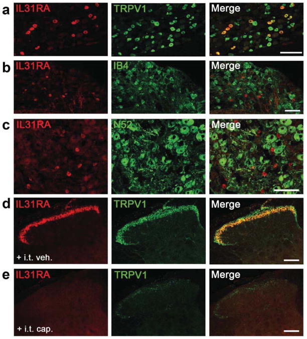
Results: Among all immune and resident skin cells examined, IL-31 was predominantly produced by TH2 and, to a significantly lesser extent, mature dendritic cells. Cutaneous and intrathecal injections of IL-31 evoked intense itch, and its concentrations increased significantly in murine atopy-like dermatitis skin. Both human and mouse dorsal root ganglia neurons express IL-31RA, largely in neurons that coexpress transient receptor potential cation channel vanilloid subtype 1 (TRPV1). IL-31-induced itch was significantly reduced in TRPV1-deficient and transient receptor channel potential cation channel ankyrin subtype 1 (TRPA1)-deficient mice but not in c-kit or proteinase-activated receptor 2 mice. In cultured primary sensory neurons IL-31 triggered Ca(2+) release and extracellular signal-regulated kinase 1/2 phosphorylation, inhibition of which blocked IL-31 signaling in vitro and reduced IL-31-induced scratching in vivo.
Conclusion: IL-31RA is a functional receptor expressed by a small subpopulation of IL-31RA(+)/TRPV1(+)/TRPA1(+) neurons and is a critical neuroimmune link between TH2 cells and sensory nerves for the generation of T cell-mediated itch. Thus targeting neuronal IL-31RA might be effective in the management of TH2-mediated itch, including atopic dermatitis and cutaneous T-cell lymphoma.
Keywords: AD; AITC; Allyl isothiocyanate; Atopic dermatitis; Cytokine; DRG; Dorsal root ganglia; ERK; Extracellular signal-regulated kinase; GRPR; Gastrin-releasing peptide receptor; HBSS; Hanks balanced salt solution; High-power field; IB4; Isolectin B4; KO; Knockout; MEK; Mas-related G protein–coupled receptor; Mitogen-activated protein kinase enzyme; Mrgpr; NPR-A; Natriuretic peptide receptor A; OSMRβ; OVA; Oncostatin M receptor β; Ovalbumin; PAR-2; Proteinase-activated receptor 2; Quantitative real-time PCR; SC; SEB; Spinal cord; Staphylococcal enterotoxin B; TG; TRPA1; TRPV1; Transient receptor channel potential cation channel ankyrin subtype 1; Transient receptor potential cation channel vanilloid subtype 1; Trigeminal ganglion; atopic dermatitis; hpf; qPCR; sensory nerve; skin; transient receptor potential channel.
Copyright © 2013 American Academy of Allergy, Asthma & Immunology. Published by Mosby, Inc. All rights reserved.
Figures


References
-
- Bilsborough J, Leung DY, Maurer M, Howell M, Boguniewicz M, Yao L, et al. IL-31 is associated with cutaneous lymphocyte antigen-positive skin homing T cells in patients with atopic dermatitis. The Journal of allergy and clinical immunology. 2006;117:418–25. - PubMed
-
- Sonkoly E, Muller A, Lauerma AI, Pivarcsi A, Soto H, Kemeny L, et al. IL-31: a new link between T cells and pruritus in atopic skin inflammation. The Journal of allergy and clinical immunology. 2006;117:411–7. - PubMed
-
- Singer EM, Shin DB, Nattkemper LA, Benoit BM, Klein RS, Didigu CA, et al. IL-31 Is Produced by the Malignant T-Cell Population in Cutaneous T-Cell Lymphoma and Correlates with CTCL Pruritus. J Invest Dermatol. 2013 - PubMed
-
- Yosipovitch G, Bernhard JD. Clinical practice. Chronic pruritus. N Engl J Med. 2013;368:1625–34. - PubMed
-
- Dillon SR, Sprecher C, Hammond A, Bilsborough J, Rosenfeld-Franklin M, Presnell SR, et al. Interleukin 31, a cytokine produced by activated T cells, induces dermatitis in mice. Nature immunology. 2004;5:752–60. - PubMed
Publication types
MeSH terms
Substances
Grants and funding
LinkOut - more resources
Full Text Sources
Other Literature Sources
Medical
Molecular Biology Databases
Research Materials
Miscellaneous
