Comparative Study
. 2013 Dec 28:13:303.
doi: 10.1186/1471-2180-13-303.

The murine lung microbiome in relation to the intestinal and vaginal bacterial communities

Affiliations
Comparative Study

The murine lung microbiome in relation to the intestinal and vaginal bacterial communities

Kenneth Klingenberg Barfod et al. BMC Microbiol. .

Abstract

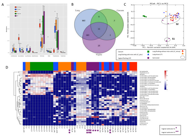
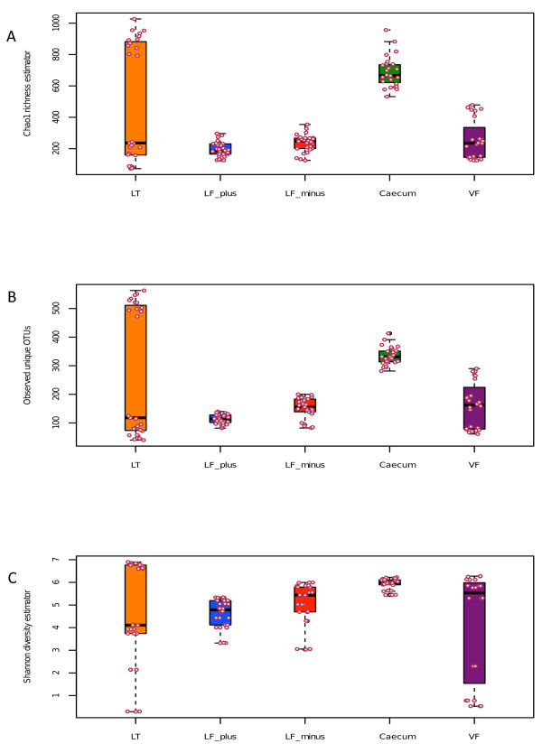
Background: This work provides the first description of the bacterial population of the lung microbiota in mice. The aim of this study was to examine the lung microbiome in mice, the most used animal model for inflammatory lung diseases such as COPD, cystic fibrosis and asthma.Bacterial communities from broncho-alveolar lavage fluids and lung tissue were compared to samples taken from fecal matter (caecum) and vaginal lavage fluid from female BALB/cJ mice.

Results: Using a customized 16S rRNA sequencing protocol amplifying the V3-V4 region our study shows that the mice have a lung microbiome that cluster separately from mouse intestinal microbiome (caecum). The mouse lung microbiome is dominated by Proteobacteria, Firmicutes, Actinobacteria, Bacteroidetes and Cyanobacteria overlapping the vaginal microbiome. We also show that removal of host tissue or cells from lung fluid during the DNA extraction step has an impact on the resulting bacterial community profile. Sample preparation needs to be considered when choosing an extraction method and interpreting data.

Conclusions: We have consistently amplified bacterial DNA from mouse lungs that is distinct from the intestinal microbiome in these mice. The gut microbiome has been extensively studied for its links to development of disease. Here we suggest that also the lung microbiome could be important in relation to inflammatory lung diseases. Further research is needed to understand the contribution of the lung microbiome and the gut-lung axis to the development of lung diseases such as COPD and asthma.


Figures

Figure 1
Figure 1
Community composition. (A) Distribution of Phyla between sample types. LF-plus bronchoalveolar lavage (BAL) fluids and LF-minus is BAL where the mouse cells have been removed. LT is lung tissue and VF is vaginal flushing, (B) Venn diagram of identified shared and unique genera from each sampling site. All the lung type samples are considered here as one. (complete list shown in Additional file 3: Table S4), (C) The PcoA plot is generated of the Bray-Curtis dissimilarity metric based on OTU counts and explains the largest variance between all samples (PCoA plot 1vs 3 and PCoA plot 2 vs. 3 are attached in Additional file 4: Figure S4), (D) Heat map of even subsampled OTU table. The dendrogram is two sited hierarchal clustered by abundance dissimilarity and the data are log transformed. Shown are only taxa, which counted for at least 0.5% of the generated sequences. The x-axis clusters the animal samples and the y-axis the taxonomical information. * marks Vaginal subcluster S1 and ** subcluster S2.
Figure 2
Figure 2
Alpha diversity plots. A: Chao1 richness estimator between sample types and individual samples (circles), LF-plus is bronchoalveolar lavage (BAL) fluids and LF-minus is BAL where the mouse cells have been removed. LT is lung tissue and VF is vaginal flushing, B: Observed unique OTUs and C: Shannon diversity estimator between sample types (s above) and individual samples (circles). The sequences (3350) were randomly even subsampled before calculating the alpha diversity. The boxplots show median, quartile, smallest and largest observations as well as outliers (circles). Significant variation is indicated by * (KW, p < 0.05).

References

    1. Beck JM, Young VB, Huffnagle GB. The microbiome of the lung. Transl Res. 2012;13:258–266. doi: 10.1016/j.trsl.2012.02.005. - DOI - PMC - PubMed
    1. Huang YJ, Nelson CE, Brodie EL, Desantis TZ, Baek MS, Liu J, Woyke T, Allgaier M, Bristow J, Wiener-Kronish JP. et al. Airway microbiota and bronchial hyperresponsiveness in patients with suboptimally controlled asthma. J Allergy Clin Immunol. 2011;13:372–381. doi: 10.1016/j.jaci.2010.10.048. - DOI - PMC - PubMed
    1. Hilty M, Burke C, Pedro H, Cardenas P, Bush A, Bossley C, Davies J, Ervine A, Poulter L, Pachter L. et al. Disordered microbial communities in asthmatic airways. PLoS One. 2010;13:e8578. doi: 10.1371/journal.pone.0008578. - DOI - PMC - PubMed
    1. Borewicz K, Pragman AA, Kim HB, Hertz M, Wendt C, Isaacson RE. Longitudinal analysis of the lung microbiome in lung transplantation. FEMS Microbiol Lett. 2013;13:57–65. doi: 10.1111/1574-6968.12053. - DOI - PMC - PubMed
    1. Turnbaugh PJ, Ley RE, Hamady M, Fraser-Liggett CM, Knight R, Gordon JI. The human microbiome project. Nature. 2007;13:804–810. doi: 10.1038/nature06244. - DOI - PMC - PubMed

Publication types

MeSH terms

Associated data

LinkOut - more resources