Review
. 2014 Apr:115:6-24.
doi: 10.1016/j.pneurobio.2013.12.002. Epub 2013 Dec 26.

Molecular dialogs between the ischemic brain and the peripheral immune system: dualistic roles in injury and repair

Affiliations
Review

Molecular dialogs between the ischemic brain and the peripheral immune system: dualistic roles in injury and repair

Chengrui An et al. Prog Neurobiol. 2014 Apr.

Abstract

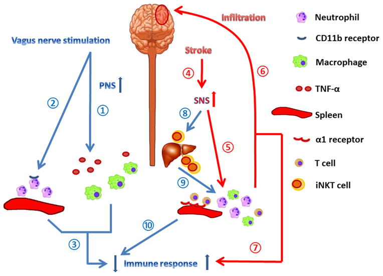
Immune and inflammatory responses actively modulate the pathophysiological processes of acute brain injuries such as stroke. Soon after the onset of stroke, signals such as brain-derived antigens, danger-associated molecular patterns (DAMPs), cytokines, and chemokines are released from the injured brain into the systemic circulation. The injured brain also communicates with peripheral organs through the parasympathetic and sympathetic branches of the autonomic nervous system. Many of these diverse signals not only activate resident immune cells in the brain, but also trigger robust immune responses in the periphery. Peripheral immune cells then migrate toward the site of injury and release additional cytokines, chemokines, and other molecules, causing further disruptive or protective effects in the ischemic brain. Bidirectional communication between the injured brain and the peripheral immune system is now known to regulate the progression of stroke pathology as well as tissue repair. In the end, this exquisitely coordinated crosstalk helps determine the fate of animals after stroke. This article reviews the literature on ischemic brain-derived signals through which peripheral immune responses are triggered, and the potential impact of these peripheral responses on brain injury and repair. Pharmacological strategies and cell-based therapies that target the dialog between the brain and peripheral immune system show promise as potential novel treatments for stroke.

Keywords: Brain repair; Cerebral ischemic injury; Inflammation; Innate immunity.


Conflict of interest statement

Conflicts of interest: None.

Figures

Figure 1
Figure 1. Immune responses mediated by the autonomic nervous system after stroke
Stimulation of the vagus nerve provides neuroprotection against acute cerebral ischemic injury, by inhibiting the release of TNF-α from macrophages ①, reducing CD11b levels on the surface of neutrophils ②, and subsequently down-regulating the peripheral immune response ③ and preventing tissue injury during inflammation. Stroke immediately elicits a fast-acting stress response ④, which activates α1 adrenergic receptors on smooth muscle cells in the spleen ⑤, leading to splenic contraction. The contraction of the spleen results in the mobilization of immune cells that infiltrate into the ischemic brain ⑥, further exacerbating ischemic brain injury and over-activating the peripheral immune response ⑦. Stroke-induced activation of the sympathetic nervous system (SNS) inhibits the normal functions of iNKT cells ⑧, which regulate T cell activation ⑨. The malfunction of iNKT cells thus contributes to immunosuppression after stroke ⑩. The discrepant actions of the SNS on immune activation versus suppression defies simplistic generalizations and may reflect the specific endpoint measured, the target organ, and the differential activation of the immune system depending upon the stage of brain injury.
Figure 2
Figure 2. Role of immune cells in tissue repair after stroke
Cellular components of the immune system participate in various aspects of post-stroke tissue repair. Microglia/macrophages engulf dead cells and debris at the site of injury ①. IL-10 and TGF-β produced by microglia/macrophages or Tregs resolve the local inflammation. Angiogenesis is enhanced by various factors released by microglia/macrophages, Tregs, and Th17 cells, such as VEGF, TGF-β, and BDNF ②. Microglia/macrophages, Tregs, and Th2 cells are also able to promote neurogenesis ③, oligodendrocyte regeneration ④, and remyelination by factors such as BDNF and TGF-β.
Figure 3
Figure 3. Innate and adaptive immune responses after stroke
In response to ischemic brain injury, a variety of danger signals are released from neurons, microglia, and astrocytes. These signals include ATP, cytokines, and chemokines. ATP can bind to P2X receptors on microglia and induce their activation and further release of inflammatory cytokines, such as TNF-α and IL-6. Activated microglia can also release chemokines, such as CCL2 and CCL3, to recruit leukocyte infiltration. The CCL2 and CCR2 axis is responsible for the recruitment of macrophages. The infiltration of macrophages has been suggested to be both detrimental in ischemic injury and protective against hemorrhagic transformation. In the ischemic brain, inflamed endothelial cells express adhesion molecules, such as ICAM-1, P-selectin, and E-selectin, which recruit leukocyte adhesion and infiltration. Following stimulation by IL-6 and TNF-α, neutrophils are activated and release MMP-9, which degrades the matrix proteins of the blood-brain barrier and stimulates peripheral leukocyte infiltration. Furthermore, brain-derived antigens can be processed by antigen presenting cells, such as dendritic cells, and presented by MHC molecules on the cell surface. Receptors on the surface of T cells can recognize the MHC and brain antigen complex. Subsequently, the adaptive immune system is activated. CD4+ T helper cells, CD8+ cytotoxic T cells, B cells, and Th17 cells all have been reported to contribute to the pathogenesis of neuroinflammation and neuronal cell death following stroke. Regulatory T cells (Tregs) and a special subset of regulatory B cells have been demonstrated to be neuroprotective through the release of IL-10. IL-35 and TGF-β are two other soluble anti-inflammatory factors that are also secreted by Tregs. Tregs can also inhibit MMP-9 production from neutrophils in a cell-cell contact-dependent manner.

References

    1. Adami C, Bianchi R, Pula G, Donato R. S100B-stimulated NO production by BV-2 microglia is independent of RAGE transducing activity but dependent on RAGE extracellular domain. Biochim Biophys Acta. 2004;1742:169–177. - PubMed
    1. Ajmo CT, Jr, Collier LA, Leonardo CC, Hall AA, Green SM, Womble TA, Cuevas J, Willing AE, Pennypacker KR. Blockade of adrenoreceptors inhibits the splenic response to stroke. Exp Neurol. 2009;218:47–55. - PMC - PubMed
    1. Ajmo CT, Jr, Vernon DO, Collier L, Hall AA, Garbuzova-Davis S, Willing A, Pennypacker KR. The spleen contributes to stroke-induced neurodegeneration. J Neurosci Res. 2008;86:2227–2234. - PMC - PubMed
    1. Albini A, Marchisone C, Del GF, Benelli R, Masiello L, Tacchetti C, Bono M, Ferrantini M, Rozera C, Truini M, Belardelli F, Santi L, Noonan DM. Inhibition of angiogenesis and vascular tumor growth by interferon-producing cells: A gene therapy approach. Am J Pathol. 2000;156:1381–1393. - PMC - PubMed
    1. Amantea D, Nappi G, Bernardi G, Bagetta G, Corasaniti MT. Post-ischemic brain damage: pathophysiology and role of inflammatory mediators. FEBS J. 2009;276:13–26. - PubMed

Publication types