. 2014 Apr;95(4):599-607.
doi: 10.1189/jlb.0613337. Epub 2013 Dec 27.

HMGB1 is an early and critical mediator in an animal model of uveitis induced by IRBP-specific T cells

Affiliations

HMGB1 is an early and critical mediator in an animal model of uveitis induced by IRBP-specific T cells

Guomin Jiang et al. J Leukoc Biol. 2014 Apr.

Abstract

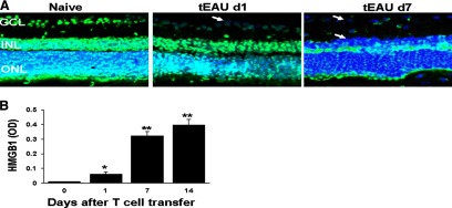
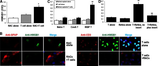
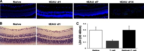
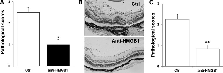
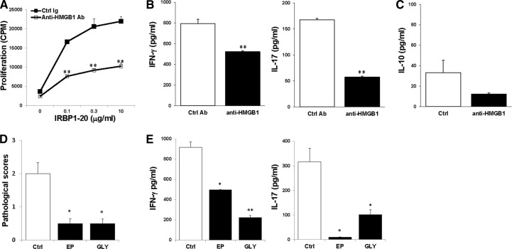
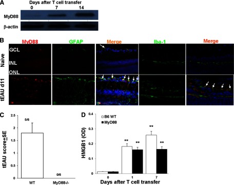
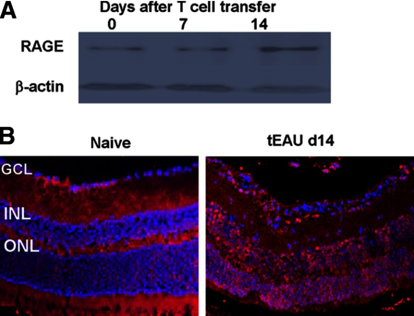
It is largely unknown how invading autoreactive T cells initiate the pathogenic process inside the diseased organ in organ-specific autoimmune disease. In this study, we used a chronic uveitis disease model in mice--EAU--induced by adoptive transfer of uveitogenic IRBP-specific T cells and showed that HMGB1, an important endogenous molecule that serves as a danger signal, was released rapidly from retinal cells into the ECM and intraocular fluid in response to IRBP-specific T cell transfer. HMGB1 release required direct cell-cell contact between retinal cells and IRBP-specific T cells and was an active secretion from intact retinal cells. Administration of HMGB1 antagonists inhibited severity of EAU significantly via mechanisms that include inhibition of IRBP-specific T cell proliferation and their IFN-γ and IL-17 production. The inflammatory effects of HMGB1 may signal the TLR/MyD88 pathway, as MyD88(-/-) mice had a high level of HMGB1 in the eye but did not develop EAU after IRBP-specific T cell transfer. Our study demonstrates that HMGB1 is an early and critical mediator of ocular inflammation initiated by autoreactive T cell invasion.

Keywords: autoimmune disease; autoreactive T cells; damage-associated molecular patterns; experimental autoimmune uveitis; immunoregulation; pathogen recognition receptors.


Figures

Figure 1.
Figure 1.. HMGB1 in retinal cells and intraocular fluid of mice after IRBP-specific T cell transfer.
(A) HMGB1 (green) was detected by immunohistochemistry in the nuclei of retinal cells from naive mice (Day 0) but was released following IRBP-specific T cell transfer; the results shown are for Days 1 and 7 (d1 and d7) post-transfer. Blue, DAPI staining of the cell nucleus; GCL, ganglion cell layer; INL, inner nuclear layer; ONL, outer nuclear layer. The arrows show loss of HMGB1 in cells in the ganglion cell layer and inner nuclear layer. (B) HMGB1 levels were determined by ELISA in the intraocular fluid of eyes from mice before receiving IRBP-specific T cells (Day 0) and on Days 1, 7, and 14 after cell transfer (six eyes/group). *P < 0.05; **P < 0.01 compared with naive mice in one-way ANOVA.
Figure 2.
Figure 2.. HMGB1 is released by cocultures of retinal cells and activated IRBP-specific T cells.
(A and B) RACs and/or activated IRBP-specific T cells (prestimulated with immunizing antigen and APCs for 2 days) were cultured for 18 h, and then culture supernatants were assayed for HMGB1 by ELISA (A) and the cells stained with the indicated fluorescent-conjugated antibody and visualized by fluorescence microscopy (B). (B) Staining is red for CD3 and GFAP, green for HMGB1, and blue for DAPI (cell nucleus). (C) Retinal explants and T cells from naive mice, Con A-stimulated T cells, or activated IRBP-specific T cells were cultured for 18 h, and then HMGB1 levels in the supernatants were measured. *P < 0.05; **P < 0.01 compared with cells cultured alone in two-way ANOVA with Fisher's least significant difference test. (D) Retinal explants and activated IRBP-specific T cells were cultured alone or together in the presence or absence of a cell insert for 18 h. **P < 0.01 compared with cells cultured alone and ††P < 0.01 compared with cells cocultured without cell insert in one-way ANOVA. (A, C, and D) The results are representative of the similar results for three separate experiments.
Figure 3.
Figure 3.. HMGB1 is actively secreted by live retinal tissue cells.
(A) TUNEL staining for apoptotic (green) cells was performed on the retina of mice before (Day 0) and on Days 1, 7, and 14 after IRBP-specific T cell transfer. The blue staining shows DAPI-stained nuclei. (B) The retinal structure of naive and Day 1 post-transfer was examined by H&E staining (original magnification, ×400). (C) Retinal explants and activated IRBP-specific T cells were cultured alone or together for 18 h as in Fig. 2D, and then LDH levels in the supernatants were measured. Values are means ± sem of two independent experiments.
Figure 4.
Figure 4.. HMGB1 antagonists significantly inhibit the development of tEAU.
B6 mice injected with IRBP-specific T cells were systemically (A and B) or locally (C) treated with neutralizing anti-HMGB1 antibody or control (Ctrl) Ig, as described in Materials and Methods and examined on Day 15. (A and C) Pathological score of each group (n=12 mice) presented as the mean ± se. *P < 0.05; **P < 0.01 compared with the group treated with control Ig using Mann-Whitney U-test. (B) Representative ocular histopathology was shown in tEAU treated with control Ig and anti-HMGB1 antibodies; H&E staining; original magnification, ×100.
Figure 5.
Figure 5.. HMGB1 antagonists inhibit IRBP-specific T cells.
T cells from mice, systemically treated with anti-HMGB1 antibodies or control Ig, on Day 15 after T cell transfer, were cultured with irradiated APCs and IRBP1–20 and then, proliferation of responding T cells (A), and levels of IFN-γ and IL-17 (B) and IL-10 (C) released in the culture supernatants were measured. Pathological scores (D) and IRBP-specific T cell cytokine production (E) in the mice treated with EP, glycyrrhizin (GLY), or PBS (Ctrl; n=15 mice/group) on Day 15 after T cell transfer were shown. *P < 0.05; **P < 0.01 compared with the control group treated with PBS.
Figure 6.
Figure 6.. Requirement of MyD88 molecule in the development of tEAU.
(A) MyD88 expression in retinas collected at Days 0, 7, or 14 after IRBP-specific T cell transfer was examined by Western blot. The loading control is β-actin. (B) MyD88 expression examined on frozen sections of retina from naive and Day 11 tEAU mice by staining with PE-conjugated anti-MyD88 antibodies (red) and FITC-conjugated anti-GFAP or anti-Iba-1 antibodies (green). Cell nuclei are stained blue with DAPI. Some double-positive cells are indicated by arrows. (C) Pathological score of WT or MyD88−/− mice (n=6/group) after adoptive transfer of IRBP-specific T cells. The incidence of tEAU was detected by pathological examination on Day 21 post-T cell transfer. (D) HMGB1 levels were determined by ELISA in the intraocular fluid of eyes from MyD88−/− mice before receiving IRBP-specific T cells (Day 0) and on Days 1 and 7 after cell transfer (six eyes/group). **P < 0.01 compared with naive mice in one-way ANOVA.
Figure 7.
Figure 7.. RAGE expression in retinas.
RAGE expression in retinas was examined by Western blot (A) as in Fig. 6A and by immunohistochemistry (B) as in Fig. 6B but using anti-RAGE antibodies.

Similar articles

Cited by

References

    1. Mochizuki M., Kuwabara T., McAllister C., Nussenblatt R. B., Gery I. (1985) Adoptive transfer of experimental autoimmune uveoretinitis in rats. Immunopathogenic mechanisms and histologic features. Invest. Ophthalmol. Vis. Sci. 26, 1–9 - PubMed
    1. Caspi R. (2008) Autoimmunity in the immune privileged eye: pathogenic and regulatory T cells. Immunol. Res. 42, 41–50 - PMC - PubMed
    1. Chan C. C., Caspi R. R., Roberge F. G., Nussenblatt R. B. (1988) Dynamics of experimental autoimmune uveoretinitis induced by adoptive transfer of S-antigen-specific T cell line. Invest. Ophthalmol. Vis. Sci. 29, 411–418 - PubMed
    1. Shao H., Liao T., Ke Y., Shi H., Kaplan H. J., Sun D. (2006) Severe chronic experimental autoimmune uveitis (EAU) of the C57BL/6 mouse induced by adoptive transfer of IRBP1–20-specific T cells. Exp. Eye Res. 82, 323–331 - PubMed
    1. Caspi R. R., Roberge F. G., McAllister C. G., el-Saied M., Kuwabara T., Gery I., Hanna E., Nussenblatt R. B. (1986) T cell lines mediating experimental autoimmune uveoretinitis (EAU) in the rat. J. Immunol. 136, 928–933 - PubMed

Publication types

LinkOut - more resources