. 2014 Feb 21;289(8):4928-40.
doi: 10.1074/jbc.M113.474049. Epub 2013 Dec 27.

The nucleolar protein Myb-binding protein 1A (MYBBP1A) enhances p53 tetramerization and acetylation in response to nucleolar disruption

Affiliations

The nucleolar protein Myb-binding protein 1A (MYBBP1A) enhances p53 tetramerization and acetylation in response to nucleolar disruption

Wakana Ono et al. J Biol Chem. .

Abstract

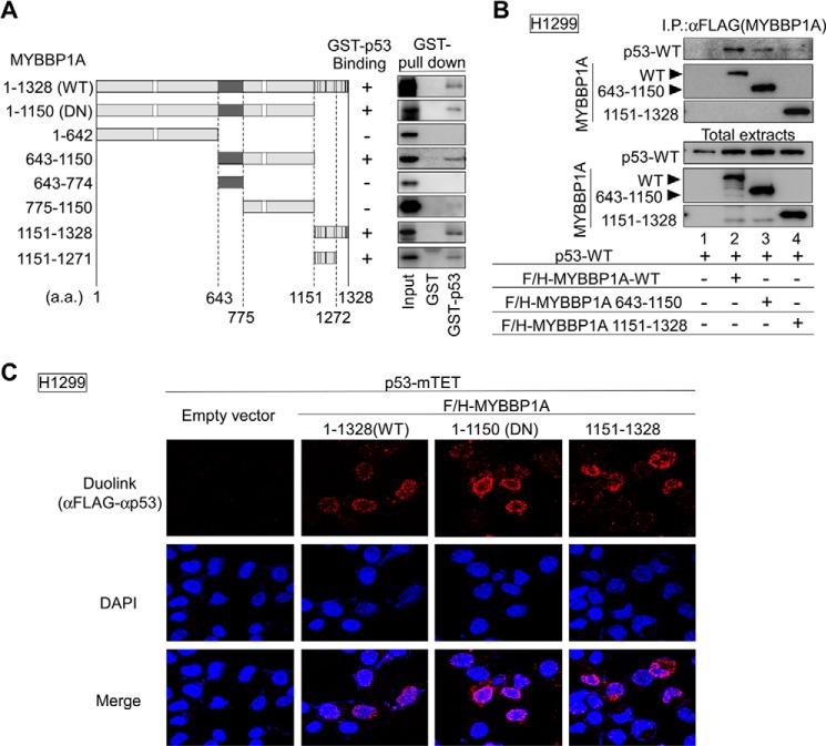
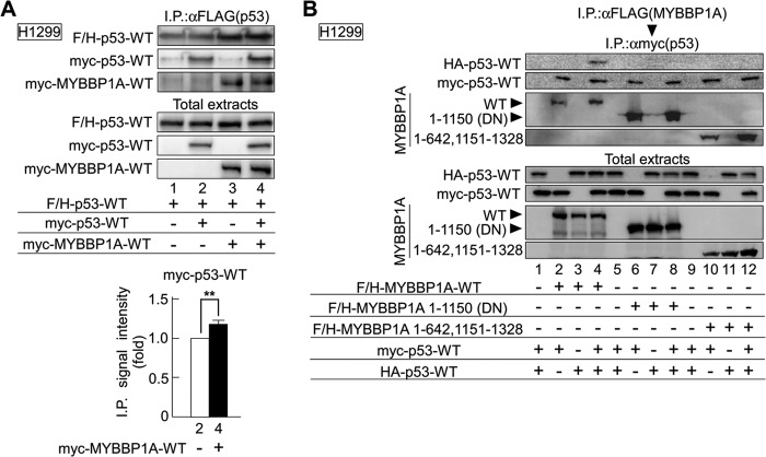
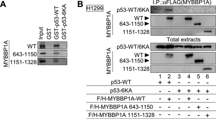
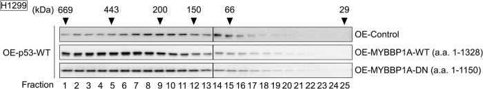
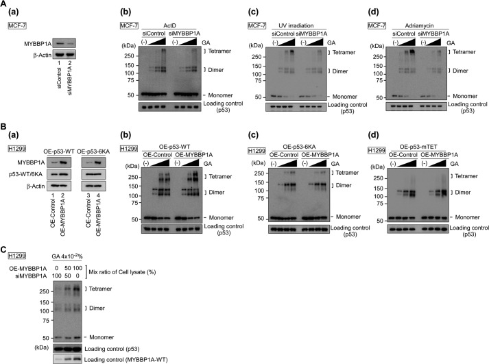
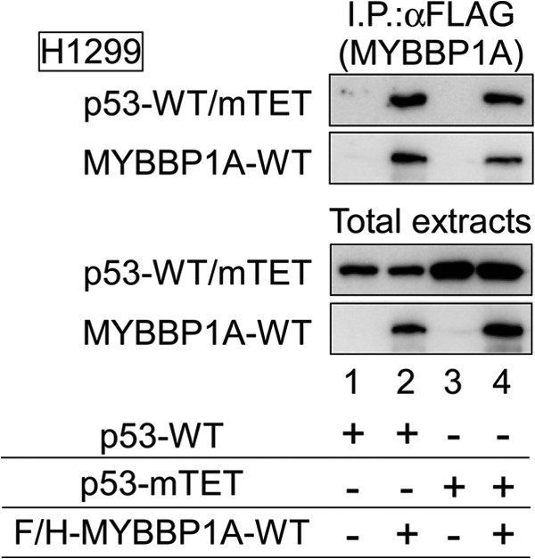
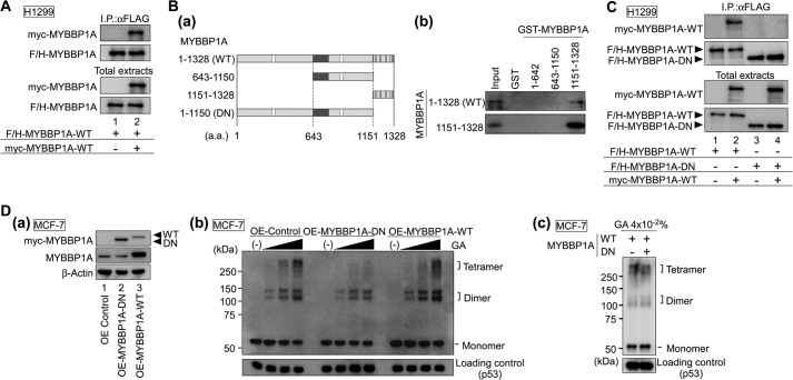
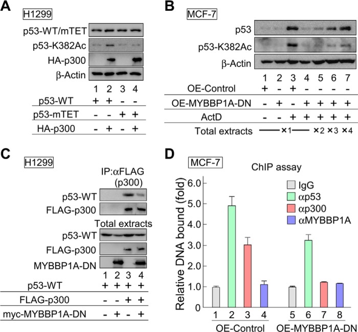
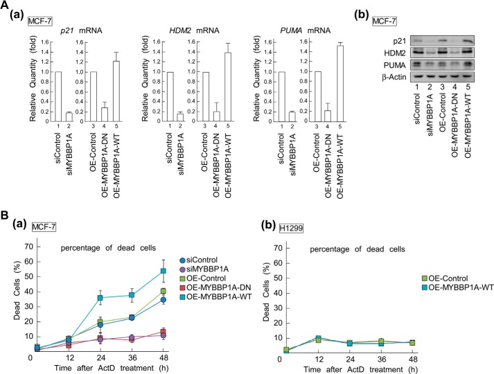
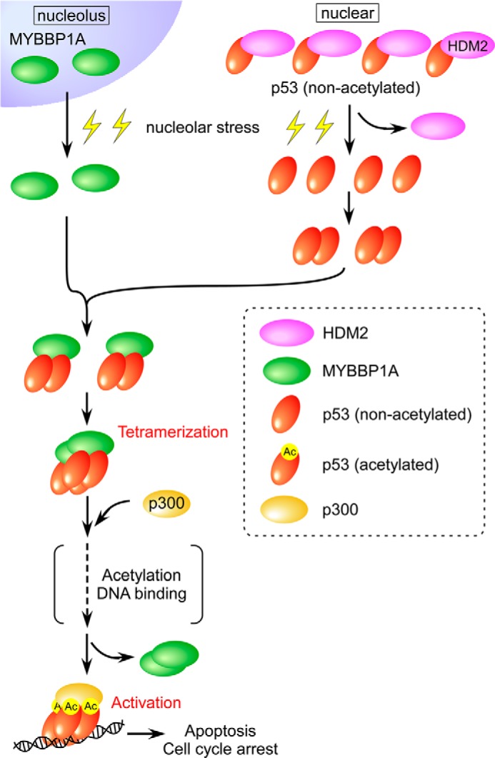
Tetramerization of p53 is crucial to exert its biological activity, and nucleolar disruption is sufficient to activate p53. We previously demonstrated that nucleolar stress induces translocation of the nucleolar protein MYBBP1A from the nucleolus to the nucleoplasm and enhances p53 activity. However, whether and how MYBBP1A regulates p53 tetramerization in response to nucleolar stress remain unclear. In this study, we demonstrated that MYBBP1A enhances p53 tetramerization, followed by acetylation under nucleolar stress. We found that MYBBP1A has two regions that directly bind to lysine residues of the p53 C-terminal regulatory domain. MYBBP1A formed a self-assembled complex that provided a molecular platform for p53 tetramerization and enhanced p300-mediated acetylation of the p53 tetramer. Moreover, our results show that MYBBP1A functions to enhance p53 tetramerization that is necessary for p53 activation, followed by cell death with actinomycin D treatment. Thus, we suggest that MYBBP1A plays a pivotal role in the cellular stress response.

Keywords: Cell Death; Nucleolus; Post-translational Modification; Stress Response; p300; p53.


Figures

FIGURE 1.
FIGURE 1.
MYBBP1A contains two p53-binding domains. A, domain structures of MYBBP1A-WT and deletion mutants of MYBBP1A (left panel). White squares, dark gray regions, and black regions show the leucine zipper-like motif, the acidic region, and the basic repeat, respectively. a.a. 1–1150, 643–1150, and 1151–1271 MYBBP1A regions directly bound to p53 (right). In vitro translated FLAG and HA (F/H)-tagged MYBBP1A-WT or its deletion mutant were incubated with GST-fused p53-WT (GST-p53) or GST proteins (GST). Bound proteins were analyzed by immunoblotting. Input, 25% of the in vitro translated MYBBP1A used in the GST pulldown assay. The deletion mutant (a.a. 1–1150) is referred to as MYBBP1A-DN (the details are provided in the text for Fig. 8, C and D). B, p53 interacted with MYBBP1A via a.a. 643–1150 or 1151–1328 regions of MYBBP1A in cells. H1299 cells were transfected with a combination of expression plasmids encoding F/H-MYBBP1A-WT, its various deletion mutants, and p53-WT, as indicated. The cells were treated with ActD for 6 h at 24 h after transfection. MYBBP1A was immunoprecipitated from the cell lysates using anti-FLAG antibodies, and the p53 association was detected by immunoblotting using anti-p53 antibody. C, MYBBP1A interacted with p53 via two independent regions in intact cells. H1299 cells transfected with the indicated plasmids were treated with ActD for 6 h. The cells were fixed and subjected to the Duolink in situ PLA using anti-FLAG and anti-p53 antibodies. The Duolink signal is shown in red. The nuclei were stained using DAPI and are shown in blue.
FIGURE 2.
FIGURE 2.
MYBBP1A binds to the p53 dimer. A, H1299 cells transfected with the indicated plasmids were treated with ActD for 6 h. p53 was immunoprecipitated from cell lysates using anti-FLAG antibodies and detected by immunoblotting using anti-p53 antibodies (top panel). Immunoprecipitated signal intensity of Myc-p53-WT in the absence or presence of Myc-MYBBP1A-WT was independently quantified three times, and the intensity of each was normalized by that of total Myc-p53-WT. The quantitative data were compared using the Student's t test (**, p < 0.01, bottom panel). B, MYBBP1A was immunoprecipitated from cell lysates using anti-FLAG (first) antibody and were eluted with FLAG peptide and immunoprecipitated again with anti-Myc (second) antibody. MYBBP1A and p53 were detected in immunoblotting precipitates.
FIGURE 3.
FIGURE 3.
MYBBP1A dissociates from p53 in a p53-CRD acetylation-dependent manner. A, a.a. 643–1150 and 1151–1328 regions of MYBBP1A did not directly bind to the p53–6KA mutant. In vitro translated F/H-MYBBP1A-WT or its deletion mutant was incubated with GST-fused p53-WT (GST-p53-WT), p53–6KA (GST-p53–6KA), or with GST. Bound proteins were analyzed as in Fig. 1A. B, p53 interacted with MYBBP1A via its lysine residues in the CRD region in cells. H1299 cells were transfected with a combination of expression plasmids encoding F/H-MYBBP1A-WT, its various deletion mutants, and p53-WT, as indicated. The cells were treated with ActD for 6 h at 24 h post-transfection. MYBBP1A was immunoprecipitated from the cell lysates using anti-FLAG antibody and analyzed as in Fig. 1B.
FIGURE 4.
FIGURE 4.
MYBBP1A enhances p53 oligomerization. Expression plasmids encoding wild-type p53 (OE-p53-WT) were transfected with that encoding EGFP (OE-Control), MYBBP1A-WT (a.a. 1–1138) (OE-MYBBP1A-WT), or MYBBP1A-DN (a.a. 1–1150) (OE-MYBBP1A-DN) into H1299 cells. The cells were treated with ActD for 6 h at 24 h post-transfection. The lysates were subjected to size exclusion chromatography. Fractions were analyzed by immunoblotting for the presence of p53. The elution positions of the molecular size markers are shown.
FIGURE 5.
FIGURE 5.
MYBBP1A promotes p53 tetramerization. A, knockdown of MYBBP1A reduced the formation of p53 tetramers. MCF-7 cells were transfected with either control siRNA (siControl) or MYBBP1A siRNA (siMYBBP1A). To validate the efficiency of MYBBP1A knockdown, protein abundance was determined by immunoblotting with anti-MYBBP1A antibody in panel a. At 48 h post-transfection, the cells were treated with ActD for 6 h in panel b, with UV irradiation in panel c, and with adriamycin for 6 h in panel d. The lysates were subjected to a protein cross-linking assay in GA at final concentrations of 0, 0.005, 0.01, or 0.02%, followed by immunoblotting. B, overexpression of MYBBP1A enhanced the formation of p53 tetramers in wild-type p53-expressing H1299 cells. Expression plasmids encoding p53-WT (OE-p53-WT), p53–6KA (OE-p53–6KA), or p53-mTET (OE-p53-mTET) were transfected into H1299 cells with that encoding EGFP (OE-Control) or F/H-MYBBP1A-WT (OE-MYBBP1A). The cells were treated with ActD for 6 h at 24 h post-transfection. To validate the efficiency of overexpression of p53-WT or p53–6KA with or without MYBBP1A, the protein abundance of p53 and MYBBP1A was determined by immunoblotting with anti-p53 or anti-MYBBP1A antibodies in panels a. The lysates were subjected to a cross-linking assay in GA at final concentrations of 0, 0.005, 0.01, or 0.02% in panels b–d. C, dose effect of MYBBP1A on p53 tetramerization. The H1299 cell lysate of OE-MYBBP1A was mixed with that of siMYBBP1A#1 at the indicated ratio and was incubated for 1 h on ice. Following this, the mixed lysates were cross-linked by GA at a final concentration of 0.04%.
FIGURE 6.
FIGURE 6.
p53-mTET interacts with MYBBP1A. H1299 cells were transfected with a combination of expression plasmids encoding F/H-MYBBP1A-WT, p53-WT, and p53-mTET, as indicated. The cells were treated with ActD for 6 h at 24 h post-transfection. MYBBP1A was immunoprecipitated from the cell lysates using anti-FLAG antibody and subjected to immunoblotting using anti-FLAG or anti-p53 antibodies.
FIGURE 7.
FIGURE 7.
MYBBP1A enhances tetramerization-dependent p53 promoter binding. H1299 cells were transfected with the indicated plasmids and treated with ActD for 6 h. Nuclear extracts were subjected to an EMSA.
FIGURE 8.
FIGURE 8.
Dimerization of MYBBP1A is necessary for p53 tetramerization. A, MYBBP1A interacted with MYBBP1A in cells. H1299 cells were transfected with a combination of expression plasmids encoding F/H-MYBBP1A-WT and Myc-MYBBP1A-WT, as indicated. The cells were treated with ActD for 6 h at 24 h post-transfection. MYBBP1A was immunoprecipitated from the cell lysates using anti-FLAG antibodies and subjected to immunoblotting using anti-Myc or anti-FLAG antibodies. B, MYBBP1A directly bound to MYBBP1A via a.a. 1151–1328 MYBBP1A region. In panel a, domain structures of full-length WT and deletion mutants of MYBBP1A are shown. White squares, dark gray regions, and black regions show the leucine zipper-like motif, the acidic region, and the basic repeat, respectively. In panel b, in vitro translated F/H-MYBBP1A was incubated with the GST-fused truncated MYBBP1A proteins, as shown in panel a. Bound proteins were then analyzed by immunoblotting. The deletion mutant (a.a. 1–1150) was referred to as MYBBP1A-DN. C, MYBBP1A-DN did not form a dimer with MYBBP1A in the cells. H1299 cells were transfected with a combination of expression plasmids encoding F/H-MYBBP1A-WT, F/H-MYBBP1A-DN, and Myc-MYBBP1A-WT, as indicated. The cells were treated with ActD for 6 h at 24 h post-transfection. MYBBP1A was immunoprecipitated from the cell lysates using anti-FLAG antibody and subjected to immunoblotting using anti-Myc or FLAG antibodies. D, MYBBP1A-DN inhibited the formation of p53 tetramers. In panel a, MCF-7 cells were transfected with the expression plasmid encoding EGFP (OE Control) or Myc-MYBBP1A (OE-MYBBP1A-DN or OE-MYBBP1A-WT). To validate the efficiency of MYBBP1A overexpression, MYBBP1A protein abundance was determined by immunoblotting with anti-Myc and anti-MYBBP1A antibodies. Note: Anti-MYBBP1A antibody does not recognize MYBBP1A-DN because MYBBP1A-DN is lacking the anti-MYBBP1A antibody epitope. In panel b, MCF-7 cells were transfected with the expression plasmid encoding EGFP (OE-Control), MYBBP1A-DN (OE-MYBBP1A-DN), or MYBBP1A-WT (OE-MYBBP1A-WT). The cells were treated with ActD for 6 h at 24 h post-transfection and were subjected to the cross-linking assay in GA at final concentrations of 0, 0.005, 0.01, or 0.02%. In panel c, MCF-7 cells were transfected with a combination of expression plasmids encoding MYBBP1A-WT and MYBBP1A-DN, as indicated. MCF-7 cells were treated with ActD for 6 h at 24 h post-transfection and were subjected to the cross-linking assay in GA at a final concentration of 0.04%.
FIGURE 9.
FIGURE 9.
MYBBP1A enhances the p53-p300 interaction and p53 acetylation by regulating p53 tetramerization. A, p53-mTET was not acetylated by overexpressing p300. The expression plasmids encoding p53-WT or p53-mTET were transfected into H1299 cells with or without p300, 24 h before treatment with ActD for 6 h. Cell lysates were analyzed by immunoblotting using anti-p53 or site-specifically acetylated p53 antibodies. B, overexpression of MYBBP1A-DN decreased Lys-382 acetylation (K382Ac) levels of p53. MCF-7 cells were transfected with the expression plasmid encoding EGFP (OE-Control) or MYBBP1A-DN (OE-MYBBP1A-DN), 24 h before treatment with ActD for 6 h. Cell lysates were analyzed by immunoblotting using anti-p53 or site-specifically acetylated p53 antibodies. ×1, ×2, ×3, and ×4 indicate the ratios of total extract volumes applied to SDS-PAGE. C, overexpression of MYBBP1A-DN decreased binding of p300 to p53. H1299 cells were transfected with a combination of expression plasmids encoding FLAG-p300, Myc-MYBBP1A-DN, and p53-WT, as indicated. The cells were treated with ActD for 6 h at 24 h after transfection. FLAG-p300 was immunoprecipitated using anti-FLAG antibody, and the immunoprecipitates were eluted using FLAG peptides and analyzed by immunoblotting using the indicated antibodies. D, overexpression of MYBBP1A-DN reduced the recruitment of p53 and p300 to the p21 promoter in ActD-treated cells. MCF-7 cells were transfected with the expression plasmid encoding EGFP (OE-Control) or FH-MYBBP1A-DN (OE-MYBBP1A-DN) for 24 h before treatment with ActD for 6 h. The CHIP assay was performed using normal rabbit IgG, anti-p53, and anti-p300 antibodies. The p53-binding region of the p21 promoter was amplified and analyzed by RT-qPCR. The values are given as the means ± standard deviation for triplicate assays.
FIGURE 10.
FIGURE 10.
MYBBP1A regulates p53 activation by enhancing tetramerization. A, overexpression of MYBBP1A-DN and knockdown of MYBBP1A reduced mRNA and protein levels of p53 target genes, p21, HDM2, and PUMA. MCF-7 cells were transfected with indicated siRNAs or plasmids for 48 or 24 h, respectively, followed by ActD treatment. In panel a, total RNAs were prepared and expression of the indicated genes was analyzed by RT-qPCR. In panel b, total extracts were prepared, and expression of the indicated genes was analyzed by immunoblotting. B, overexpression of MYBBP1A-DN and knockdown of MYBBP1A decreased the percentage of apoptosis induced by ActD treatment. p53-positive MCF-7 (panel a) and p53-null H1299 (panel b) cells were treated with the indicated siRNAs or plasmids and cultured for 24 h. Following this, the cells were treated with or without ActD at a final concentration of 5 nm. The percentage of dead cells was measured by a trypan blue exclusion assay at the indicated times after ActD treatment. The values are means ± standard deviation for triplicate assays.
FIGURE 11.
FIGURE 11.
Proposed model for the role of MYBBP1A in p53 tetramerization following nucleolar stress. The details are provided in the text.

Similar articles

Cited by

References

    1. Vogelstein B., Lane D., Levine A. J. (2000) Surfing the p53 network. Nature 408, 307–310 - PubMed
    1. Levine A. J. (1997) p53, the cellular gatekeeper for growth and division. Cell 88, 323–331 - PubMed
    1. Prives C., Hall P. A. (1999) The p53 pathway. J. Pathol. 187, 112–126 - PubMed
    1. Michael D., Oren M. (2003) The p53-Mdm2 module and the ubiquitin system. Semin. Cancer Biol. 13, 49–58 - PubMed
    1. Brooks C. L., Gu W. (2006) p53 ubiquitination. Mdm2 and beyond. Mol. Cell 21, 307–315 - PMC - PubMed

Publication types

MeSH terms