. 2014 May;59(5):1984-1997.
doi: 10.1002/hep.26976. Epub 2014 Apr 1.

Hepatocyte-specific high-mobility group box 1 deletion worsens the injury in liver ischemia/reperfusion: a role for intracellular high-mobility group box 1 in cellular protection

Affiliations

Hepatocyte-specific high-mobility group box 1 deletion worsens the injury in liver ischemia/reperfusion: a role for intracellular high-mobility group box 1 in cellular protection

Hai Huang et al. Hepatology. 2014 May.

Abstract

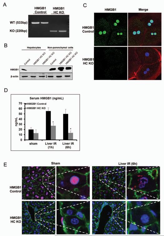
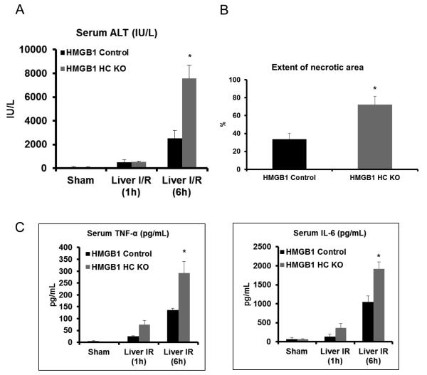
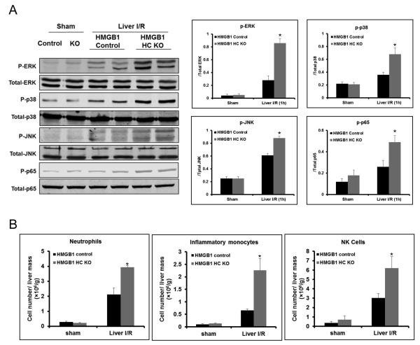
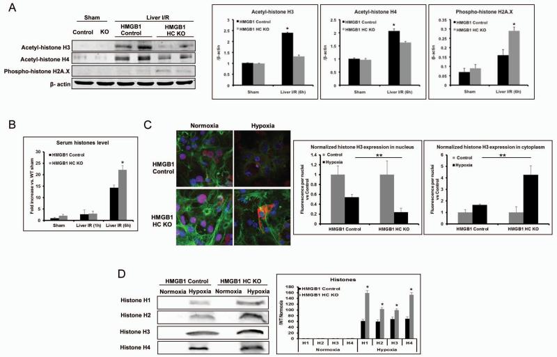
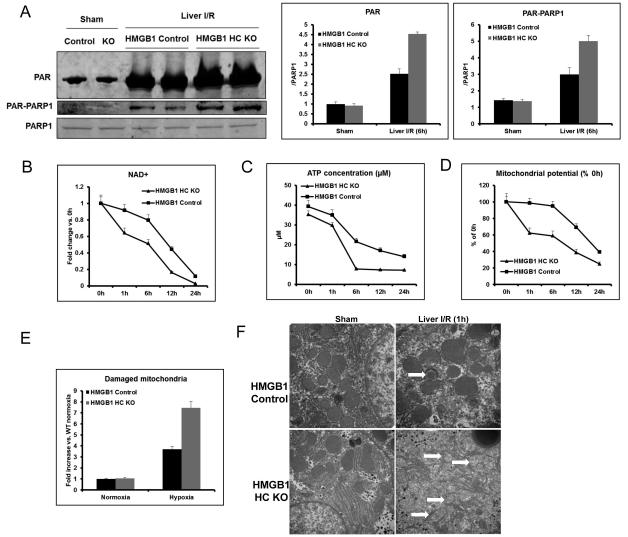
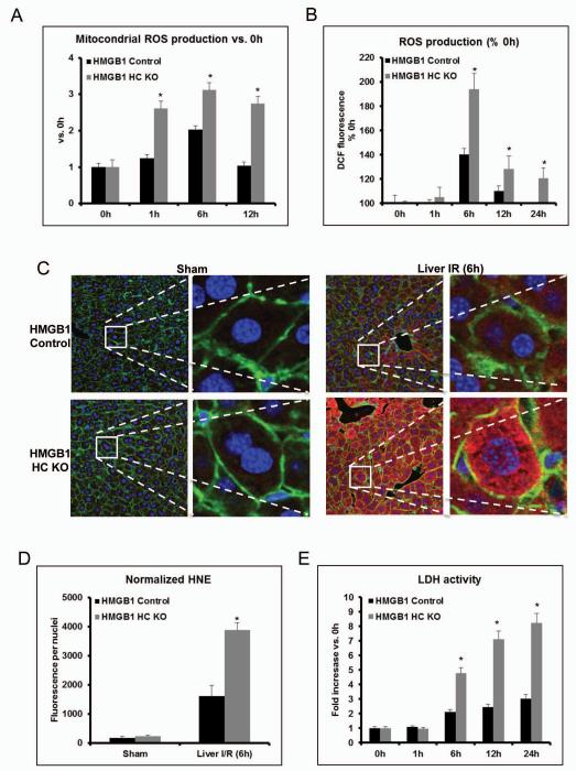
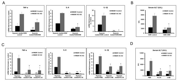
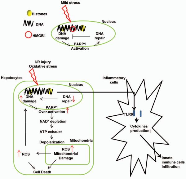
High-mobility group box 1 (HMGB1) is an abundant chromatin-associated nuclear protein and released into the extracellular milieu during liver ischemia-reperfusion (I/R), signaling activation of proinflammatory cascades. Because the intracellular function of HMGB1 during sterile inflammation of I/R is currently unknown, we sought to determine the role of intracellular HMGB1 in hepatocytes after liver I/R. When hepatocyte-specific HMGB1 knockout (HMGB1-HC-KO) and control mice were subjected to a nonlethal warm liver I/R, it was found that HMGB1-HC-KO mice had significantly greater hepatocellular injury after I/R, compared to control mice. Additionally, there was significantly greater DNA damage and decreased chromatin accessibility to repair with lack of HMGB1. Furthermore, lack of hepatocyte HMGB1 led to excessive poly(ADP-ribose)polymerase 1 activation, exhausting nicotinamide adenine dinucleotide and adenosine triphosphate stores, exacerbating mitochondrial instability and damage, and, consequently, leading to increased cell death. We found that this was also associated with significantly more oxidative stress (OS) in HMGB1-HC-KO mice, compared to control. Increased nuclear instability led to a resultant increase in the release of histones with subsequently more inflammatory cytokine production and organ damage through activation of Toll-like receptor 9.

Conclusion: The lack of HMGB1 within hepatocytes leads to increased susceptibility to cellular death after OS conditions.


Figures

Figure 1
Figure 1. Confirmation of specificity of HMGB1 knockout (HMGB1-HC-KO)
(A) RT-PCR was used to determine mRNA levels of HMGB1 within isolated hepatocytes from control and HMGB1-HC-KO mice. (B) HMGB1 protein levels within isolated hepatocytes or non-parenchymal cells from control and HMGB1-HC-KO mice were assessed by Western blot analysis. Figure is representative of three experiments with similar results. (C) Immunofluorescent stain of HMGB1 within cultured hepatocytes from control and HMGB1-HC-KO mice (magnification ×400). Images are representative of three experiments with similar results. Green, HMGB1; blue, nuclei; red, F-actin. (D) Serum HMGB1 ELISA after 1h or 6h of reperfusion. *P<0.05 when compared against control. (E) Immunofluorescent stain of HMGB1 within from sections of normal liver and liver 6h after I/R in control and HMGB1-HC-KO mice (magnification ×400). Images are representative liver sections from six mice per group. Red, HMGB1; blue, nuclei; green, F-actin.
Figure 2
Figure 2. Cellular specific role of HMGB1 on hepatocellular injury after I/R
(A) Serum ALT levels were analyzed in control and HMGB1-HC-KO mice after either sham laparotomy or 1h of ischemia and 1h or 6h of reperfusion. Data represent the mean ± SE (n = 6 mice per group). *P < 0.05 vs. HMGB1 control. (B) Quantification of necrotic hepatocytes in H&E-stained liver sections (Supplemental Figure 2B) from HMGB1-HC-KO and control mice 6h after reperfusion. The graph is representative of liver sections from six mice per group. *P < 0.05 vs. control. (C) Serum levels of TNF-α and IL-6 obtained from HMGB1-HC-KO and control mice at 6h after reperfusion were measured by ELISA and compared to the sham group. Data represent the mean ± SE (n = 6 mice per group) *P<0.05 vs. control.
Figure 3
Figure 3. Lack of HMGB1 in hepatocytes mediates inflammatory signaling and regulates innate immune cells in liver I/R
(A) Mitogen-activated protein kinase activation and phosphorylation at serine 536 of the p65 subunit of NF-κB were determined by Western blot and quantitative densitometry analysis of the protein expressions in sham-treated mice and mice that underwent ischemia and 1h of reperfusion. Hepatic protein lysates from ischemic lobes were obtained; each lane represents a separate animal. The blots shown are representative of three experiments with similar results. *P < 0.05 vs. control. (B) Flow cytometry analysis with a quantitative evaluation of NPCs in homogenized ischemia liver lobes in HMGB1-HC-KO and control mice. Fold change of cell numbers of neutrophils, inflammatory monocytes cells, and nature killer (NK) cells. Data represent the mean ± SE (n = 6 mice per group). *P < 0.05 compared to sham mice. Each experiment was repeated a minimum of three times.
Figure 4
Figure 4. Lack of HMGB1 in hepatocytes leads to less DNA repair and more DNA damage, with subsequently more cell death and histone release
(A) Acetylation of histone H3 and H4, and phosphorylation of H2A.X were determined by Western blot and quantitative densitometry analysis in sham-treated mice and mice that underwent ischemia and 6h of reperfusion. Hepatic protein lysates from ischemic lobes were obtained; each lane represents a separate animal. The blots shown are representative of three experiments with similar results. *P < 0.05 vs. control. (B) Serum histone levels were assessed after I/R using ELISA. Data represent the mean ± SE (n = 6 mice per group). *P < 0.05 compared to control. (C) The translocation of histone H3 in cultured hepatocytes from HMGB1-HC-KO or control mice that were stimulated with either hypoxia or normoxic for 12h was visualized and observed under confocal microscope (magnification ×400). Green, actin; blue, nuclei; red, histone H3. Quantitation of nucleic or cytoplasmic histone H3 in cultured hepatocytes from HMGB1-HC-KO or control mice was measured using the analytical software MetaMorph™ and normalized to nuclei within the sample field. *P < 0.05; hypoxia vs. normoxia control, **P < 0.05; HMGB1-HC-KO vs. control. (D) Cultured hepatocytes from HMGB1-HC-KO or control mice were exposed to hypoxia. Media were subjected to Western blot and quantitative densitometry analysis of histone H1, H2, H3, and H4. The blots shown are representative of three experiments with similar results. *P < 0.05 vs. control.
Figure 5
Figure 5. Lack of HMGB1 in hepatocytes induces PARP-1 over-activation, damages mitochondria by exhausting NAD+ and ATP
(A) Total PAR formation and Poly (ADP-ribosylation) of PARP-1 was determined by Western blot and quantitative densitometry analysis of the protein expressions in sham-treated mice and mice that underwent ischemia and 6h of reperfusion. Hepatic protein lysates from ischemic lobes were obtained; each lane represents a separate animal. The blots shown are representative of three experiments with similar results. *P < 0.05 vs. control. (B) Time course of NAD+ level in cultured hepatocytes from HMGB1-HC-KO or control mice during hypoxia (n=4-6 for each point). *P < 0.05 vs. control. (C) Time course of ATP level in cultured hepatocytes from HMGB1-HC-KO or control mice during hypoxia (n=4-6 for each point). *P < 0.05 vs. control. (D) Time course of mitochondrial potential in cultured hepatocytes from HMGB1-HC-KO or control mice during hypoxia (n=4-6 for each point). *P < 0.05 vs. control. (E) HMGB1 KO or control hepatocytes were cultures under normoxia or 12h hypoxia. The cells were then stained with 200nM MitoTracker Green and MitoTracker Deep Red (Invitrogen) for 45min at 37°C then analyzed using flow cytometry for mitochondrial damage. *P < 0.05 vs. control. (F) Mitochondrial ultrastructure from livers of HMGB1-HC-KO or control mice following sham or 1h reperfusion were imaged by transmission electron microscope (magnification ×50000). *, P<0.05; results are representative of 3 separate independent experiments. White arrows point to damaged mitochondrial cristae.
Figure 6
Figure 6. Lack of HMGB1 in hepatocytes mediates mitochondrial and cellular ROS production
(A) Flow cytometry of cultured hepatocytes from HMGB1-HC-KO or control mice that were stimulated with hypoxia and stained with MitoSOX to determine mitochondrial ROS production. The bar graph represents pooled data from three experiments. 0h represents normoxia. *P < 0.05 compared to PBS treatment. (B) Cellular ROS production as detected by the DCF-DA assay in cultured whole hepatocytes, which were obtained from either HMGB1-HC-KO or HMGB1 control mice. 0h represents normoxia. *P < 0.05 compared to control. (C) Representative 4-HNE staining Green, actin; blue, nuclei; 4-HNE, red and (D) quantification of 4-hydroxy-2-nonenal (4-HNE) adducts (4-HNE normalized per nuclei in the field) in ischemic liver of HMGB1-HC-KO or control mice. *P < 0.05 compared to control. (E) Time course of LDH activity in cultured hepatocytes from HMGB1-HC-KO or control mice during hypoxia (n=4-6 for each point). *P < 0.05 vs. control.
Figure 7
Figure 7. Addition of TLR9 antagonists and PARP-1 inhibitors both protect HMGB1-HC-KO mice from liver I/R injury
(A) TNF-α, IL-6 and IL-1β mRNA expression was determined in non-parenchymal cells cultured overnight with media from hypoxic HMGB1 KO or control hepatocytes. Non-parenchymal cells were treated with TLR9 antagonist, ODN2088, or ODN control. Results are expressed as the relative increase of mRNA expression compared with PBS treatment. Data represent the mean ± SE and are representative of three experiments with similar results. *P < 0.05 compared to ODN control-treated HMGB1-HC-KO group. **P < 0.05 compared to ODN control-treated HMGB1 Control group. (B) Serum ALT levels in HMGB1-HC-KO or control mice after 6h of reperfusion that were treated with ODN2088 or ODN control. Data represent the mean ± SE (n = 6 mice per group). *P < 0.05 compared to ODN control-treated HMGB1-HC-KO group. **P < 0.05 compared to ODN control-treated HMGB1 Control group. (C) TNF-α, IL-6 and IL-1β mRNA expression was determined in non-parenchymal cells cultured overnight with media from hypoxic HMGB1 KO or control hepatocytes. Hepatocytes were pre-treated with the PARP-1 inhibitor, 3-AB or PJ-34, or negative control, PBS. Results are expressed as the relative increase of mRNA expression compared with PBS treatment. Data represent the mean ± SE and are representative of three experiments with similar results. *P < 0.05 compared to PBS-treated HMGB1-HC-KO group. **P < 0.05 compared to PBS-treated HMGB1 Control group. (D) Serum ALT levels in HMGB1-HC-KO or control mice after 6h of reperfusion that were treated with the PARP-1 inhibitor, 3-AB or PJ-34, or PBS. Data represent the mean ± SE (n = 6 mice per group). *P < 0.05 compared to PBS-treated HMGB1-HC-KO group. **P < 0.05 compared to PBS-treated HMGB1 Control group.
Figure 8
Figure 8
Schematic representation of the intracellular role of HMGB1 during liver I/R injury. During liver I/R injury, HMGB1 deletion in hepatocytes leads to excessive DNA damage, which initiates over-activation of PARP-1, subsequently exhausting both NAD+ and ATP reserves, damaging mitochondria by depolarization of the mitochondrial membrane. Damaged mitochondria are associated with more mitochondrial and cellular ROS production, eventually leading to more cell stress and death. Excessive cell death further propagates the inflammatory response and infiltration of innate immune cells in liver I/R by released histones.

Similar articles

Cited by

References

    1. Fondevila C, Busuttil RW, Kupiec-Weglinski JW. Hepatic ischemia/reperfusion injury--a fresh look. Experimental and Molecular Pathology. 2003;74:86–93. - PubMed
    1. Huang H, Evankovich J, Yan W, Nace G, Zhang L, Ross M, Liao X, et al. Endogenous histones function as alarmins in sterile inflammatory liver injury through toll-like receptor 9. Hepatology. 2011 - PMC - PubMed
    1. Tsung A, Klune JR, Zhang X, Jeyabalan G, Cao Z, Peng X, Stolz DB, et al. HMGB1 release induced by liver ischemia involves Toll-like receptor 4 dependent reactive oxygen species production and calcium-mediated signaling. J Exp Med. 2007;204:2913–2923. - PMC - PubMed
    1. Tsung A, Sahai R, Tanaka H, Nakao A, Fink MP, Lotze MT, Yang H, et al. The nuclear factor HMGB1 mediates hepatic injury after murine liver ischemia-reperfusion. The Journal of experimental medicine. 2005;201:1135–1143. - PMC - PubMed
    1. Javaherian K, Liu JF, Wang JC. Nonhistone proteins HMG1 and HMG2 change the DNA helical structure. Science. 1978;199:1345–1346. - PubMed

Publication types