. 2014 Feb;3(2):141-8.
doi: 10.5966/sctm.2013-0146. Epub 2013 Dec 27.

Accurate prediction of drug-induced liver injury using stem cell-derived populations

Affiliations

Accurate prediction of drug-induced liver injury using stem cell-derived populations

Dagmara Szkolnicka et al. Stem Cells Transl Med. 2014 Feb.

Abstract

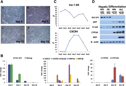
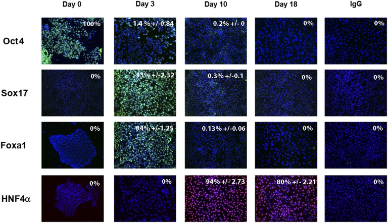
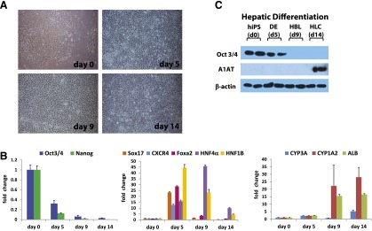
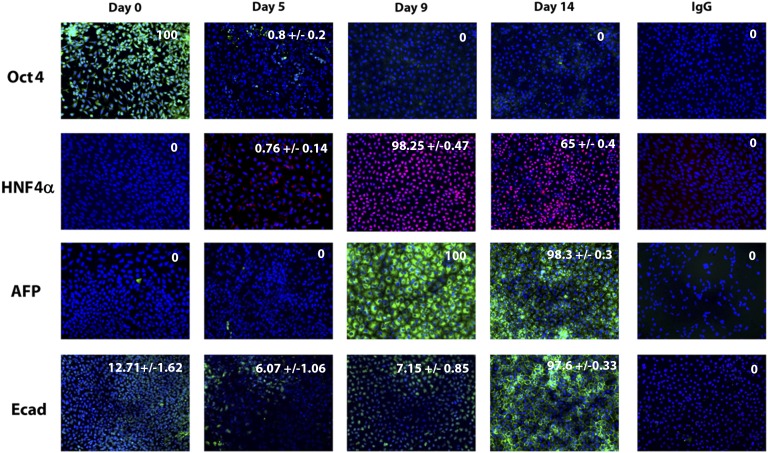
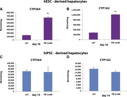
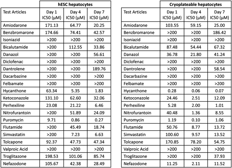
Despite major progress in the knowledge and management of human liver injury, there are millions of people suffering from chronic liver disease. Currently, the only cure for end-stage liver disease is orthotopic liver transplantation; however, this approach is severely limited by organ donation. Alternative approaches to restoring liver function have therefore been pursued, including the use of somatic and stem cell populations. Although such approaches are essential in developing scalable treatments, there is also an imperative to develop predictive human systems that more effectively study and/or prevent the onset of liver disease and decompensated organ function. We used a renewable human stem cell resource, from defined genetic backgrounds, and drove them through developmental intermediates to yield highly active, drug-inducible, and predictive human hepatocyte populations. Most importantly, stem cell-derived hepatocytes displayed equivalence to primary adult hepatocytes, following incubation with known hepatotoxins. In summary, we have developed a serum-free, scalable, and shippable cell-based model that faithfully predicts the potential for human liver injury. Such a resource has direct application in human modeling and, in the future, could play an important role in developing renewable cell-based therapies.

Keywords: Cytochrome P450; Drug metabolism; Drug toxicity; Human embryonic stem cells; Liver; iPSC.


Figures

Figure 1.
Figure 1.
Driving efficient hepatocellular differentiation using a serum-free system. Human embryonic stem cells were differentiated in a stagewise fashion toward the hepatocyte lineage. (A): Phase-contrast imaging demonstrated the cells were undergoing sequential morphological changes during transit from stem cell (day 0) (Aa) to definitive endoderm (day 3) (Ab) to hepatoblast (day 10) (Ac) to hepatocyte (day 18) (Ad). (B): The quantitative polymerase chain reaction (qPCR) gene expression analysis demonstrated that changes in morphology were paralleled by changes in pluripotent (Oct4, Nanog), definitive endoderm (Sox17, CXCR4, Nodal), and hepatic (HNF4α, HNF1β, CYP3A, and 1A2) transcripts in line with human development. (C): Cell surface marker expression was also significantly altered during cellular differentiation, as assessed by flow cytometry. The downregulation of TRA 1-60, marking onset of cellular differentiation at day 6, and the increase in CXCR4 staining confirmed definitive endoderm specification at days 3 and 4. (D): Western blotting was used to assess protein expression of a number of differentiation markers. As the cells differentiated toward the hepatocyte lineage, we detected key changes in Oct4, AFP, albumin, and E-cadherin gene expression. Images were taken at ×10 magnification. The qPCRs were run in triplicates. Error bars represent the standard deviation. Abbreviations: d, day; DE, definitive endoderm; HBC, hepatocyte; HBL, hepatoblast; hES, human embryonic stem cells.
Figure 2.
Figure 2.
Stagewise human embryonic stem cell (hESC) differentiation to the hepatocyte lineage. hESCs were differentiated in a stagewise fashion toward the hepatocyte lineage. Immunocytochemistry showing downregulation of pluripotent marker Oct4, as the cells expressed endodermal transcripts (Sox17, Foxa1). Upon hepatic specification, HNF4α expression increased. Immunoglobulin G controls demonstrated the specificity of immunostaining. The percentage of positive cells is provided in the top right of each panel. This was calculated from four random fields of view and is quoted as ±standard error. The images were taken at ×20 magnification. Abbreviations: Foxa1, Forkhead box protein 1; HNF4α, hepatocyte nuclear factor 4α; Oct4, octamer 4; Sox 17, SRY-box containing gene 17.
Figure 3.
Figure 3.
Translating new model to human induced pluripotent stem cells (iPSCs). iPSCs were differentiated in a stagewise fashion toward the hepatocyte lineage. (A): Imaging showing sequential morphological changes during iPSC differentiation. The light microscopy demonstrated transition from iPSC (day 0), to definitive endoderm (day 5), to hepatoblast (day 9), and to hepatocyte (day 14). (B): The quantitative polymerase chain reaction (qPCR) gene expression analysis demonstrated that changes in morphology were paralleled by changes in pluripotent (Oct4, Nanog), definitive endoderm (Sox17, CXCR4, Foxa 2), and hepatic (HNF4α, HNF1β, CYP3A, CYP1A2, ALB) transcripts, which is in line with human development. (C): Western blotting was used to assess protein expression of a number of differentiation markers. As the cells differentiated toward hepatic lineage, Oct4 protein expression was reduced and α1 antitrypsin gene expression increased. Images were taken at ×10 magnification. The qPCRs were run in triplicates. Error bars represent the standard deviation. Abbreviations: d, day; DE, definitive endoderm; HBC, hepatocyte; HBL, hepatoblast; hIPS, human induced pluripotent cells.
Figure 4.
Figure 4.
Stagewise induced pluripotent stem cell (iPSC) differentiation to the hepatocyte lineage. iPSCs were differentiated in a stagewise fashion toward the hepatocyte lineage. Immunocytochemistry shows downregulation of pluripotent marker Oct4 during cellular differentiation. Upon hepatic specification, HNF4α, AFP, and E-cad expression increased. Immunoglobulin G controls demonstrated the specificity of immunostaining. The percentage of positive cells is provided in the top right of each panel. This was calculated from four random fields of view and is quoted as ±standard error. The images were taken at ×20 magnification. Abbreviations: AFP, a-fetoprotein; Ecad, E cadherin; HNF4α, hepatocyte nuclear factor 4α; Oct4, octamer 4.
Figure 5.
Figure 5.
Identification of mature hepatocyte populations for scale-up and drug toxicity testing. Pluripotent stem cells were differentiated to hepatocytes as previously described. (A, B): hESC hepatocytes were induced with 1 mM phenobarbital drug for 48 hours prior to analysis of CYP3A and CYP1A2 function. (C, D): iPSC hepatocytes were induced with 1 mM phenobarbital drug for 48 hours prior to analysis of CYP3A and CYP1A2 function. The activity of CYP3A and CYP1A2 was analyzed using Promega pGlo technology, and activity was measured on a luminometer (POLARstar Optima). Units of activity are expressed as relative light units (RLU)/milliliter (ml)/milligram (mg) of protein (RLU/ml/mg). Levels of significance were measured by Student's t test. A p value <.01 is denoted as ∗∗, n = 3. Abbreviations: ctrl, control; hESC, human embryonic stem cell; hiPSC, human induced pluripotent stem cell; PB, phenobarbital.
Figure 6.
Figure 6.
IC50 for 20 human hepatotoxicants cultured with hESC-derived and cryopreserved human hepatocytes for 1, 4, or 7 days. A comparison of hESC-derived and cryoplateable human hepatocytes exposed to 20 known human hepatotoxicants for 1, 4, or 7 days. Cellular ATP levels were measured and used to calculate IC50 (the concentration of the compound resulting in 50% toxicity). Wells were plated in triplicate for each concentration and each compound tested. If a compound failed to generate an IC50 with the maximum concentration tested (200 µM), it was expressed as >200. In general, toxicity increased with time of exposure. All compounds eventually identified as toxic (i.e., an IC50 < 200 µM) in primary hepatocyte cultures were also toxic in hESC hepatocytes. Abbreviation: hESC, human embryonic stem cell.

References

    1. British Association for the Study of the Liver. A Time to Act: Improving Liver Health and Outcomes in Liver Disease. Available at http://www.bsg.org.uk/attachments/1004_National%20Liver%20Plan%202009.pdf. Accessed August 1, 2014.
    1. Adam R, Cailliez V, Majno P, et al. Normalised intrinsic mortality risk in liver transplantation: European Liver Transplant Registry study. Lancet. 2000;356:621–627. - PubMed
    1. Faber W, Seehofer D, Puhl G, et al. Donor age does not influence 12-month outcome after orthotopic liver transplantation. Transplant Proc. 2011;43:3789–3795. - PubMed
    1. Dhawan A, Strom SC, Sokal E, et al. Human hepatocyte transplantation. Methods Mol Biol. 2010;640:525–534. - PubMed
    1. Boulter L, Govaere O, Bird TG, et al. Macrophage-derived Wnt opposes Notch signaling to specify hepatic progenitor cell fate in chronic liver disease. Nat Med. 2012;18:572–579. - PMC - PubMed

Publication types

MeSH terms