Review
. 2014 Jan;15(1):28-45.
doi: 10.1002/embr.201338025. Epub 2013 Dec 27.

Ubiquitin in the immune system

Affiliations
Review

Ubiquitin in the immune system

Julia Zinngrebe et al. EMBO Rep. 2014 Jan.

Erratum in

  • EMBO Rep. 2014 Mar;15(3):322

Abstract

Ubiquitination is a post-translational modification process that has been implicated in the regulation of innate and adaptive immune responses. There is increasing evidence that both ubiquitination and its reversal, deubiquitination, play crucial roles not only during the development of the immune system but also in the orchestration of an immune response by ensuring the proper functioning of the different cell types that constitute the immune system. Here, we provide an overview of the latest discoveries in this field and discuss how they impact our understanding of the ubiquitin system in host defence mechanisms as well as self-tolerance.


Figures

Figure 1
Figure 1. Different ubiquitin linkage types and their role in immune signalling
Ubiquitin, a small protein of 76 amino acids, can be attached to a substrate protein or to a ubiquitin molecule that is already attached to a substrate, with the latter resulting in an inter-ubiquitin linkage. Attachment to substrates will typically occur through an isopeptide bond between the ε-amino group of a lysine residue (K) within the substrate protein and the C-terminal carboxyl group of glycine 76 (G76) of ubiquitin. Inter-ubiquitin linkages are usually between the ε-amino group of one of the seven internal lysine residues (K6, K11, K27, K29, K33, K48 and K63) of the substrate-associated acceptor ubiquitin and the carboxyl group of G76 of the incoming ubiquitin. Another type of inter-ubiquitin linkage can be formed between the α-amino group of the N-terminal methionine 1 (M1) of the substrate-associated ubiquitin and the carboxyl group of G76 of the incoming ubiquitin. The resulting linkage type is called M1- or linear linkage. Different inter-ubiquitin linkage types fulfil different functions in immune signalling. The functions currently attributed to the different linkage types are summarised in the respective boxes. Removal of ubiquitin is carried out by DUBs, which have recently been shown to specifically cleave certain linkage types, for example CYLD cleaves K63- and M1–linkages.
Figure 2
Figure 2. Ubiquitination and deubiquitination are involved in multiple PRR signalling complexes
(A) Multiple E3 ligases have been shown to be required for maximal gene activation in TLR3, TLR4, RIG-I and NOD2 signalling. Stimulation of TLR3 in the endosome results in recruitment of TRIF and the E3 ligases TRAF3 and TRAF6. TRAF3 enables further recruitment of the TBK1/IKKε kinase complex resulting in IRF3 activation and subsequent IFN production. TRAF6 mediates recruitment of PELI-1 via K63-linkages. PELI-1 in turn attaches K63-polyUb chains to RIP1. In addition, TRAF6 is required for recruitment of TAB/TAK and IKK kinase complexes, resulting in NF-κB activation. The E3 ligases cIAP1 and cIAP2 are also recruited to the complex. Similarly, stimulation of TLR4 by LPS results in receptor complex formation on the plasma membrane. This complex consists of MyD88, IRAK1/4, TRAF3/6 and cIAP1/2. cIAP1/2-mediated K48-poly-ubiquitin linkages on TRAF3 result in its proteasomal degradation and the formation of a secondary cytoplasmic complex. LUBAC has been suggested to regulate TLR signalling. RIG-I activation upon virus recognition is driven by TRIM25- and RNF135-mediated K63-poly-ubiquitination and oligomerisation of RIG-I. Activated RIG-I binds to and activates the adaptor protein IPS-1 in the mitochondrial membrane, which induces the recruitment of TRADD, FADD and RIP1 and subsequent recruitment of TRAF2/3/5/6 and cIAP1/2, resulting in TBK1/IKKε-mediated IRF3 and TRAF2/5/6-mediated NF-κB activation. NOD2 is an intracellular receptor that detects bacterially derived muramyl dipeptide (MDP), leading to the formation of a complex consisting of RIP1, TRAF2/6, cIAP1/2 and XIAP, and RIP2. Ubiquitination of RIP2 is a key event in NOD2 signalling, which results in the recruitment of LUBAC. (B) LUBAC, although a positive modulator in most SCs was shown to negatively regulate RIG-I-signalling by preventing TRIM25/RIG-I interaction or by linear ubiquitination of NEMO and prevention of TRAF3 recruitment. RIP1 poly-ubiquitination has both a positive and negative regulation on RIG-I-signalling, as it is needed for its recruitment as well as cleavage by caspase-8 at the complex. RNF125 catalyzes the formation of K48-linked polyUb chains in both RIG-I and IPS-1, thereby inducing their proteasomal degradation. Several DUBs regulate these signalling complexes. USP25 binding to MyD88 results in removal of K48-linkages from, and consequently stabilisation of, TRAF3. TRAF3 is then recruited to a TLR4/TRIF/TRAM complex leading to IRF3 activation. A20 and CYLD have been shown to modulate these receptors although the exact mechanisms are still poorly understood. In NOD-2 signalling, A20 is known to deubiquitinate RIP2. OTULIN has been described to selectively hydrolyse M1-linkages, thereby preventing NOD2 signalling under both basal and stimulated conditions.
Figure 3
Figure 3. Different linkage types in the TNFR1-signalling complex orchestrate the TNF signalling output
Crosslinking of TNFR1 by TNF results in trimerisation of TNFR1 and recruitment of TRADD and RIP1 via their DDs. This results in subsequent recruitment of TRAF2 and in turn cIAP1/2. cIAP1/2 place different lysine-linked ubiquitin chains (K11, K48 and K63) on various components of the TNF-RSC. This activity is required for recruitment of LUBAC, which subsequently places linear ubiquitin linkages (M1) on RIP1, NEMO and possibly other components. Both cIAP1/2 and LUBAC may also create K63-M1-hybrid chains on RIP1. The different ubiquitin linkages placed by cIAPs and LUBAC enable and orchestrate the physiologically required gene-activatory capacity of this complex by ensuring exact positioning of both the IKK and TAK/TAB complexes. Different ubiquitin linkages are indicated in different colours. The exact positions of these chains have not yet been identified, and the precise lengths and linkage sequences of these chains remain to be established.
Figure 4
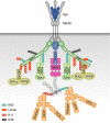
Figure 4. Ubiquitin and immune system modulation
Cells of both the innate and adaptive arms of the immune system are crucial for immune reactivity and self-tolerance. Defects in the expression of E3 ligases or DUBs can lead to deregulation of these processes which can resulting in autoimmunity or autoinflammation. Autoimmune disorders are characterised by disturbed function of mTEC and DCs (impairment in their presentation of self-peptides to T cells; highlighted in red) or by perturbed peripheral tolerance, which will lead to the development of self-reactive T and/or B cells (highlighted in red), production of autoantibodies and subsequent tissue destruction. During autoinflammation, the innate immune cells are hyperactive (highlighted in red) and release pro-inflammatory cytokines, including IL-1β and TNF, which will result in the development of auto-reactive inflammation.

References

    1. Kho Y. A tagging-via-substrate technology for detection and proteomics of farnesylated proteins. Proc Natl Acad Sci USA. 2004;101:12479–12484. - PMC - PubMed
    1. Mann M, Jensen ON. Proteomic analysis of post-translational modifications. Nat Biotechnol. 2003;21:255–261. - PubMed
    1. Ciechanover A, Elias S, Heller H, Hershko A. ‘Covalent affinity’ purification of ubiquitin-activating enzyme. J Biol Chem. 1982;257:2537–2542. - PubMed
    1. Hershko A, Heller H, Elias S, Ciechanover A. Components of ubiquitin-protein ligase system. Resolution, affinity purification, and role in protein breakdown. J Biol Chem. 1983;258:8206–8214. - PubMed
    1. Kirisako T. A ubiquitin ligase complex assembles linear polyubiquitin chains. EMBO J. 2006;25:4877–4887. - PMC - PubMed

Publication types

LinkOut - more resources