. 2014 Feb 15;306(4):L351-60.
doi: 10.1152/ajplung.00264.2013. Epub 2013 Dec 27.

Inducible HSP70 regulates superoxide dismutase-2 and mitochondrial oxidative stress in the endothelial cells from developing lungs

Affiliations

Inducible HSP70 regulates superoxide dismutase-2 and mitochondrial oxidative stress in the endothelial cells from developing lungs

Adeleye J Afolayan et al. Am J Physiol Lung Cell Mol Physiol. .

Abstract

Superoxide dismutase 2 (SOD-2) is synthesized in the cytosol and imported into the mitochondrial matrix, where it is activated and functions as the primary antioxidant for cellular respiration. The specific mechanisms that target SOD-2 to the mitochondria remain unclear. We hypothesize that inducible heat shock protein 70 (iHSP70) targets SOD-2 to the mitochondria via a mechanism facilitated by ATP, and this process is impaired in persistent pulmonary hypertension of the newborn (PPHN). We observed that iHSP70 interacts with SOD-2 and targets SOD-2 to the mitochondria. Interruption of iHSP70-SOD-2 interaction with 2-phenylethylenesulfonamide-μ (PFT-μ, a specific inhibitor of substrate binding to iHSP70 COOH terminus) and siRNA-mediated knockdown of iHSP70 expression disrupted SOD-2 transport to mitochondria. Increasing intracellular ATP levels by stimulation of respiration with CaCl2 facilitated the mitochondrial import of SOD-2, increased SOD-2 activity, and decreased the mitochondrial superoxide (O2(·-)) levels in PPHN pulmonary artery endothelial cells (PAEC) by promoting iHSP70-SOD-2 dissociation at the outer mitochondrial membrane. In contrast, oligomycin, an inhibitor of mitochondrial ATPase, decreased SOD-2 expression and activity and increased O2(·-) levels in the mitochondria of control PAEC. The basal ATP levels and degree of iHSP70-SOD-2 dissociation were lower in PPHN PAEC and lead to increased SOD-2 degradation in cytosol. In normal pulmonary arteries (PA), PFT-μ impaired the relaxation response of PA rings in response to nitric oxide (NO) donor, S-nitroso-N-acetyl-penicillamine. Pretreatment with Mito-Q, a mitochondrial targeted O2(·-) scavenger, restored the relaxation response in PA rings pretreated with PFT-μ. Our observations suggest that iHSP70 chaperones SOD-2 to the mitochondria. Impaired SOD-2-iHSP70 dissociation decreases SOD-2 import and contributes to mitochondrial oxidative stress in PPHN.

Keywords: nitric oxide; oxidative stress; persistent pulmonary hypertension of the newborn; pulmonary artery endothelium; vasodilation.


Figures

Fig. 1.
Fig. 1.
Colocalization of superoxide dismutase-2 (SOD)-2 and inducible heat shock protein 70 (iHSP70) in the lung tissue was assessed using double labeling and immunofluorescence method. A: FITC (A,A, green fluorescence) was used to label SOD-2 and red fluorescence for iHSP70 (A,B), and the merged picture represented by yellow color (A,C) indicates SOD2-iHSP70 colocalization. SOD-2 colocalizes with iHSP70 in the alveoli, pulmonary arteries (PA) and pulmonary veins (PV), and airway epithelial cells (A). iHSP70 expression and SOD2-iHSP70 association were increased in persistent pulmonary hypertension of the newborn (PPHN). The SOD2-iHSP70 association, evaluated by ratio of iHSP70/SOD-2, was higher in PA endothelial cells (PAEC) cytosol (n = 5, P < 0.05) (B) and PA homogenates (n = 4, P < 0.05) (C) but not in whole lung (n = 4, P = 0.85) in PPHN. D: iHSP70 expression was increased in PPHN PA homogenates (n = 4, P < 0.05). *P < 0.05 from corresponding controls.
Fig. 2.
Fig. 2.
Effect of inhibition of SOD2-iHSP70 interaction by 2-phenylethylenesulfonamide-μ (PFT-μ) on mitochondrial SOD-2 levels (A), iHsp70 expression in cytosol (C), and effect of siRNA knockdown of iHSP70 on mitochondrial SOD-2 levels (C). Proinhibitin-2 was used as loading control for mitochondrial and β-actin for cytosolic fractions, respectively. A: incubation of control PAEC with 5 μM PFT-μ for 18 h decreased SOD-2 levels in the mitochondria compared with untreated PAEC (n = 5, P < 0.01). B: PFT-μ did not change the basal expression of iHSP70 in treated PAEC (n = 5, P = 0.7). C: siRNA knockdown of iHSP70 expression decreased SOD-2 protein levels in the mitochondria of control cells (n = 3, P < 0.05). D: localization and quantification of SOD-2 in the mitochondria of intact cell following iHSP70 knockdown was assessed using double labeling and immunofluorescence method. Mitotracker red was used to label the mitochondria (a and d), FITC (green fluorescence) for SOD-2 (b and e), and merged pictures are represented by yellow color for localization of SOD-2 in the mitochondria (c and f). iHSP70 knockdown decreased fluorescence signals (e) and localization of SOD-2 to the mitochondria (f) when compared with negative control (b and c), (n = 3, P < 0.05). E: siRNA decreased iHSP70 expression by 60% (n = 3, P < 0.01), #P < 0.05 from control. IOD, integrated optical density.
Fig. 3.
Fig. 3.
A: iHSP70 regulates mitochondrial O2·− levels. Mitochondrial O2·− level was assessed by mitochondrial-targeted hydro-ethidine (mito-HE, MitoSOX) fluorescence as shown in the representative photomicrographs. 1 μM Antimycin A (AA) (a complex III inhibitor) increased Mito-HE fluorescence signals (positive control, II) when compared with untreated control (I); however, 100 U/ml PEG-SOD decreased the fluorescence signals in Antimycin A-treated cell (n = 4, P < 0.05, III). Treatment of control PAEC with 10 μM phorbol 12-myristate 13-acetate (PMA) (NADPH oxidase agonist) has no effect on MitoSOX fluorescence signals (negative control, IV). A: PFT-μ decreased iHSP70-SOD2 association when compared with control (P < 0.05, n = 3). Interruption of SOD2-iHSP70 interaction by 5 μM PFT-μ (VI), siRNA knockdown of iHSP70 expression (VII), and inhibition of mitochondrial ATPase activity by 10 μM oligomycin (VIII) increased Mito-HE fluorescence signals compared with control (V). The baseline Mito-HE fluorescence signal is higher PPHN (IX), when compared with control (V); however, increasing de novo ATP synthesis by treating PPHN PAEC with 10 μM CaCl2 decreased Mito-HE signals (X) (n = 4, P < 0.05). B and C: summary of data shown in A as mean ± SE. *,**P < 0.05 from control.
Fig. 4.
Fig. 4.
Oxidation of Mito-HE by O2·− was also measured by high-performance liquid chromatography as the level of 2-OH-Mito-E+ to verify MitoSOX fluorescence. A: interruption of SOD2-iHSP70 interaction by 5 μM PFT-μ, siRNA knockdown of iHSP70, and inhibition of mitochondrial ATPase activity with 10 μM oligomycin increased 2-OH-Mito-E+ in PAEC. The baseline level of 2-OH-Mito-E+ in PPHN is higher than control. However, 10 μM CaCl2 decreased 2-OH-Mito-E+ levels in PPHN PAEC (n = 3, P < 0.05). B: effects of intracellular ATP and interruption of SOD2-iHSP70 interaction on H2O2 production. H2O2 release from mitochondria was measured using Amplex red-horseradish peroxidase reagent in intact mitochondria, isolated from PAEC. H2O2 detected was corrected against mitochondrial protein content. Interruption of SOD2-iHSP70 interaction by PFT-μ, depletion of iHSP70 protein by siRNA knockdown, and inhibition of mitochondrial ATPase activity by oligomycin decreased H2O2 formation and release from the mitochondria of control PAEC. C: H2O2 in PPHN is lower than control; however, treatment of PPHN PAEC with 10 μM CaCl2 for 30 min increased H2O2 release from mitochondria of PPHN PAEC (n = 3, P < 0.05), **P < 0.05 from untreated PPHN, *P < 0.05 from control.
Fig. 5.
Fig. 5.
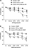
Interruption of SOD2-iHSP70 interaction attenuated ṄO-mediated vasodilator response of control PA. Data are means ± SE from 12 PFT-μ-treated rings and 12 untreated rings. The basal ring tension after preconstriction with norepinephrine was normalized to 100%. Disruption of iHSP70-SOD2 interaction by PFT-μ decreased normal PA relaxation response to ATP (A) and to ṄO donor, S-nitroso-N-acetyl-penicillamine (SNAP, B). Pretreatment with Mito-Q improved the responses to ATP (A) and SNAP (B) in PFT-μ-treated rings. *P < 0.05 from PFT-μ alone.
Fig. 6.
Fig. 6.
Effect of ATP on SOD-2 activity in PAEC. SOD-2 activity was measured by a colorimetric method and corrected for protein concentration. A: ATP increased SOD-2 activity in a dose-dependent manner in PPHN-PAEC lysates (n = 6, P < 0.05). B: incubation of control PAEC with 10 μM oligomycin for 2 h attenuated SOD-2 activity; however, 10−5 M ATP restored SOD-2 activity in oligomycin-treated PAEC. C: interruption of SOD2-iHSP70 interaction by 5 μM PFT-μ (n = 5, P < 0.05). D: siRNA knockdown of iHSP70 expression (n = 4, P < 0.05) decreased SOD-2 activity and ATP responses in control PAEC. *P < 0.05 from control, *#&P < 0.05 from ATP105M, *¶P < 0.05 from control and oligomycin-treated control cells.
Fig. 7.
Fig. 7.
Effects of ATP on SOD-2 interactions with chaperone in the cytosol and mitochondria of PAEC. iHSP70-SOD2 association in the cytosolic and mtHSP70-SOD2 in the mitochondrial fractions was assessed by immunoprecipitation, and the results are represented by the ratio of iHSP70 or mt-HSP70 to SOD-2. A: exogenous ATP (10 μM) decreased iHSP70-SOD2 association after addition to cell lysates in the cytosolic fraction of PPHN PAEC (n = 5, P < 0.05). B: in the mitochondrial fractions, exogenous ATP increased mtHSP70-SOD2 association in PPHN (n = 4, P < 0.05). The effect of ATP on SOD-2 import was assessed by Western blots to quantify changes in SOD-2 protein level in the mitochondria after ATP treatment and corrected against proinhibitin-2, the loading control for mitochondrial protein. C: ATP increased SOD-2 levels in the mitochondria of PPHN PAEC (n = 5, P < 0.05). D: incubation of control PAEC with 10 μM oligomycin for 2 h decreased SOD-2 levels in the mitochondria when compared with ATP-treated samples and untreated controls (n = 5, P < 0.05). #&P < 0.005 from control and oligomycin-treated control.
Fig. 8.
Fig. 8.
Coupling of mitochondrial respiration and ATP synthesis is depressed and increases SOD-2 degradation in PPHN. A: ATP levels were lower, and the levels of ADP and AMP were higher in PPHN-PAEC when compared with control PAEC (n = 3, P < 0.05). #φπP < 0.05 from control. B: representative immunoblots of ubiqitinated SOD-2 and summary data for ratio of ubiquitinated to total SOD2 are shown for control and PPHN PAEC. SOD-2 degradation was assessed by quantifying ubiqitinated fraction of total SOD-2 protein by immunoprecipitation. Poly-ubiqitinated SOD-2 is higher in PPHN PAEC than controls (n = 3, P < 0.05), #P < 0.05 from control.

References

    1. Abman SH, Chatfield BA, Hall SL, McMurtry IF. Role of endothelium-derived relaxing factor during transition of pulmonary circulation at birth. Am J Physiol Heart Circ Physiol 259: H1921–H1927, 1990 - PubMed
    1. Afolayan AJ, Eis A, Teng RJ, Bakhutashvili I, Kaul S, Davis JM, Konduri GG. Decreases in manganese superoxide dismutase expression and activity contribute to oxidative stress in persistent pulmonary hypertension of the newborn. Am J Physiol Lung Cell Mol Physiol 303: L870–L879, 2012 - PMC - PubMed
    1. Azem A, Oppliger W, Lustig A, Jeno ÈP, Feifel B, Schatz G, Horst M. The mitochondrial hsp70 chaperone system, Effect of adenine nucleotides, peptide substrate and mGrpE on the oligomeric state of mhsp70. J Biol Chem 272: 20901–20906, 1997 - PubMed
    1. Beddoe T, Lithgow T. Delivery of nascent polypeptides to the mitochondrial surface. Biochim Biophys Acta 1592: 35–39, 2002 - PubMed
    1. Bhatt DP, Chen X, Geiger JD, Rosenberger TA. A sensitive HPLC-based method to quantify adenine nucleotides in primary astrocyte cell cultures. J Chromatogr B Analyt Technol Biomed Life Sci 889: 110–115, 2012 - PMC - PubMed

Publication types

MeSH terms