

The periductal channels of the endolymphatic duct, hydrodynamic implications
- PMID: 24376120
- PMCID: PMC4034528
- DOI: 10.1177/0194599813516420
The periductal channels of the endolymphatic duct, hydrodynamic implications
Abstract
Objective: To describe the anatomy of a small network of channels surrounding the human endolymphatic duct.
Study design: Archival temporal bone sections and a surgical specimen were studied using a variety of techniques.
Setting: Temporal bone laboratory of the House Research Institute.
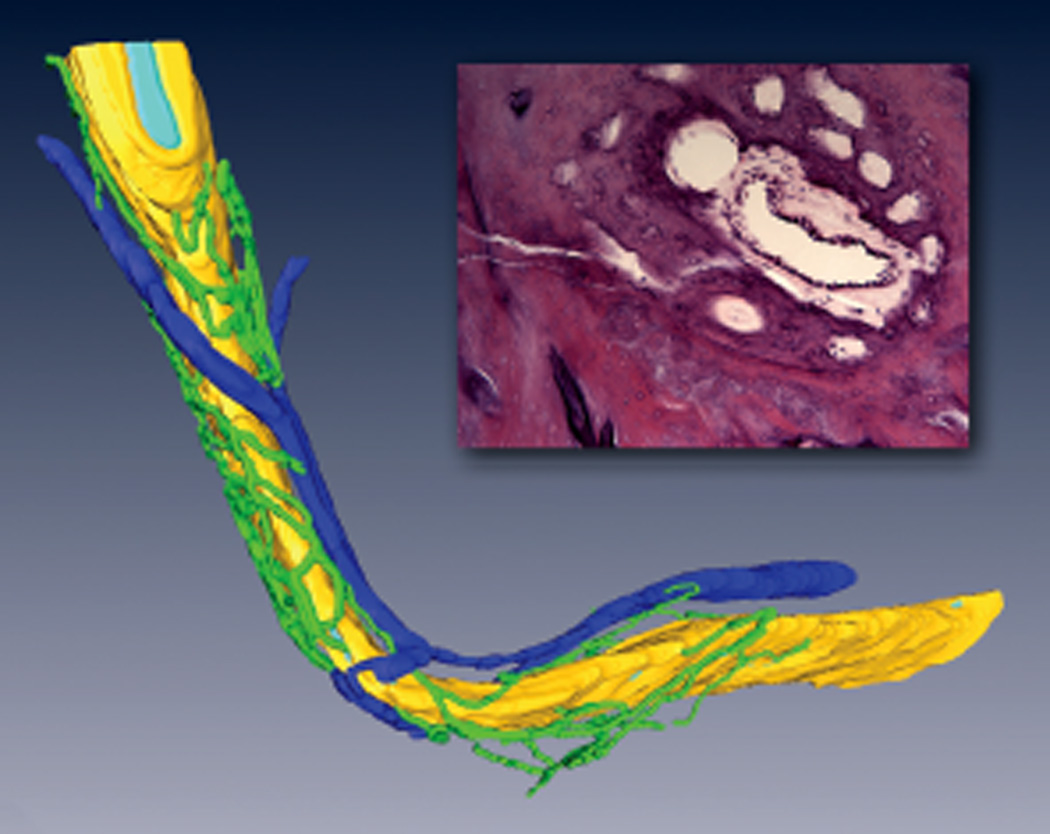
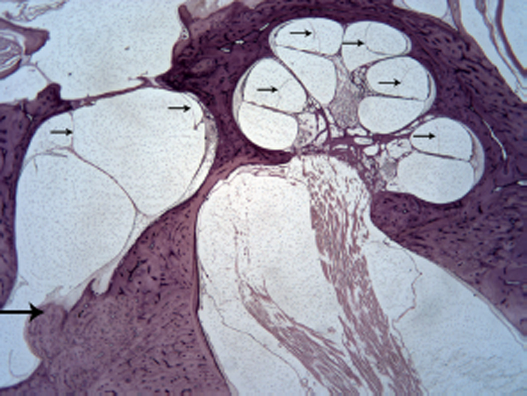
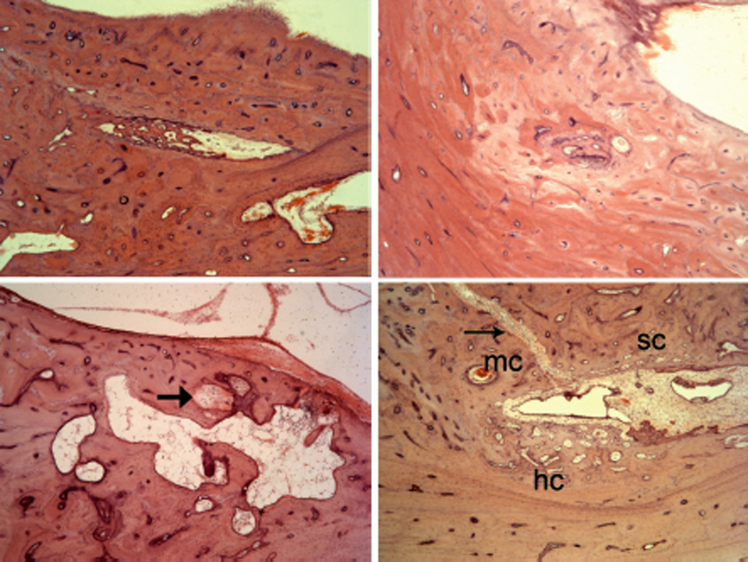
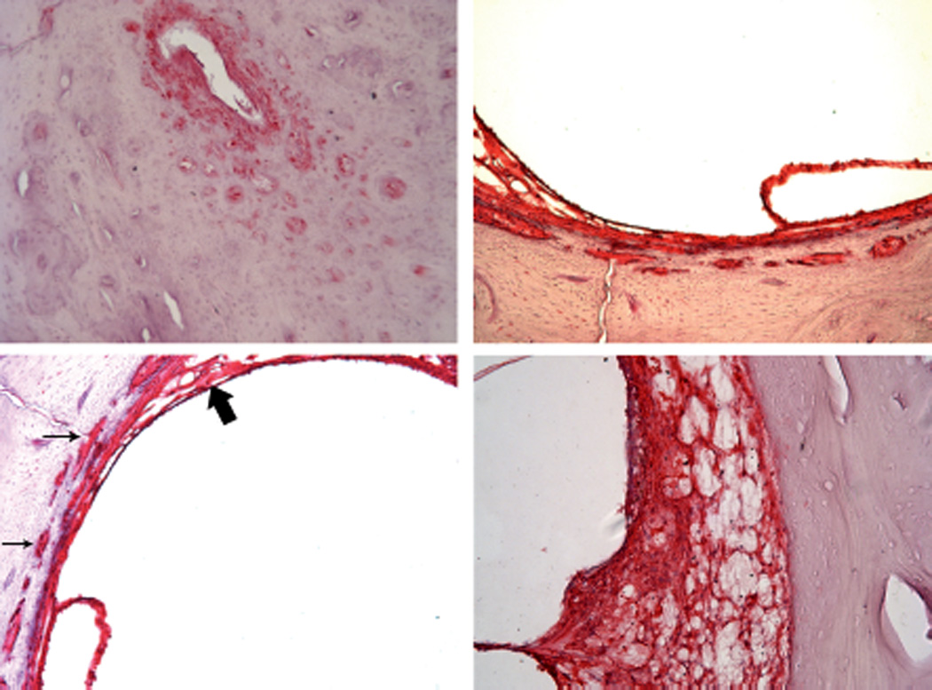
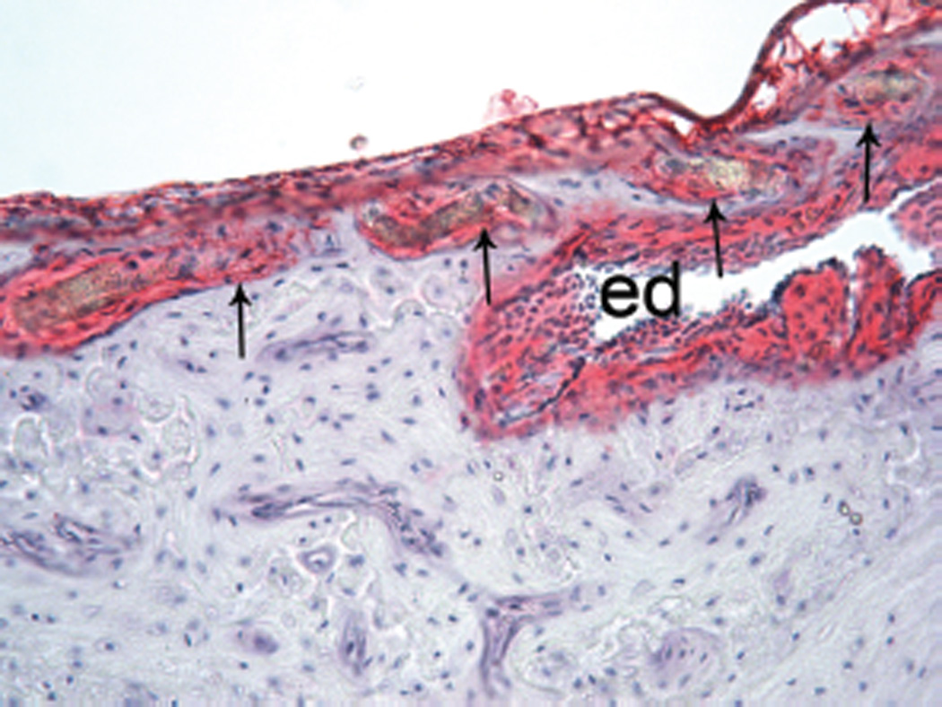
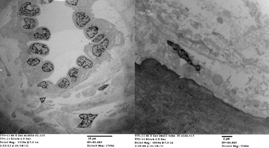
Subjects and methods: Archival temporal bone sections were examined by light microscopy, 3D reconstruction, and immunohistochemical labeling. A surgical specimen was examined using electron microscopy. Sections from temporal bones with blocked endolymphatic ducts or amputated sacs were examined for the manifestations of endolymphatic hydrops.
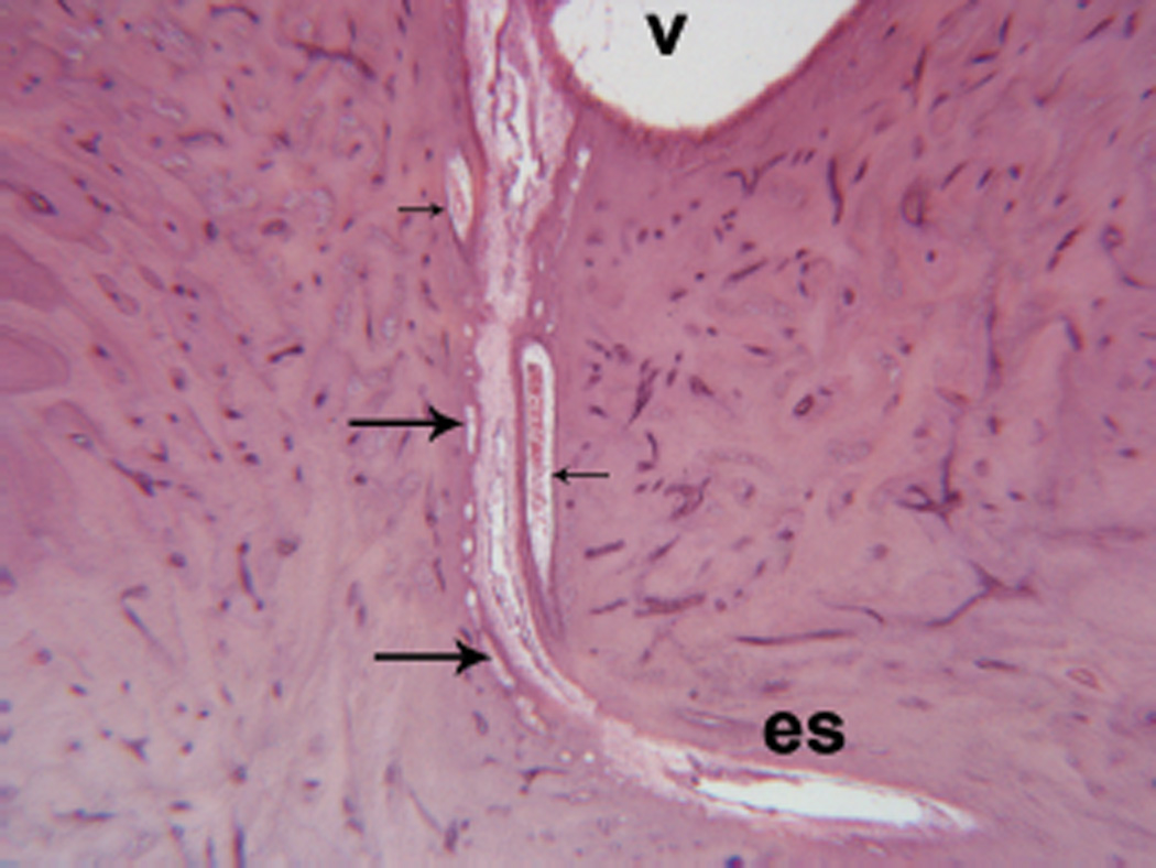
Results: Peri-endolymphatic duct channels were found to extend from the proximal cisternal part of the endolymphatic sac to the supporting tissue of the saccule and utricle. Tissue in the channels, as seen by conventional and electron microscopy, is continuous with and identical with the tissue surrounding the endolymphatic duct. Tissue in the channels labels with the S100 antibody similar to the spiral ligament and supporting tissue of the vestibular end organs and suggests a neural crest origin, as did the presence of melanocytes. Obstruction of the endolymphatic duct resulted in endolymphatic hydrops whereas amputation of the sac did not.
Conclusion: Endolymph is probably absorbed in the endolymphatic duct. The peri-endolymphatic duct channels that extend from the proximal sac to the supporting tissue of the saccule label with the S100 antibody and contain melanocytes suggest a neural crest origin and involvement in fluid and potassium hydrodynamics similar to those described for the similarly staining spiral ligament of the cochlea.
Keywords: channels; endolymphatic duct.
Figures

References
-
- Ng M, Linthicum FH., Jr Periductal vessels of the endolymphatic duct. Acta Otolaryngol (Stockh) 1992;112:649–657. - PubMed
-
- Somdas MA, Li PM, Whiten DM, Eddington DK, Nadol JB., Jr Quantitative evaluation of new bone and fibrous tissue in the cochlea following cochlear implantation in the human. Audiol Neurootol. 2007;12:277–284. - PubMed
Publication types
MeSH terms
Grants and funding
LinkOut - more resources
Full Text Sources
Other Literature Sources
