. 2013 Dec 20;8(12):e80288.
doi: 10.1371/journal.pone.0080288. eCollection 2013.

IL-10 is significantly involved in HSP70-regulation of experimental subretinal fibrosis

Affiliations

IL-10 is significantly involved in HSP70-regulation of experimental subretinal fibrosis

Yang Yang et al. PLoS One. .

Erratum in

  • PLoS One. 2014;9(3):e91197

Abstract

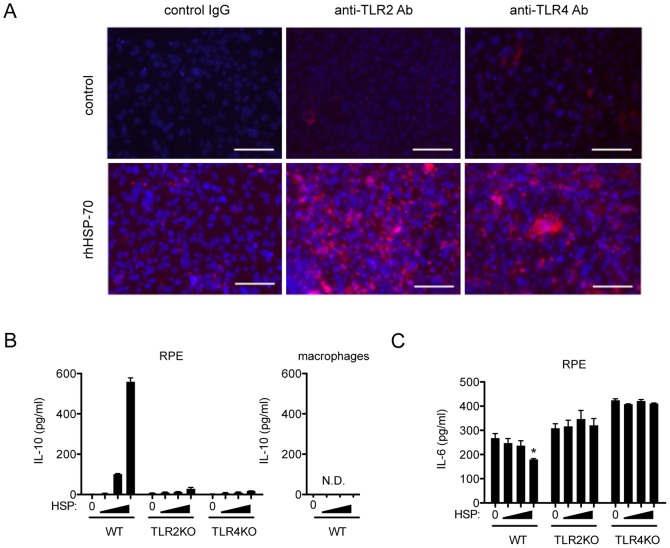
Subretinal fibrosis is directly related to severe visual loss, especially if occurs in the macula, and is frequently observed in advanced age-related macular degeneration and other refractory eye disorders such as diabetic retinopathy and uveitis. In this study, we analyzed the immunosuppressive mechanism of subretinal fibrosis using the novel animal model recently demonstrated. Both TLR2 and TLR4 deficient mice showed significant enlargement of subretinal fibrotic area as compared with wild-type mice. A single intraocular administration of heat shock protein 70 (HSP70), which is an endogenous ligand for TLR2 and TLR4, inhibited subretinal fibrosis in wild-type mice but not in TLR2 and TLR4-deficient mice. Additionally, HSP70 induced IL-10 production in eyes from wild-type mice but was impaired in both TLR2- and TLR4-deficient mice, indicating that HSP70-TLR2/TLR4 axis plays an immunomodulatory role in subretinal fibrosis. Thus, these results suggest that HSP70-TLR2/TLR4 axis is a new therapeutic target for subretinal fibrosis due to prognostic CNV.


Conflict of interest statement

Competing Interests: The authors have declared that no competing interests exist.

Figures

Figure 1
Figure 1. Both TLR2 and TLR4 signaling pathways are significantly involved in the formation of subretinal fibrosis.
Subretinal fibrotic models were generated as previously described . Briefly, laser photocoagulation (wave length 532 nm, 0.1 s, spot size 75 um, power 200 mW) was performed to the retina to make subretinal bubble and rupture Bruch membrane in WT mice. PECs (4×10⁴) from indicated mice collected as Materials and Methods without any stimulation were inoculated into the subretinal space of indicated mice. (A) PECs from each WT, TLR2KO and TLR4KO mice were inoculated into subretinal space of WT mice (n = 5). (B) PECs from WT mice were inoculated into subretinal space of WT, TLR2KO and TLR4KO mice. (n = 5) (C) With neutralizing antibody anti-TLR2, anti-TLR4 antibody, control IgG or without any reagent (naïve), PECs from WT mice were inoculated into subretinal space of WT mice. After 7 days, eyes were enucleated, and choroidal flatmounts were prepared and stained with anti-GFAP antibody. The areas of subretinal fibrosis were measured by ImageJ. *p<0.05 versus control; double asterisks, **p<0.005 versus control. (n = 5) Data represents mean ± SEM. (D) Histological cross sections from (C) were stained with anti-GFAP antibody. Representative images were shown. Scale bars, 500 µm. Results are represents as mean ± SEM.
Figure 2
Figure 2. Role of HSP70 in the formation of subretinal fibrosis.
(A) 0, 24, 48 and 72 hours after PEC inoculation, total RNA was extracted from the eyes and the amounts of HSP70 mRNA were assessed by quantitative real-time RT-PCR. (B) 72 hours after PEC inoculation following intravitreal HSP70 injection, eyes were enucleated, then histological cross sections at the site of subretinal fibrosis were stained with control IgG, anti-TLR2 and anti-TLR4 antibody, respectively. Representative images were shown. Scale bars, 100 µm. (C) Indicated hours after PECs inoculation, total RNA was extracted from eyes of each WT (WT PEC into lasered WT mice), TLR2KO (TLR2KO PEC into lasered TLR2KO) and TLR4KO (TLR4KO PEC into lasered TLR4KO) mice. The amount of HSP70 mRNA were evaluated by quantitative real-time PCR (n = 9). (D) Recombinant human HSP70 or control PBS was injected into vitreous cavity of WT, TLR2KO and TLR4KO mice 2 hours after PEC inoculation. After 7 days, eyes were enucleated, and choroidal flatmounts were prepared and stained with anti-GFAP antibody. The area of subretinal fibrosis were measured by ImageJ. (n = 5) (E) Representative images of choroidal flatmount stained with anti-GFAP antibody. Scale bars, 500 µm. Results are represents as mean ± SEM.
Figure 3
Figure 3. Implications of IL-10 and its possible association of HSP70 in the formation of subretinal fibrosis.
(A) PECs from WT mice were inoculated into the subretinal space of WT mice. 0, 24, 72 hours after, total RNA was extracted from the eyes and the amounts of IL-10 mRNA were assessed by quantitative real-time RT-PCR. (n = 6) (B) PECs from WT were inoculated to each WT, TLR2 and TLR4 mice. 48 hours after PEC inoculation, total RNA was extracted from the eyes of each mice and the amount of IL-10 mRNA was assessed by quantitative real-time RT-PCR. (n = 6) (C) 2 hours after PEC inoculation, each control IgG, anti-TLR2 and anti-TLR4 neutralizing antibody was injected into vitreous cavity of WT mice. After 48 hours, total RNA was extracted from the eyes and the amounts of IL-10 mRNA were asessed by quantitative real-time RT-PCR. (n = 5) (D) 2 hours after PEC inoculation into subretinal space of WT mice, recombinant human HSP70 or control PBS was injected into vitreous cavity. After 48 hours, eyes were enucleated, total protein was extracted from the retina-choroid-RPEs and amounts of IL-10 were quantified by ELISA (n = 12). Results are represents as mean ± SEM.
Figure 4
Figure 4. IL-10 is responsible for subretinal formation.
(A) Neutralizing anti-IL-10 antibodies were inoculated with PECs from WT to subretinal space of WT mice. (B) 2 hours after PEC inoculation, recombinant IL-10 was intravitreally injected. After 7 days, eyes were enucleated, and choroidal flatmounts were prepared and stained with anti-GFAP antibody. The areas of subretinal fibrosis were measured by ImageJ. Results are represents as mean ± SEM.
Figure 5
Figure 5. HSP70 induces IL-10 production by cultured RPEs but not by macrophages.
(A) Cultured RPE cells in the presence or absence of rhHSP70 were stained with WT, anti-TLR2 and anti-TLR4 antibody, respectively. Representative images were shown. Scale bars, 100 µm. (B) RPE cells from each WT, TLR2 and TLR4 (left) and macrophages (right) were stimulated with 0.1, 0.3 and 1 ng/ml of recombinant human Hsp70 for 48hours, then supernatants were harvested and the levels of IL-10 were quantified by ELISA. (C) IL-6 was quantified by ELISA from same supernatants from (Figure B, right) RPE cells. Each experiment was representative of at least two experiments with similar results. Results are represents as mean ± SEM.
Figure 6
Figure 6. The effects of human Hsp70 on IL-10 production in subretinal fibrotic model are not due to the contamination of LPS.
(A) 2 hours after PECs-inoculation, 50 ng (25 µg/ml, 2 µl) of lipopolysaccharides (LPS) or PBS was administered into vitreous cavity in WT mice. After 7 days, eyes were enucleated, choroidal flatmounts were prepared and stained with anti-GFAP antibody. The areas of subretinal fibrosis were measured by ImageJ. Results are represents as mean ± SEM. (B) Histological cross sections from (A) were stained with anti-GFAP antibody. Representative images were shown. Scale bars, 100 µm.
Figure 7
Figure 7. Suppressive effect of HSP70 and IL-10 on α-SMA expression in the RPE cells cultured with PECs.
RPE cells were prepared from C57BL/6 mice and cultured approximately 2 weeks until becoming confluent in a 24 well plate. PECs were added to the primary culture in the absence or presence of HSP70 (1 ng/ml) or IL-10 (10 ng/ml). 48 hours later, cells were stained by FITC-conjugated α-SMA and PE-conjugated anti-F4/80 antibody at 4°C for 24 hours. All samples were counterstained with DAPI, mounted, and subjected to fluorescence microscopy. Representative images of α-SMA-stained RPE cells (green) and F4/80-stained macrophages (red) on the dish were shown.
Figure 8
Figure 8. Possible mechanism of HSP70 in the regulation of subretinal formation.
Intraocular expression of HSP70 activates TLR2 and TLR4 signaling cascade in RPEs but not in macrophages, results in the regulation of subretinal formation via production of IL-10.

Similar articles

Cited by

References

    1. Bressler NM (2004) Age-related macular degeneration is the leading cause of blindness… JAMA. 291: 1900–1901. - PubMed
    1. Miller JW (2011) Treatment of age-related macular degeneration: Beyond VEGF. Jpn J Ophthalmol 54: 523–528. - PubMed
    1. Stramer BM, Mori R, Martin P (2007) The inflammation-fibrosis link? A Jekyll and Hyde role for blood cells during wound repair. J Invest Dermatol 127: 1009–1017. - PubMed
    1. van Eden W, van der Zee R, Prakken B (2005) Heat-shock proteins induce T-cell regulation of chronic inflammation. Nat Rev Immunol 5: 318–330. - PubMed
    1. Ludwig D, Stahl M, Ibrahim ET, Wenzel BE, Drabicki D, et al. (1999) Enhanced intestinal expression of heat shock protein 70 in patients with inflammatory bowel diseases. Dig Dis Sci 44: 1440–1447. - PubMed

MeSH terms

LinkOut - more resources