

A meta-analysis of anti-vascular endothelial growth factor remedy for macular edema secondary to central retinal vein occlusion
- PMID: 24376538
- PMCID: PMC3871640
- DOI: 10.1371/journal.pone.0082454
A meta-analysis of anti-vascular endothelial growth factor remedy for macular edema secondary to central retinal vein occlusion
Abstract
Background: Central retinal vein occlusion (CRVO) associates with severe vision outcome and no proven beneficial treatment. Our meta-analysis intended to appraise the efficacy and safety of anti-vascular endothelial growth factor (anti-VEGF) agents in macular edema (ME) following CRVO.
Methods: Data were collected and analyzed by Review Manager 5.2.1. We employed a random-effects model to eliminate between-study heterogeneity. Nfs (called fail-safe number) was calculated to evaluate the publication bias.
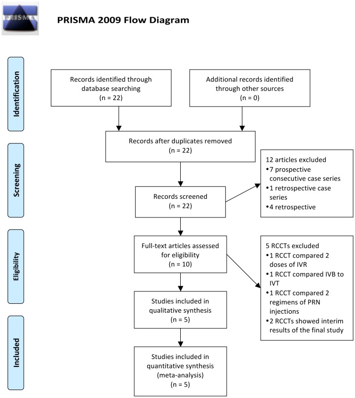
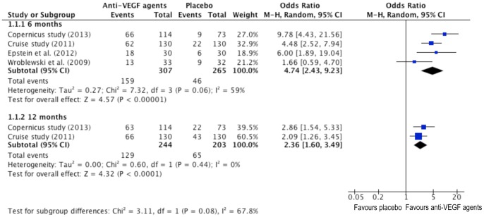
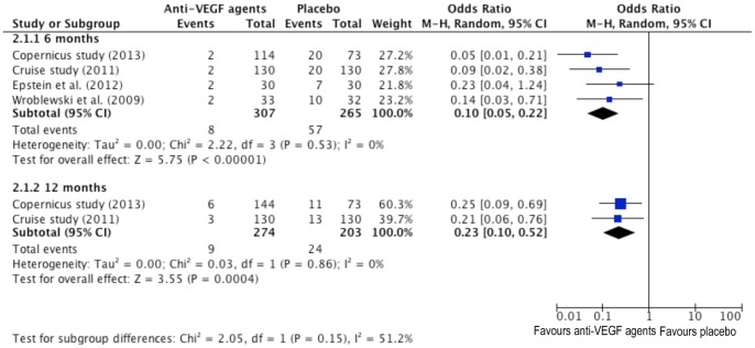
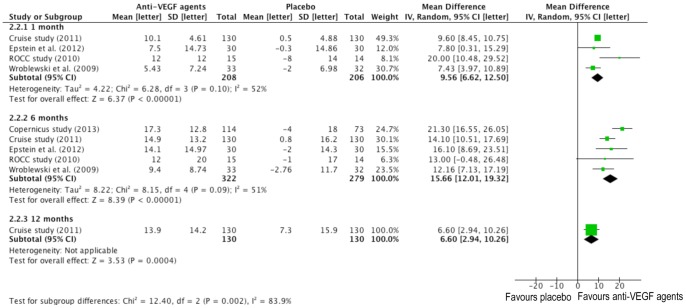
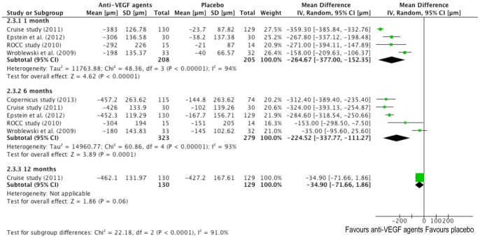
Results: We included 5 trials consisting 323 cases and 281 controls. Primary outcomes showed that overall comparison of anti-VEGF agents with placebo control yielded a 374% and 136% increased tendency for a gain of 15 letters or more on Early Treatment Diabetic Retinopathy Study (ETDRS) chart (95% confidence interval [95% CI]: 2.43-9.23; P<0.00001; I(2) = 59%, 95% CI: 1.60-3.49; P<0.0001; I(2) = 0%, respectively) at 6 and 12 months. Secondary outcomes showed that a 90% and 77% decreased risk at 6 and 12 months for a loss of 15 letters or more. The overall mean difference showed a statistically significance in best-corrected visual acuity (BCVA) on each time point. However, changes of central retinal thickness (CRT) lost significance at 12 months after 6-month as-needed treatment. The incidence of adverse events (AEs) had no statistical difference between anti-VEGF and placebo groups. Subgroup analyses indicated that patients receiving Aflibercept got the highest tendency to gain 15 letters or more (OR = 9.78; 95% CI: 4.43-21.56; P<0.00001). Age controlled analysis suggested a weaken tendency of BCVA improvement in age over 50 (MD = 12.26; 95% CI: 7.55-16.98; P<0.00001). Subgroup analysis by clinical classification showed a strengthen difference of BCVA changes at 6 months in ischemic type (MD = 19.65 letters, 95% CI: 13.15 to 26.14 letters, P<0.00001).
Conclusions: Our results showed that anti-VEGF agents were superior to placebo in CRVO-ME treatment with no statistically significant AEs, especially in younger people and for ischemic type.
Conflict of interest statement
Figures
References
-
- Orth DH, Patz A (1978) Retinal branch vein occlusion. Surv Ophthalmol 22: 357–376. - PubMed
-
- Branch Vein Occlusion Study Group (1986) Argon laser scatter photocoagulation for prevention of neovascularization and vitreous hemorrhage in branch vein occlusion: a randomized clinical trial. Arch Ophthalmol 104: 34–41. - PubMed
-
- McAllister IL (2012) Central retinal vein occlusion: a review. Clin Experiment Ophthalmol 40: 48–58. - PubMed
-
- Hayreh SS (2005) Prevalent misconceptions about acute retinal vascular occlusive disorders. Prog Retin Eye Res 24: 493–519. - PubMed
Publication types
MeSH terms
Substances
LinkOut - more resources
Full Text Sources
Other Literature Sources
Research Materials
