. 2013 Dec 23;8(12):e82914.
doi: 10.1371/journal.pone.0082914. eCollection 2013.

Cyclic AMP-dependent resuscitation of dormant Mycobacteria by exogenous free fatty acids

Affiliations

Cyclic AMP-dependent resuscitation of dormant Mycobacteria by exogenous free fatty acids

Margarita Shleeva et al. PLoS One. .

Erratum in

  • PLoS One. 2014;9(5):e97206

Abstract

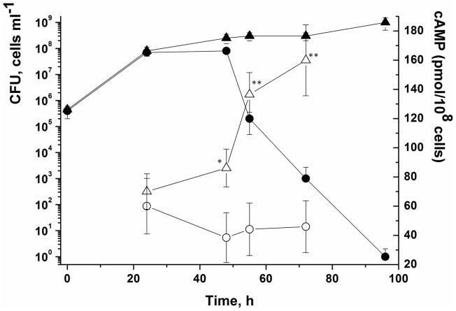
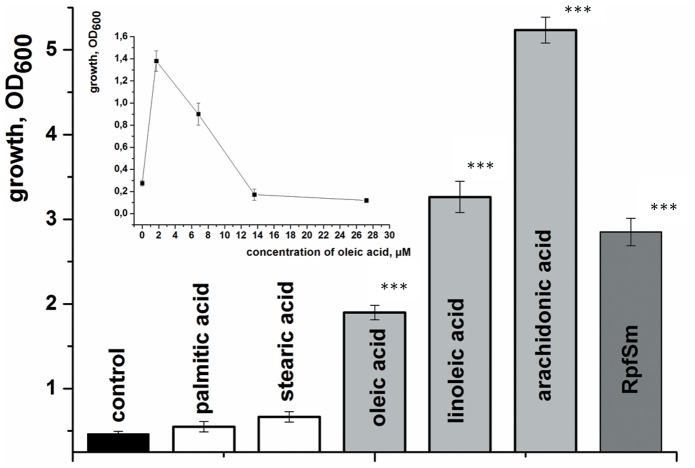
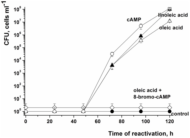
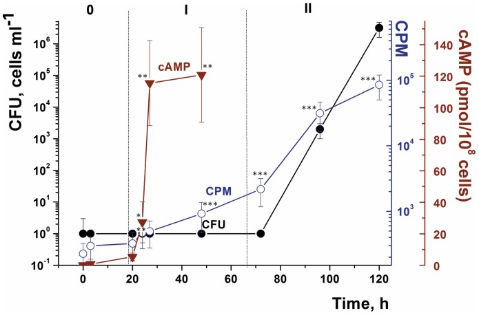
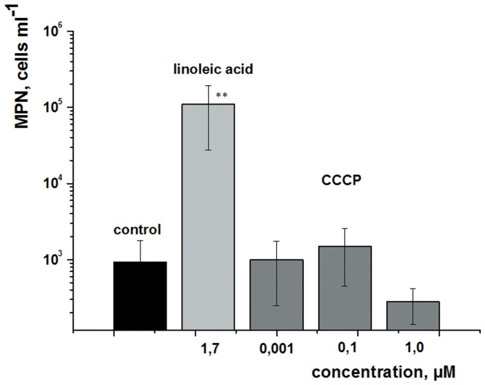
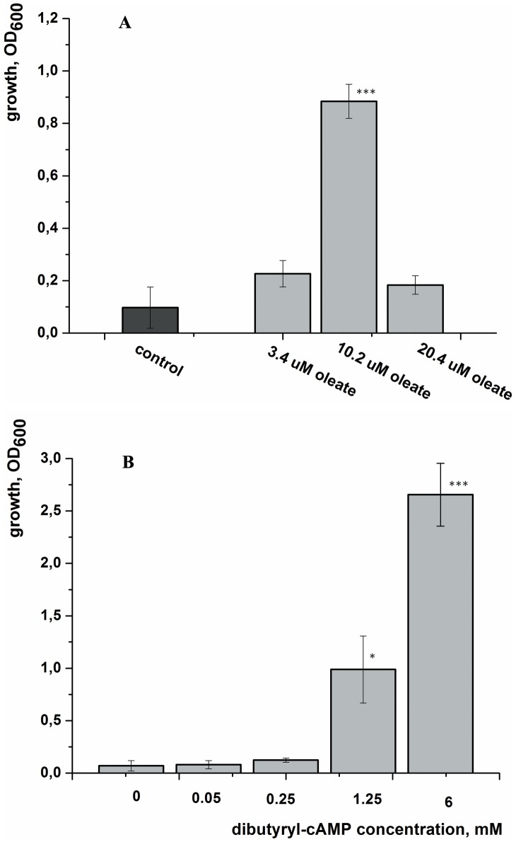
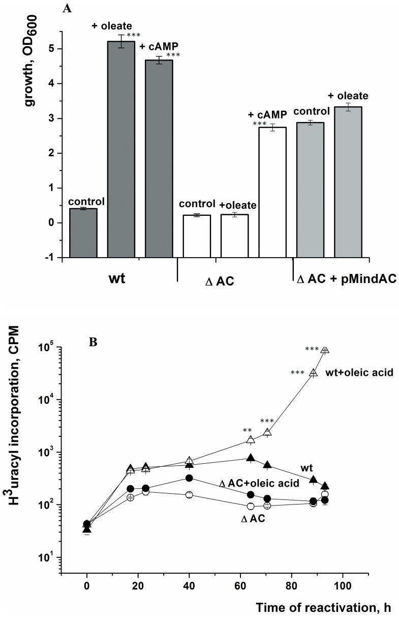
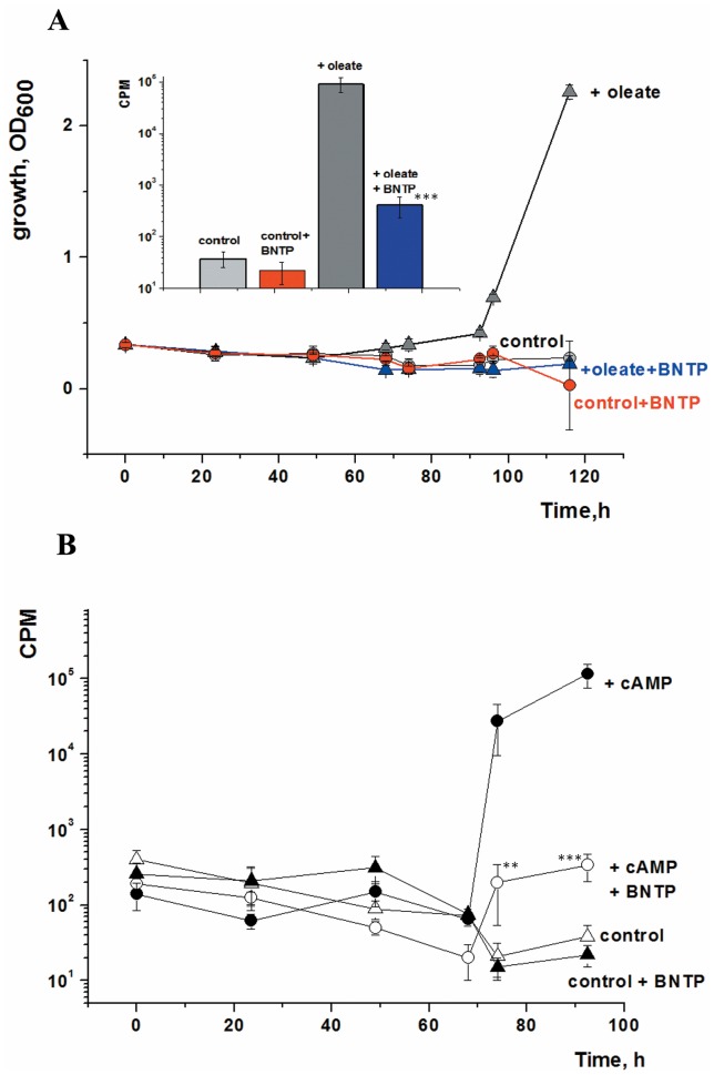
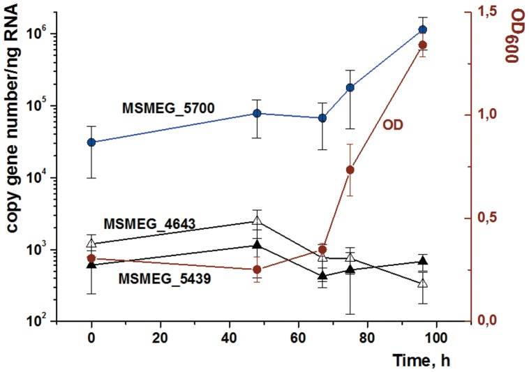
One third of the world population carries a latent tuberculosis (TB) infection, which may reactivate leading to active disease. Although TB latency has been known for many years it remains poorly understood. In particular, substances of host origin, which may induce the resuscitation of dormant mycobacteria, have not yet been described. In vitro models of dormant ("non-culturable") cells of Mycobacterium smegmatis (mc(2)155) and Mycobacterium tuberculosis H37Rv were used. We found that the resuscitation of dormant M. smegmatis and M. tuberculosis cells in liquid medium was stimulated by adding free unsaturated fatty acids (FA), including arachidonic acid, at concentrations of 1.6-10 µM. FA addition enhanced cAMP levels in reactivating M. smegmatis cells and exogenously added cAMP (3-10 mM) or dibutyryl-cAMP (0.5-1 mM) substituted for FA, causing resuscitation of M. smegmatis and M. tuberculosis dormant cells. A M. smegmatis null-mutant lacking MSMEG_4279, which encodes a FA-activated adenylyl cyclase (AC), could not be resuscitated by FA but it was resuscitated by cAMP. M. smegmatis and M. tuberculosis cells hyper-expressing AC were unable to form non-culturable cells and a specific inhibitor of AC (8-bromo-cAMP) prevented FA-dependent resuscitation. RT-PCR analysis revealed that rpfA (coding for resuscitation promoting factor A) is up-regulated in M. smegmatis in the beginning of exponential growth following the cAMP increase in lag phase caused by FA-induced cell activation. A specific Rpf inhibitor (4-benzoyl-2-nitrophenylthiocyanate) suppressed FA-induced resuscitation. We propose a novel pathway for the resuscitation of dormant mycobacteria involving the activation of adenylyl cyclase MSMEG_4279 by FAs resulted in activation of cellular metabolism followed later by increase of RpfA activity which stimulates cell multiplication in exponential phase. The study reveals a probable role for lipids of host origin in the resuscitation of dormant mycobacteria, which may function during the reactivation of latent TB.


Conflict of interest statement

Competing Interests: The authors have declared that no competing interests exist.

Figures

Figure 1
Figure 1. Hyper-expression of the MSMEG_4279 adenylyl cyclase abolishes the transition of M. smegmatis cells to the NC state.
M. smegmatis cells were inoculated into modified (lacking potassium ions) Hartman’s–de Bont (mHdeB) medium and incubated with strong aeration 200 rpm) at 37°C. Strains harbouring pMind (empty plasmid vector) or pMindAc (adenylyl cyclase hyper-expression plasmid) are denoted with circles and triangles, respectively. Samples were withdrawn periodically for CFU determination (closed symbols) and intracellular cAMP content (open symbols). This experiment was repeated three times with similar results; the error bars represent the standard error of the mean. Asterisks indicate significant difference in cAMP concentration between pMindAC cells and pMind cells by Student’s t-test.
Figure 2
Figure 2. Fatty acid-induced resuscitation of M. smegmatis NC cells.
NC cells were resuspended in reactivation medium to an initial OD600 ∼ 0.3 and resuscitated in batch format. The OD600 was measured after 5 d of resuscitation. Palmitic, stearic, oleic, linoleic and arachidonic acids were added at their optimum concentrations (4 µM, 4 µM 3.5 µM, 1.7 µM and 1.6 µM, respectively). The insert shows the concentration-dependence of oleic acid-mediated resuscitation. Each point represents the OD600 measurement after 5 d of resuscitation. This experiment was repeated three times with similar results; the error bars represent the standard error of the mean. Asterisks indicate that the results are significantly different from the control by Student’s t-test.
Figure 3
Figure 3. cAMP stimulates the resuscitation of M. smegmatis NC cells.
NC cells were obtained and resuscitated in batch format. The OD600 was measured after 5 d of resuscitation. The concentrations of oleic acid, linoleic acid, cAMP and 8-bromo-cAMP were 2 µM, 1.7 µM, 3 mM and 2 mM, respectively. This experiment was repeated three times with similar results; the error bars represent the standard error of the mean.
Figure 4
Figure 4. Comparison of the effects of linoleic acid and the uncoupler CCCP on the resuscitation of M. smegmatis NC cells.
The resuscitation of NC cells was performed in MPN format. Each dilution was supplemented with linoleic acid or CCCP at the concentrations indicated. The ordinate shows the number of potentially viable (resuscitated) cells per ml of the initial NC population. This experiment was repeated twice with similar results; the error bars represent the standard error of the mean. Asterisks indicate that the result is significantly different from the control by Student’s t-test.
Figure 5
Figure 5. Intracellular cAMP levels and 3H-uracil incorporation during oleic acid-induced resuscitation of M. smegmatis NC cells.
NC cells were obtained and resuscitated in batch mode. Oleic acid was added at a concentration of 3.5 µM. The intracellular level of cAMP was estimated after cells had been harvested and disrupted as described in the Materials and Methods. For samples taken during the first 48 h of resuscitation, metabolic activity was determined using 3H-uracil incorporation (denoted CPM on the Figure axis) as detailed in Materials and Methods. Dotted lines divide the overall process into three phases: 0 - true lag, I - metabolic activation, II - cell multiplication. This experiment was repeated three times with similar results. Error bars represent the standard error of the mean. Asterisks indicate that the results are significantly different from the values at zero time by Student’s t-test.
Figure 6
Figure 6. Fatty acid- and dibutyryl-cAMP-induced resuscitation of M. tuberculosis NC cells.
NC cells were obtained inoculated to an initial OD600 = 0.2 and resuscitated in batch format. The OD600 was measured after resuscitation for 20 d (A) or 25 d (B). This experiment was repeated two times with similar results; the error bars represent the standard error of the mean. Asterisks indicate that the results are significantly different from the control by Student’s t-test.
Figure 7
Figure 7. Involvement of the adenylyl cyclase encoded by MSMEG_4279 in the oleic acid-induced resuscitation of NC cells.
NC cells were obtained and resuscitated in batch mode. In part A, the OD600 was measured after resuscitation for 5 d for the wild type and the complemented strain, ΔAC(pMindAc), and after 11 d for the ΔAC knock-out strain. Oleic acid and cAMP were added at concentrations of 3.5 µM and 3 mM, respectively. In part B, samples were taken at intervals over the first four days from cultures of the wild type and the mutant, both with and without oleic acid, for the estimation of cellular metabolic activity (uracil incorporation). Asterisks in Fig. 6A indicate that the results are significantly different from the control for each strain by Student’s t-test. Asterisks in Fig. 6B indicate significant difference between wt cells and wt+oleic acid by Student’s t-test.
Figure 8
Figure 8. Oleic acid-mediated resuscitation of M. smegmatis NC cells is Rpf-dependent.
NC cells were resuscitated in batch mode in the presence and the absence of both oleic acid (3.5 µM) (A) or cAMP (3.0 mM) (B) and the Rpf inhibitor, BNPT (1 µg/ml) in all four possible combinations. BNPT was added at zero time and again after 48 h of incubation. Samples were withdrawn periodically for OD600 determination (A) and estimation of metabolic activity using 3H-uracil incorporation (B) The insert to part A shows the level of uracil incorporation in the four cultures measured after 92 h of incubation. The error bars represent the standard error of the mean. Asterisks indicate significance between 3H-uracil incorporation by cells incubated in the presence of oleate vs both oleate and BNTP (A, insert) or in the presence of cAMP vs both cAMP and BNTP (B) by Student’s t-test.
Figure 9
Figure 9. Expression profiles of three M. smegmatis rpf genes during oleic acid-mediated resuscitation of NC cells.
NC cells were resuscitated in batch mode. Oleic acid was added at a concentration of 3.5 µM and the initial culture density was adjusted to give an OD600 = 0.3. RNA was isolated from cells withdrawn from the culture at different time points. Quantitative RT-PCR was performed using equal amounts of RNA (50 ng) as described in Materials and Methods. Each point is the mean of nine measurements (three technical replicates of the three biological replicates). The average OD600 values of the three replicate cultures are also shown. Error bars represent the standard error of the mean. Significance between expression level of MSMEG_5700 after 67 h of incubation was demonstrated by ANOVA (P<0,05).

References

    1. Lillebaek T, Kok-Jensen A, Viskum K (2002) Bacillarity at autopsy in pulmonary tuberculosis. Mycobacterium tuberculosis is often disseminated. Apmis 110(9): 625–9. - PubMed
    1. Chao MC, Rubin EJ (2010) Letting sleeping dos Lie: does dormancy play a role in tuberculosis? Annu Rev Microbiol 64: 293–311. - PubMed
    1. Wayne LG, Hayes LG (1996) An in vitro model for sequential study of shiftdown of Mycobacterium tuberculosis through two stages of nonreplicating persistence. Infect Immun 64(6): 2062–9. - PMC - PubMed
    1. Shleeva MO, Bagramyan K, Telkov MV, Mukamolova GV, Young M, et al. (2002) Formation and resuscitation of “non-culturable” cells of Rhodococcus rhodochrous and Mycobacterium tuberculosis in prolonged stationary phase. Microbiology 148(5): 1581–91. - PubMed
    1. Dhillon J, Lowrie DB, Mitchison DA (2004) Mycobacterium tuberculosis from chronic murine infections that grows in liquid but not on solid medium. BMC Infect Dis 17 4: 51. - PMC - PubMed

Publication types

MeSH terms