

Clinical forms of canine visceral Leishmaniasis in naturally Leishmania infantum-infected dogs and related myelogram and hemogram changes
- PMID: 24376612
- PMCID: PMC3871677
- DOI: 10.1371/journal.pone.0082947
Clinical forms of canine visceral Leishmaniasis in naturally Leishmania infantum-infected dogs and related myelogram and hemogram changes
Abstract
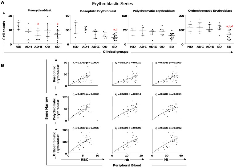
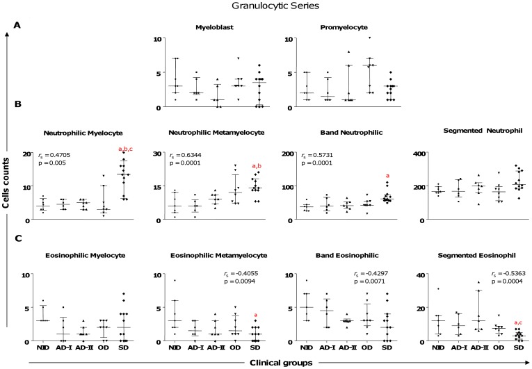
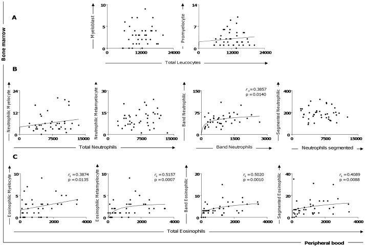
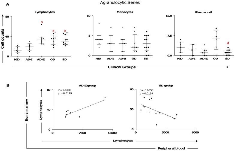
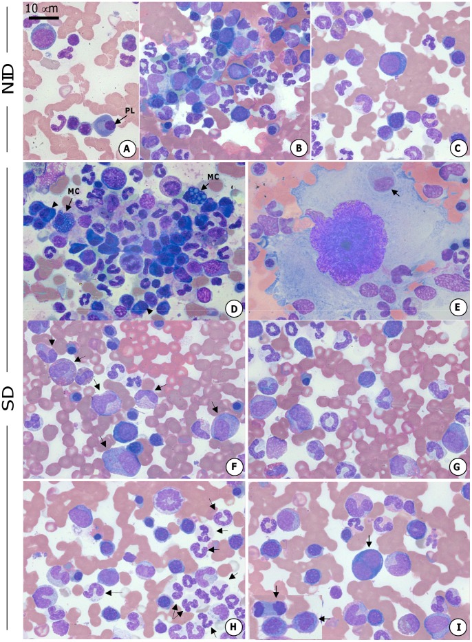
Hematological analysis has limited applications for disease diagnosis in Leishmania infantum-infected dogs, but it can be very important in evaluating the clinical forms of the disease and in understanding the evolution of canine visceral leishmaniasis (CVL) pathogenesis. Recently, we demonstrated that alterations in leucopoiesis and erythropoiesis are related to clinical status and bone marrow parasite density in dogs naturally infected by L. infantum. To further characterize these alterations, we evaluated the association between the hematological parameters in bone marrow and peripheral blood alterations in groups of L. infantum-infected dogs: asymptomatic I (AD-I: serum negative/PCR+), asymptomatic II (AD-II: serum positive), oligosymptomatic (OD), and symptomatic (SD). Results were compared with those from noninfected dogs (NID). The SD group was found to present a decrease in erythropoietic lineage with concomitant reductions in erythrocytes, hemoglobin, and hematocrit parameters, resulting in anemia. The SD group also had increased neutrophils and precursors and decreased band eosinophils and eosinophils, leading to peripheral blood leucopenia. In the AD-II group, lymphocytosis occurred in both the peripheral blood and the bone marrow compartments. The SD group exhibited lymphocytosis in the bone marrow, with lymphopenia in the peripheral blood. In contrast, the AD-I group, showed no significant changes suggestive of CVL, presenting normal counts in bone marrow and peripheral blood. Our results showed for the first time that important changes in hematopoiesis and hematological parameters occur during ongoing CVL in naturally infected dogs, mainly in symptomatic disease. Taken together, our results based on myelogram and hemogram parameters enable better understanding of the pathogenesis of the anemia, lymphocytosis, and lymphopenia, as well as the leucopenia (eosinopenia and monocytopenia), that contribute to CVL prognosis.
Conflict of interest statement
Figures
Similar articles
-
Association between mast cells, tissue remodelation and parasite burden in the skin of dogs with visceral leishmaniasis.Vet Parasitol. 2017 Aug 30;243:260-266. doi: 10.1016/j.vetpar.2017.05.028. Epub 2017 Jul 9. Vet Parasitol. 2017. PMID: 28807304
-
Isotype patterns of immunoglobulins: hallmarks for clinical status and tissue parasite density in Brazilian dogs naturally infected by Leishmania (Leishmania) chagasi.Vet Immunol Immunopathol. 2006 Aug 15;112(3-4):102-16. doi: 10.1016/j.vetimm.2006.02.001. Epub 2006 Apr 18. Vet Immunol Immunopathol. 2006. PMID: 16621021
-
Systemic and compartmentalized immune response in canine visceral leishmaniasis.Vet Immunol Immunopathol. 2009 Mar 15;128(1-3):87-95. doi: 10.1016/j.vetimm.2008.10.307. Epub 2008 Oct 17. Vet Immunol Immunopathol. 2009. PMID: 19054576 Review.
-
Phenotypic features of circulating leucocytes as immunological markers for clinical status and bone marrow parasite density in dogs naturally infected by Leishmania chagasi.Clin Exp Immunol. 2006 Nov;146(2):303-11. doi: 10.1111/j.1365-2249.2006.03206.x. Clin Exp Immunol. 2006. PMID: 17034583 Free PMC article.
-
Elucidating the role played by bone marrow in visceral leishmaniasis.Front Cell Infect Microbiol. 2023 Oct 4;13:1261074. doi: 10.3389/fcimb.2023.1261074. eCollection 2023. Front Cell Infect Microbiol. 2023. PMID: 37860064 Free PMC article. Review.
Cited by
-
Clinical, hematological and biochemical alterations in hamster (Mesocricetus auratus) experimentally infected with Leishmania infantum through different routes of inoculation.Parasit Vectors. 2016 Mar 31;9:181. doi: 10.1186/s13071-016-1464-y. Parasit Vectors. 2016. PMID: 27030128 Free PMC article.
-
Canine Leishmaniasis in an Endemic Area for Human Leishmaniasis in Nicaragua.J Trop Med. 2022 Aug 29;2022:5774296. doi: 10.1155/2022/5774296. eCollection 2022. J Trop Med. 2022. PMID: 36072825 Free PMC article.
-
Circulating Biomarkers of Immune Activation, Oxidative Stress and Inflammation Characterize Severe Canine Visceral Leishmaniasis.Sci Rep. 2016 Sep 6;6:32619. doi: 10.1038/srep32619. Sci Rep. 2016. PMID: 27595802 Free PMC article.
-
Characterization and Proteomic Analysis of Plasma EVs Recovered from Healthy and Diseased Dogs with Canine Leishmaniosis.Int J Mol Sci. 2023 Mar 13;24(6):5490. doi: 10.3390/ijms24065490. Int J Mol Sci. 2023. PMID: 36982564 Free PMC article.
-
Prognostic Factors and Life Expectancy in Canine Leishmaniosis.Vet Sci. 2020 Sep 4;7(3):128. doi: 10.3390/vetsci7030128. Vet Sci. 2020. PMID: 32899831 Free PMC article.
References
-
- Nunes CM, Pires MM, da Silva KM, Assis FD, Goncalves Filho J, et al. (2010) Relationship between dog culling and incidence of human visceral leishmaniasis in an endemic area. Vet Parasitol 170: 131–133. - PubMed
-
- Giunchetti RC, Mayrink W, Genaro O, Carneiro CM, Correa-Oliveira R, et al. (2006) Relationship between canine visceral leishmaniosis and the Leishmania (Leishmania) chagasi burden in dermal inflammatory foci. J Comp Pathol 135: 100–107. - PubMed
-
- Coura-Vital W, Reis AB, Reis LE, Braga SL, Roatt BM, et al... (2013) Canine visceral leishmaniasis: Incidence and risk factors for infection in a cohort study in Brazil. Vet Parasitol In press. - PubMed
Publication types
MeSH terms
LinkOut - more resources
Full Text Sources
Other Literature Sources
