. 2013 Dec 23;8(12):e83247.
doi: 10.1371/journal.pone.0083247. eCollection 2013.

In vitro activity of the antifungal azoles itraconazole and posaconazole against Leishmania amazonensis

Affiliations

In vitro activity of the antifungal azoles itraconazole and posaconazole against Leishmania amazonensis

Sara Teixeira de Macedo-Silva et al. PLoS One. .

Abstract

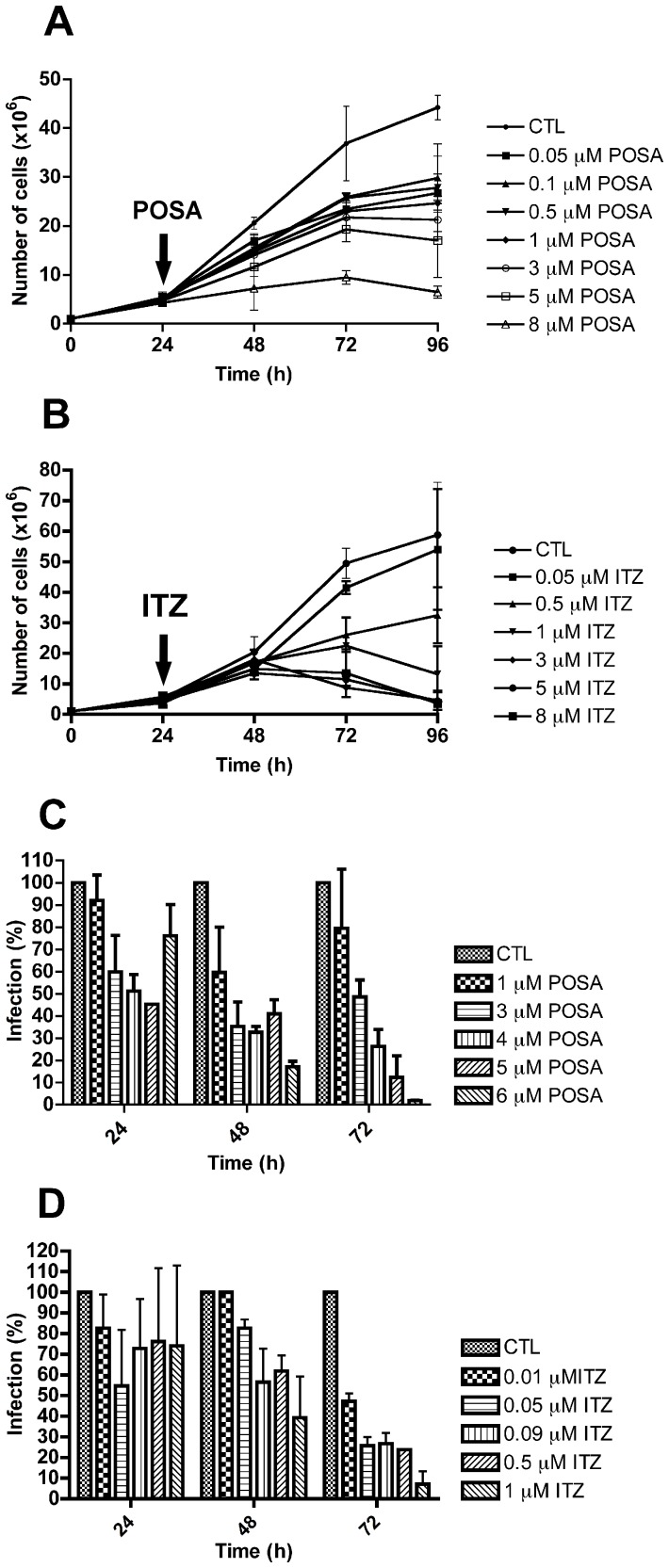
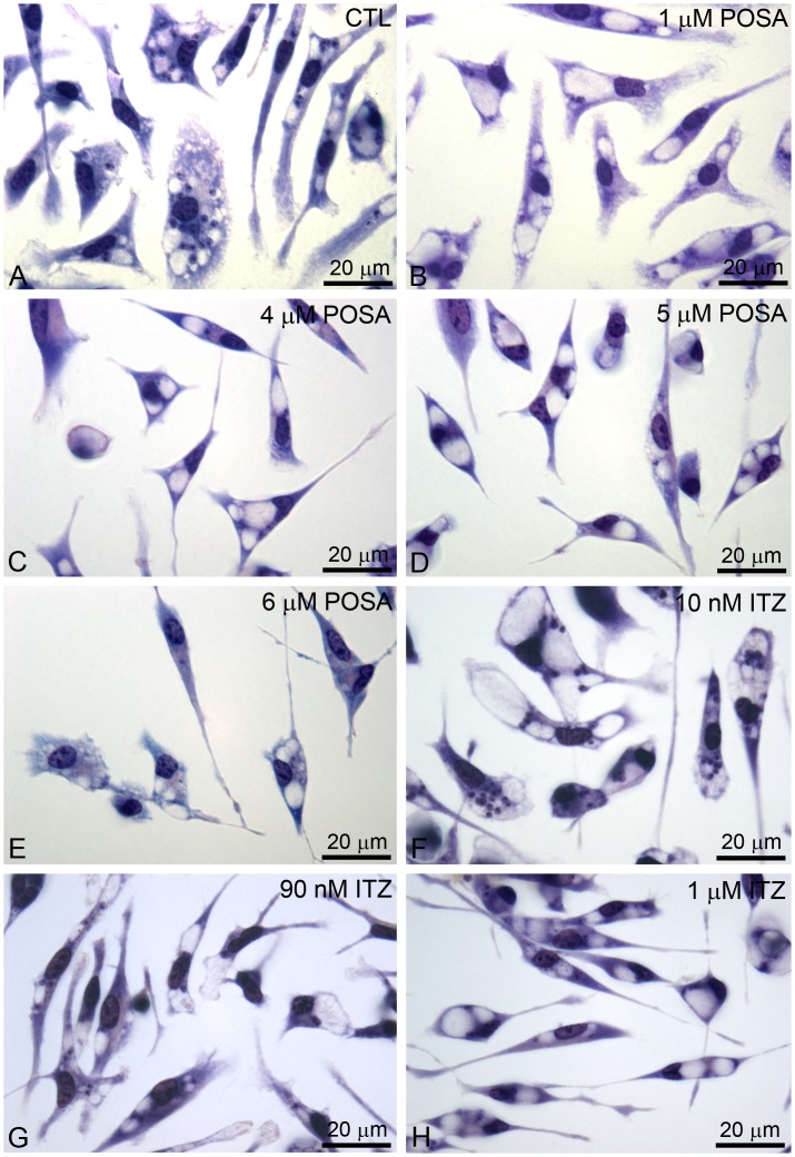
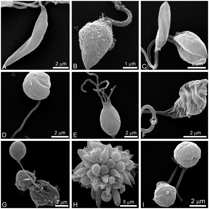
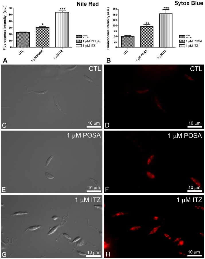
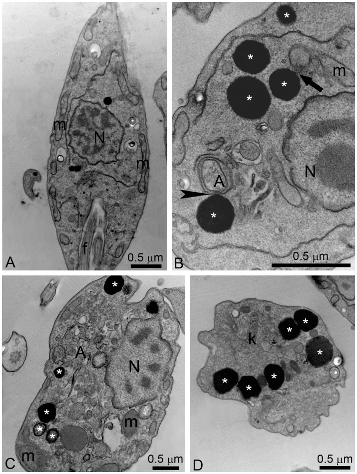
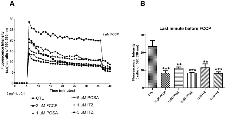
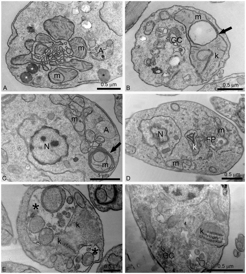
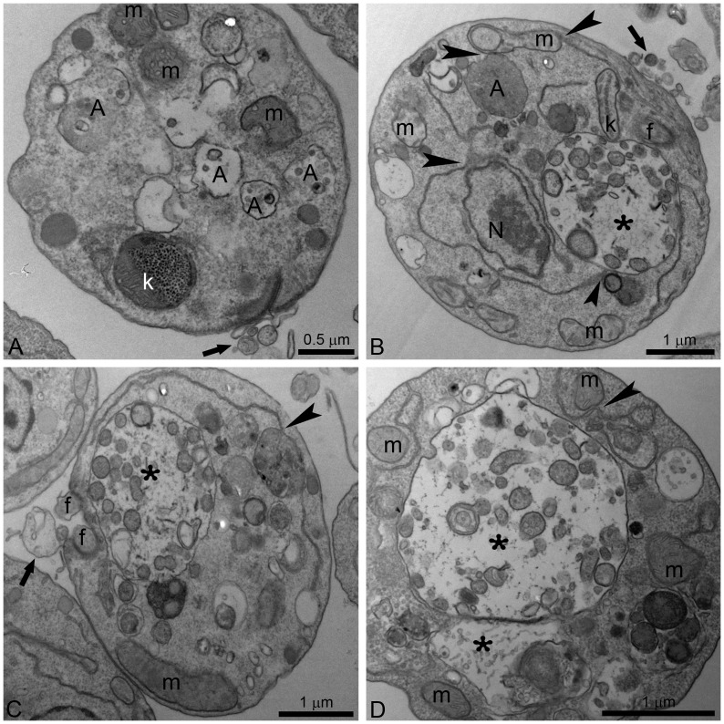
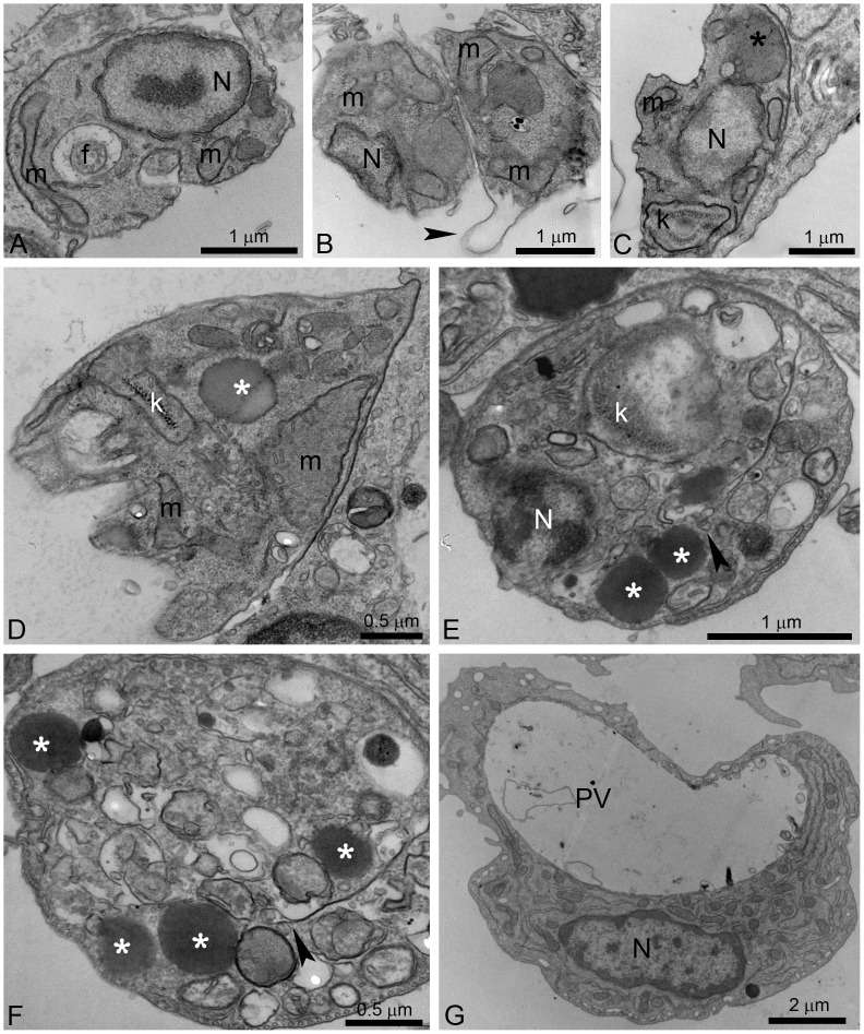
Leishmaniasis, caused by protozoan parasites of the Leishmania genus, is one of the most prevalent neglected tropical diseases. It is endemic in 98 countries, causing considerable morbidity and mortality. Pentavalent antimonials are the first line of treatment for leishmaniasis except in India. In resistant cases, miltefosine, amphotericin B and pentamidine are used. These treatments are unsatisfactory due to toxicity, limited efficacy, high cost and difficult administration. Thus, there is an urgent need to develop drugs that are efficacious, safe, and more accessible to patients. Trypanosomatids, including Leishmania spp. and Trypanosoma cruzi, have an essential requirement for ergosterol and other 24-alkyl sterols, which are absent in mammalian cells. Inhibition of ergosterol biosynthesis is increasingly recognized as a promising target for the development of new chemotherapeutic agents. The aim of this work was to investigate the antiproliferative, physiological and ultrastructural effects against Leishmania amazonensis of itraconazole (ITZ) and posaconazole (POSA), two azole antifungal agents that inhibit sterol C14α-demethylase (CYP51). Antiproliferative studies demonstrated potent activity of POSA and ITZ: for promastigotes, the IC50 values were 2.74 µM and 0.44 µM for POSA and ITZ, respectively, and for intracellular amastigotes, the corresponding values were 1.63 µM and 0.08 µM, for both stages after 72 h of treatment. Physiological studies revealed that both inhibitors induced a collapse of the mitochondrial membrane potential (ΔΨm), which was consistent with ultrastructural alterations in the mitochondrion. Intense mitochondrial swelling, disorganization and rupture of mitochondrial membranes were observed by transmission electron microscopy. In addition, accumulation of lipid bodies, appearance of autophagosome-like structures and alterations in the kinetoplast were also observed. In conclusion, our results indicate that ITZ and POSA are potent inhibitors of L. amazonensis and suggest that these drugs could represent novel therapies for the treatment of leishmaniasis, either alone or in combination with other agents.


Conflict of interest statement

Competing Interests: The authors have declared that no competing interests exist.

Figures

Figure 1
Figure 1. Antiproliferative effects of posaconazole and itraconazole on Leishmania amazonensis. L. amazonensis promastigotes (A, B) and intracellular amastigotes (C, D) were treated with posaconazole (POSA) (A, C) or itraconazole (ITZ) (B, D) to evaluate the parasite growth.
The arrows indicate the time of the addition of the drugs at the indicated concentrations. The results were plotted as the mean of three independent experiments and the bars represent the standard deviation.
Figure 2
Figure 2. Light microscopy of murine macrophages infected with L. amazonensis amastigotes.
(A) Control culture with many amastigotes inside parasitophorous vacuoles. (B–H) After 72 h of treatment with different concentrations of POSA and ITZ, a significant reduction in the number of parasites and the presence of several empty parasitophorous vacuoles was observed.
Figure 3
Figure 3. Scanning electron microscopy (SEM) of L. amazonensis promastigotes.
Control parasites (A) and promastigotes that were treated with different concentrations of POSA and ITZ for 48 h (B–I) were observed by SEM. (B, C) 1 µM ITZ; (D–F) 1 µM POSA; (G) 5 µM ITZ; (H, I) 5 µM POSA. The images show dramatic alterations in promastigote shape (B–I), a promastigote with four flagella (E), and profound changes in the plasma membrane (B, C, F).
Figure 4
Figure 4. Analysis of lipid body accumulation and plasma membrane integrity in L. amazonensis promastigotes.
(A–B) Quantitative fluorimetric analysis using Nile Red (A) and Sytox Blue (B). Fluorescence intensity is expressed as arbitrary units (A.U.). The results were plotted as mean of three independent experiments and the bars represent the standard deviation. *p<0.01; **p<0.05; ***p<0.0001. (C–H) Differential interference contrast (DIC) microscopy (C, E, G) and fluorescence microscopy using Nile Red (D, F, H) of control L. amazonensis promastigotes and promastigotes treated with 1 µM POSA or ITZ for 48 h. The images demonstrate an accumulation of lipid bodies that are randomly distributed throughout the cytoplasm, confirming the increase in the fluorescence intensity observed in Fig. 4A.
Figure 5
Figure 5. Ultrathin sections of control L. amazonensis promastigotes (A) and promastigotes treated with ITZ (B–D).
(A) Control promastigotes; (B–C) 5 µM ITZ; (D) 1 µM ITZ. The images show the presence of several electron-dense lipid bodies (asterisks), which sometimes appear near the plasma membrane, the endoplasmic reticulum and mitochondrion profiles (arrow), and autophagosomes (arrowhead). A, autophagosome; f, flagellum; k, kinetoplast; m, mitochondrion; N, nucleus.
Figure 6
Figure 6. Evaluation of mitochondrial transmembrane electric potential (ΔΨm) in L. amazonensis promastigotes using the JC-1 fluorochrome.
(A) Values of ΔΨm were evaluated over 36 min, before the addition of 2 µM FCCP to abolish the mitochondrial potential. Two concentrations of POSA and ITZ were used (1 and 5 µM) for 48 h of treatment. The ΔΨm values are expressed as the ratio of the reading at 590 nm (aggregate) to the reading at 530 nm (monomer). (B) Analysis of ΔΨm at the last minute before the addition of 2 µM FCCP. The data suggest that similar alterations in ΔΨm are induced by POSA, ITZ, and FCCP. The experiments were performed three times, each time in triplicate, and the figures shown are representative of these experiments. **p<0.05; ***p<0.0001.
Figure 7
Figure 7. Ultrathin sections of L. amazonensis promastigotes treated with different concentrations of ITZ and POSA.
(A, B) 1 µM ITZ; (C, D) 1 µM POSA; (E) 3 µM POSA for 48 h; (F) 5 µM POSA for 72 h. Several alterations were observed in the mitochondrion-kinetoplast complex such as: intense disorganization and swelling (A, B, D); alterations in the mitochondrion membranes and the appearance of circular cristae (B, C, arrows); changes in the structure of the kinetoplast (B, D, E, F); and the presence of autophagosomes (A, C, D). In Fig. 7E, two large vacuoles containing membranes and portions of the cytoplasm were observed (asterisks). FP, flagellar pocket; GC, Golgi complex; k: kinetoplast; m, mitochondrion; N: nucleus, A: autophagosome.
Figure 8
Figure 8. Ultrathin sections of L. amazonensis promastigotes.
Promastigotes were treated with 1 µM POSA (A, B), and 3 µM POSA (C, D) for 48 h. All images show the presence of small and large vacuoles containing several vesicles, membrane profiles and portions of the cytoplasm (asterisks). The endoplasmic reticulum appears in close association with the nucleus, the mitochondrion and autophagosomes (B–D, arrowheads). In Fig. 8A, changes in kinetoplast structure and vesiculation of the inner mitochondrial membrane were observed. N: nucleus; k: kinetoplast; m: mitochondrion; f: flagellum; A: autophagosome; FP; flagellar pocket.
Figure 9
Figure 9. Ultrathin sections of L. amazonensis intracellular amastigotes.
Control intracellular amastigotes (A) and treated amastigotes with ITZ and POSA (B–G) were observed. (B, C) 500 nM ITZ; (D) 1 µM ITZ; (E–G) 6 µM POSA. Different ultrastructural alterations were observed: mitochondrial swelling (B, D); detachment of the plasma membrane (B, arrowhead); presence of a large megasome (C, black asterisk), lipid bodies (D, E, G, white asterisks) and many vacuoles in the cytoplasm (D, E, G); changes in kinetoplast structure (C, E); and a cell with an empty parasitophorous vacuole (PV) (G). f, flagellum; m, mitochondrion; N: nucleus; PV: parasitophorous vacuole, A: autophagosome, k: kinetoplast.

Similar articles

Cited by

References

    1. Alvar J, Vélez ID, Bern C, Herrero M, Desjeux P, et al. (2012) Leishmaniasis worldwide and global estimates of its incidence. PloS One 7: e35671. - PMC - PubMed
    1. Herwaldt BL (1999) Leishmaniasis. Lancet 354: 1191–1199. - PubMed
    1. Grimaldi G Jr, McMahan-Pratt D (1991) Leishmaniasis and its etiologic agents in the New World: an overview. Prog Clin Parasitol 2: 73–118. - PubMed
    1. Liew FY, O’Donnell CA (1993) Immunology of leishmaniasis. Adv Parasitol 32: 161–259. - PubMed
    1. Barral A, Pedral-Sampaio D, Grimaldi Jr G, Momen H, McMahon-Pratt D, et al. (1991) Leishmaniasis in Bahia, Brazil: evidence that Leishmania amazonensis produces a wide spectrum of clinical disease. Am J Trop Med Hyg 44: 536–546. - PubMed

Publication types

MeSH terms

LinkOut - more resources