. 2013 Dec 23;8(12):e83519.
doi: 10.1371/journal.pone.0083519. eCollection 2013.

Induction of paclitaxel resistance by ERα mediated prohibitin mitochondrial-nuclear shuttling

Affiliations

Induction of paclitaxel resistance by ERα mediated prohibitin mitochondrial-nuclear shuttling

Pei Dong et al. PLoS One. .

Abstract

Paclitaxel is a drug within one of the most promising classes of anticancer agents. Unfortunately, clinical success of this drug has been limited by the insurgence of cellular resistance. To address this, Paclitaxel resistance was modeled in an in vitro system using estrogen treated prostate cancer cells. This study demonstrates that emerging resistance to clinically relevant doses of Paclitaxel is associated with 17-β-estradiol (E2) treatment in PC-3 cells, but not in LNCaP cells. We found that small interfering RNA mediated knockdown of ERα lead to a decrease in E2 induced Paclitaxel resistance in androgen-independent cells. We also showed that ERα mediated the effects of estrogen, thereby suppressing androgen-independent cell proliferation and mediating Paclitaxel resistance. Furthermore, E2 promoted Prohibitin (PHB) mitochondrial-nucleus translocation via directly mediation of ERα, leading to an inhibition of cellular proliferation by PHB. Additionally, restoration of Paclitaxel sensitivity by ERα knockdown could be overcome by PHB overexpression and, conversely, PHB knockdown decreased E2 induced Paclitaxel resistance. These findings demonstrate that PHB lies downstream of ERα and mediates estrogen-dependent Paclitaxel resistance signaling cascades.


Conflict of interest statement

Competing Interests: The authors have declared that no competing interests exist.

Figures

Figure 1
Figure 1. E2 inhibits Paclitaxel induced androgen-independent prostate cancer cell death.
(A-D) 100 nM of E2 was added to the media of (A and B) LNCaP and (C and D) PC3 cells for 96h, followed by addition of Paclitaxel at the indicated concentrations for 24h. The cells were stained with Hoechst 33258 (5 µg/ml) to visualize nuclei and propidium iodide (PI) (0.2 µg/ml) to detect membrane damage (B and D). Cell death was quantified by scoring the number of PI positive cells relative to the total number cell nuclei in the same visual field (A and C). The values represent the mean ± S.E. of at least three independent experiments. * denotes p<0.05, ** denotes p<0.01, and *** denotes p<0.001.
Figure 2
Figure 2. ERα mediates the estrogen induced Paclitaxel resistance of PC3 cells.
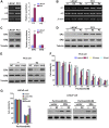
(A-D) LNCaP and PC3 cells were treated with or without E2 (100 nM) for 96h, Paclitaxel (Pa 50 nM) for 24h. (A and B) Representative and quantification (A right) of mRNA expression levels of erα and erβ. Total mRNA was extracted and RT-PCR was performed with primers specific to erα, erβ and β-actin. (C and D) Representative and quantification (C right) of protein expression levels of ERα and ERβ. Total protein was extracted and analyzed by Western blot with antibodies specific to ERα, ERβ and Tubulin. (E) Efficacy and specificity of sierα and sierβ knockdown. PC3 cells were treated with (right) or without (left) 100 nM of E2 for 96h, then transfected with the indicated siRNAs or negative control siRNA (NC). After 24h, expression of ERα or ERβ was monitored using Western blotting. (F) PC3 cells were treated with 100nM of E2 for 96h, then transfected with the indicated siRNAs or NC siRNA respectively. Twenty four hours post-transfection, Paclitaxel was added to the media at the indicated concentrations for 24h and the level of cell death was quantified as in Figure 1. (G) LNCaP cells were treated with 100 nM of E2 for 96h, then transfected with vector or ERα expression plasmids. Twenty four hours post-transfection, 50 nM of Paclitaxel was added to the media for 24h and the level of cell death was quantified (left) as in Figure 1 and then the expression of ERα was monitored using Western blotting (right). The values represent the mean ± S.E. of at least three independent experiments. * denotes p<0.05, ** denotes p<0.01, and *** denotes p<0.001.
Figure 3
Figure 3. Estrogen activates ERα, suppressing PC3 cell proliferation and mediating its Paclitaxel resistance.
E2 was added to the media of (A) LNCaP cells or (B) PC3 cells at the indicated concentrations for 96h, and cell proliferation was quantified. (C) PC3 cells were transfected with the indicated siRNAs or NC siRNA. 24h after transfection, 100 nM E2 was added to the media for 96h as indicated, and cell proliferation was quantified. The values represent the mean ± S.E. of at least three independent experiments. * denotes p<0.05; ** denotes p<0.01; *** denotes p<0.001.
Figure 4
Figure 4. E2 promotes PHB mitochondrial-nuclear translocation, thus inhibiting cell proliferation.
(A) E2 was added to the media of LNCaP or PC3 cells at the indicated concentrations for 96h. Total cell protein was extracted and analyzed by Western blot using antibodies specific to PHB and Tubulin. (B) Efficacy and specificity of PHB siRNA is shown. PC3 cells were transfected with PHB siRNA or NC RNA. Twenty four hours post-transfection, 100 nM of E2 was added to the media for 96h and the expression of PHB was analyzed by Western blot. (C) PC3 cells were treated as in B, and the levels of cell proliferation were quantified. (D) 100 nM of E2 was added to the media for 96h, followed by transfection of PC3 cells with PHB siRNA or NC RNA. Twenty four hours post-transfection, Paclitaxel was added to the media at the indicated concentrations for 24h, and the level of cell death was quantified as described in Figure 1. (E) 100 nM of E2 was added to the media for 96h, and then LNCaP cells were transfected with either vector or PHB expression plasmids. Twenty four hours post-transfection, 50 nM of Paclitaxel was added to the media for 24h and the level of cell death was quantified as described in Figure 1. (F) E2 promoted PHB mitochondrial-nucleus translocation. 100 nM of E2 was added to the media for 96h, then PC3 or LNCaP cell mitochondria (M) and nuclei (N) were separated and analyzed by Western blot using PHB, Histone H1 (nucleus marker), VDAC (mitochondrial marker) and Tubulin (cytoplasm marker) antibodies. T (total cell lysates). Results are representative of three independent experiments. The values represent the mean ± S.E. of at least three independent experiments. * denotes p<0.05; ** denotes p<0.01; *** denotes p<0.001.
Figure 5
Figure 5. ERα directly mediated PHB mitochondrial-nuclear shuttling.
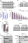
(A and B) ERα mediated PHB mitochondrial-nucleus translocation. PC3 cells were transfected with siRNAs specific to (A) erα or (B) erβ, or NC RNA. Twenty four hours post-transfection, 100 nM of E2 was added to the media for 96h. The PC3 cell mitochondria (M) and nuclei (N) were separated and analyzed by Western blot using PHB, ERα Histone H1 (nucleus marker), VDAC (mitochondrial marker) and Tubulin (cytoplasm marker) antibodies. T (total cell lysates). (C) ERα directly associates with PHB. 100 nM of E2 was added to the media for 96h, then PC3 cell lysates were immunoprecipitated (IP) using PHB antibody and analyzed by Western blot (WB) using the indicated antibodies (left panel). PC3 lysates were immunoprecipitated with ERα antibody, and PHB and ERα levels were analyzed by Western blot (middle panel). Equal amounts of total input PHB and ERα (Input) were used for immunoprecipitations for each condition (right). (D) ERα directly associates with PHB in mitochondria. PC3 cells were treated as in C, then the PC3 cell mitochondria were separated and immunoprecipitated as in C. (E) ERα directly associates with PHB in nucleus. PC3 cells were treated as in C, then PC3 cell nuclei were separated and immunoprecipitated as in C. Results are representative of three independent experiments.
Figure 6
Figure 6. PHB acts downstream of ERα to mediate resistance to Paclitaxel.
100α or PHB expression plasmids, or NC RNA. Twenty four hours post-transfection, Paclitaxel was added to the media at the indicated concentrations for 24h, and the level of cell death was quantified as described in Figure 1. The values represent the mean ± S.E. of at least three independent experiments. * denotes p<0.05; ** denotes p<0.01; *** denotes p<0.001.

Similar articles

Cited by

References

    1. Seruga B, Tannock IF (2011) Chemotherapy-based treatment for castration-resistant prostate cancer. J Clin Oncol 29: 3686–3694. - PubMed
    1. Rodriguez-Antona C (2010) Pharmacogenomics of paclitaxel. Pharmacogenomics 11: 621–623. - PubMed
    1. Doyle-Lindrud S (2012) Managing side effects of the novel taxane cabazitaxel in castrate-resistant prostate cancer. Clin J Oncol Nurs 16: 286–291. - PubMed
    1. Devlin HL, Mudryj M (2009) Progression of prostate cancer: multiple pathways to androgen independence. Cancer Lett 274: 177–186. - PubMed
    1. Powell E, Shanle E, Brinkman A, Li J, Keles S, et al. (2012) Identification of estrogen receptor dimer selective ligands reveals growth-inhibitory effects on cells that co-express ERalpha and ERbeta. PLoS One 7: e30993. - PMC - PubMed

Publication types

MeSH terms

LinkOut - more resources