. 2013 Dec 23;8(12):e83607.
doi: 10.1371/journal.pone.0083607. eCollection 2013.

Next-generation sequencing of lung cancer EGFR exons 18-21 allows effective molecular diagnosis of small routine samples (cytology and biopsy)

Affiliations

Next-generation sequencing of lung cancer EGFR exons 18-21 allows effective molecular diagnosis of small routine samples (cytology and biopsy)

Dario de Biase et al. PLoS One. .

Abstract

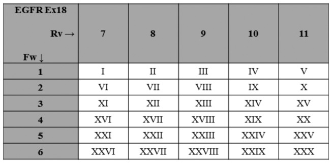
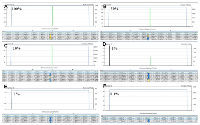
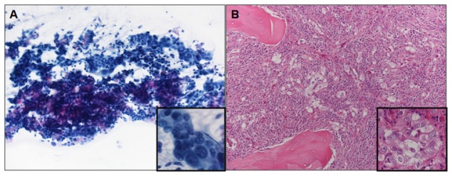
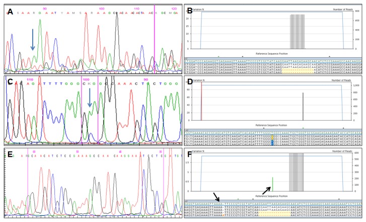
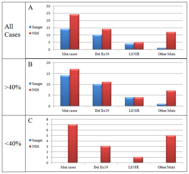
Selection of lung cancer patients for therapy with tyrosine kinase inhibitors directed at EGFR requires the identification of specific EGFR mutations. In most patients with advanced, inoperable lung carcinoma limited tumor samples often represent the only material available for both histologic typing and molecular analysis. We defined a next generation sequencing protocol targeted to EGFR exons 18-21 suitable for the routine diagnosis of such clinical samples. The protocol was validated in an unselected series of 80 small biopsies (n=14) and cytology (n=66) specimens representative of the material ordinarily submitted for diagnostic evaluation to three referral medical centers in Italy. Specimens were systematically evaluated for tumor cell number and proportion relative to non-neoplastic cells. They were analyzed in batches of 100-150 amplicons per run, reaching an analytical sensitivity of 1% and obtaining an adequate number of reads, to cover all exons on all samples analyzed. Next generation sequencing was compared with Sanger sequencing. The latter identified 15 EGFR mutations in 14/80 cases (17.5%) but did not detected mutations when the proportion of neoplastic cells was below 40%. Next generation sequencing identified 31 EGFR mutations in 24/80 cases (30.0%). Mutations were detected with a proportion of neoplastic cells as low as 5%. All mutations identified by the Sanger method were confirmed. In 6 cases next generation sequencing identified exon 19 deletions or the L858R mutation not seen after Sanger sequencing, allowing the patient to be treated with tyrosine kinase inhibitors. In one additional case the R831H mutation associated with treatment resistance was identified in an EGFR wild type tumor after Sanger sequencing. Next generation sequencing is robust, cost-effective and greatly improves the detection of EGFR mutations. Its use should be promoted for the clinical diagnosis of mutations in specimens with unfavorable tumor cell content.


Conflict of interest statement

Competing Interests: The authors have declared that no competing interests exist.

Figures

Figure 1
Figure 1. Multiple Identifier (MID) grid scheme.
To ensure that in a given 454 next generation sequencing run a specific target sequence is associated with a unique pair of MID we used grid schemes; the MID pairs for EGFR exon 18 are illustrated; roman numerals indicate individual patient samples. Ex, Exon; Fw, Forward; Rv, Reverse.
Figure 2
Figure 2. Analytical sensitivity and definition of threshold for mutational call.
A-F) the polymorphic c.2470 G>A substitution was observed with the following dilutions of DNA not carrying the polymorphism: 1:1 (A), 1:2 (B), 1:10 (C), 1:100 (D), but not at 1:1000 (F). At 1:100 dilution the c.2470 G>A substitution was observed when the total number of reads allowed to detect at least 10 reads with the c.2470 G>A change (this means at least 1,000 total reads) (D). The c.2470 G>A substitution was not observed when the total number of reads was not sufficient to detect at least 10 reads with the c.2470 G>A change (E).
Figure 3
Figure 3. Microscopic evaluation of tumor cellularity in non-small cell lung carcinoma samples.
A) cytology specimen from a 72 year old woman with adenocarcinoma metastatic to a mediastinal lymph node (May Grumwald Giemsa, 200X magnification, inset 600X); the proportion of neoplastic cells in the sample is 35%; DNA analysis was wild type after Sanger sequencing, but NGS showed two EGFR mutations (G721W, R831H) (case 57 of Table 5). B) biopsy specimen from a 65 year old man with adenocarcinoma metastatic to bone (vertebral body) (Hematoxylin and Eosin, 200X magnification, inset 600X); the proportion of neoplastic cells in the sample is 5%; DNA analysis was wild type after Sanger sequencing, but NGS showed the L858R EGFR mutation (case 80 of Table 5).
Figure 4
Figure 4. Results of sequencing analysis of EGFR gene after Sanger (A-C-E) and Next Generation (B-D-F) sequencing.
A-B) an exon 19 deletion detected by both Sanger (A) and NGS (B); the percentage of mutated alleles identified by NGS was >20%. C-D) the L858R detected by both Sanger (C) and NGS (D); the percentage of mutated alleles identified by NGS was >20%. E-F) an exon 19 deletion detected by NGS (F) but not by the Sanger method (E); the percentage of mutated alleles identified by NGS was <20%. Blue arrows in A and in C indicate the starting nucleotide of deletion and the mutated nucleotide, respectively; black arrows in F indicate homopolymer stretches (e.g. four adenines).
Figure 5
Figure 5. Schematic illustrating mutations detected by Sanger and Next Generation sequencing versus the proportion of neoplastic cells in the sample.
NGS, Next Generation Sequencing; Mut cases, cases with any mutation; Del Ex19, deletion in exon 19; Other muts, mutations other than exon 19 deletion or L858R.
Figure 6
Figure 6. Tumor cell number in cases with multiple vs.
single EGFR mutation (A) and in cases where NGS identified mutations not seen after Sanger sequencing (discrepant cases) (B). Numbers refer to the median number of neoplastic cells in the sample; bars indicate the smallest and highest number of neoplastic cells; the p-value was obtained with the student T test.

References

    1. Lynch TJ, Bell DW, Sordella R, Gurubhagavatula S, Okimoto RA et al. (2004) Activating mutations in the epidermal growth factor receptor underlying responsiveness of non-small-cell lung cancer to gefitinib. N Engl J Med 350: 2129-2139. doi:10.1056/NEJMoa040938. PubMed: 15118073. - DOI - PubMed
    1. Paez JG, Jänne PA, Lee JC, Tracy S, Greulich H et al. (2004) EGFR mutations in lung cancer: correlation with clinical response to gefitinib therapy. Science 304: 1497-1500. doi:10.1126/science.1099314. PubMed: 15118125. - DOI - PubMed
    1. Fukuoka M, Wu YL, Thongprasert S, Sunpaweravong P, Leong SS et al. (2011) Biomarker analyses and final overall survival results from a phase III, randomized, open-label, first-line study of gefitinib versus carboplatin/paclitaxel in clinically selected patients with advanced non-small-cell lung cancer in Asia (IPASS). J Clin Oncol 29: 2866-2874. doi:10.1200/JCO.2010.33.4235. PubMed: 21670455. - DOI - PubMed
    1. Mitsudomi T, Morita S, Yatabe Y, Negoro S, Okamoto I et al. (2010) Gefitinib versus cisplatin plus docetaxel in patients with non-small-cell lung cancer harbouring mutations of the epidermal growth factor receptor (WJTOG3405): an open label, randomised phase 3 trial. Lancet Oncol 11: 121-128. doi:10.1016/S1470-2045(09)70364-X. PubMed: 20022809. - DOI - PubMed
    1. Mok TS, Wu YL, Thongprasert S, Yang CH, Chu DT et al. (2009) Gefitinib or carboplatin-paclitaxel in pulmonary adenocarcinoma. N Engl J Med 361: 947-957. doi:10.1056/NEJMoa0810699. PubMed: 19692680. - DOI - PubMed

Publication types

MeSH terms